doi:10.1046/j.1432-1033.2002.03339.x
Eur. J. Biochem. 269, 6204–6211 (2002) (cid:2) FEBS 2002
Requirement of caspase and p38MAPK activation in zinc-induced apoptosis in human leukemia HL-60 cells
Masuo Kondoh1,2, Emi Tasaki2, Saeko Araragi2, Masufumi Takiguchi2, Minoru Higashimoto2, Yoshiteru Watanabe1 and Masao Sato2 1Department of Pharmaceutics and Biopharmaceutics, Showa Pharmaceutical University, Machida, Tokyo; 2Faculty of Pharmaceutical Sciences, Tokushima Bunri University, Tokushima, Japan
levels, and N,N,N¢,N¢-tetrakis (2-pyridylmethyl)ethylenedi- amine, a cell-permeable Zn chelator, inhibited DNA ladder formation induced by Py/Zn treatment (1 lM Py and 25 lM Zn). Py/Zn treatment activated the caspases, as assessed by the proteolysis of poly(ADP-ribose) polymerase (PARP), which is a substrate of caspase, and activated p38 mitogen- activated protein kinase (p38MAPK), which is a transducer of apoptotic stimuli to the apparatus of the apoptosis execu- tion. Z-Asp-CH2-DCB, a broad-spectrum inhibitor of caspase, attenuated proteolysis of PARP and DNA ladder formation by Py/Zn, indicating that apoptosis induced by Py/Zn is mediated by caspase activation. The p38MAPK- specific inhibitor SB203580 also inhibited induction of apoptosis by Py/Zn. Although SB203580 suppressed the proteolysis of PARP, Z-Asp-CH2-DCB did not inhibit the phosphorylation of p38MAPK, raising the possibility that apoptosis triggered by Py/Zn might be mediated by the p38MAPK/caspase pathway.
Keywords: pyrithione; apoptosis; zinc; caspase; p38MAPK.
Zinc (Zn), an endogenous regulator of apoptosis, and has abilities both to induce apoptosis and inhibit the induction of apoptosis via the modulation of caspase activity. Due to the multifunctions of Zn, the intracellular Zn level is strictly regulated by a complex system in physiological and patho- logical conditions. The commitment of Zn to the regulation of apoptosis is not fully understood. In the present study, we investigated the role of intracellular Zn level in the induction of apoptosis in human leukemia cells (HL-60 cells) using a Zn ionophore [pyrithione (Py)]. Treatment of HL-60 cells with Zn for 6 h in the presence of Py (1 lM) exhibited cytotoxicity in a Zn dose-dependent manner (25–200 lM). Necrotic cells, assayed by trypan blue permeability, increased in number in a Zn dose-dependent fashion (50– 100 lM), but the appearance of apoptotic cells, assayed by formation of a DNA ladder and terminal deoxynucleo- tidyltransferase-mediated dUTP-biotin nick end-labeling method, peaked at 25 lM, suggesting the dependence of intracellular Zn level on the execution of apoptosis. In fact, treatment with Py resulted in increases in intracellular Zn
Despite differences in their morphology and most of their biochemical features, apoptosis and necrosis frequently coexist following insult [8–11].
There are two major mechanisms of cellular death: necrosis and apoptosis. Necrotic cell death is an unregulated process resulting from severe damage to the cell and is characterized by ATP depletion, cell swelling, lysis, and the release of intracellular contents resulting in tissue inflammation [1–3]. In contrast, apoptosis is a highly regulated, energy-depend- ent event that leads to the elimination of excess or damaged cells from tissues. There are numerous pathological and physiological stimuli of apoptotic cell death, including death factors, reactive oxygen species, and genotoxic agents [4–7].
Various cellular functions are influenced by essential trace-elements such as the divalent cations zinc (Zn). The physiological concentrations of these cations are strictly regulated, and the maintenance of discrete subcellular pools of Zn is critical for the functional and structural integrity of cells and contributes to a number of important biological processes, including not only gene expression, DNA synthesis and enzymatic catalysis but also regulation of apoptosis. For example, Zn is present in presynaptic vesicles of central excitatory neurons and is released by synaptic activity or membrane depolarization [12–17]. Exposure to excessive Zn is neurotoxic to cortical neurons, and this toxicity has been shown to be mediated by Zn influx through glutamate receptor- or voltage-gated Ca2+ channels [18–20]. Zn also regulates apoptosis in thymo- cytes. A high concentration of zinc (0.5–5 mM) inhibited glucocorticoid-induced apoptosis in mouse thymocytes [21], but Zn itself induced apoptosis at a low concentra- tion (80–200 lM) in mouse thymocytes [22]. Schrantz et al. [23] reported that Zn (at concentration of 10 lM to 50 lM) inhibited manganese-induced apoptosis, dependent on the inhibition of caspase-3 activation, but that higher concentrations of Zn (50–100 lM) did not prevent
Correspondence to M. Sato, Faculty of Pharmaceutical Sciences, Tokushima Bunri University, Yamashiro-cho 180, Tokushima 770-8514, Japan. Fax: +81 88 6553051, Tel.: +81 88 6229611 ext. 5611, E-mail: msato@ph.bunri-u.ac.jp Abbreviations: ERK, extracellular signal-regulated kinase; ICP-MS, inductively coupled plasma mass spectrometry; JNK, c-Jun NH2- terminal kinases; MAPK, mitogen-activated protein kinases; MTT, 3-(4,5-dimethyl-2-thiazolyl)-2,5-diphenyl-2H-tetrazolium bromide; PARP, poly(ADP-ribose) polymerase; Py, pyrithione; TPEN, N,N,N¢,N¢-tetrakis-(2-pyridylmethyl)ethylenediamine; TUNEL, terminal deoxynucleotidyltransferase-mediated dUTP-biotin nick end-labeling. (Received 9 August 2002, revised 20 September 2002, accepted 30 October 2002)
Zn and apoptosis (Eur. J. Biochem. 269) 6205
(cid:2) FEBS 2002
manganese-mediated apoptosis but rather increased cell death in human Burkitt lymphoma B cells.
MA, USA). An antibody for poly(ADP-ribose) poly- merase (PARP) was purchased from BD-PharMingen. Anti-p38MAPK and a phosphorylated form of p38MAPK antibodies were purchased from New England BioLabs (Beverly, MA, USA).
Cell culture
HL-60 cells, a human leukemia cell lines, were cultured in RPMI 1640 containing 10% fetal bovine serum in a 5% CO2 atmosphere.
Cytotoxicity of Py/Zn in HL-60 cells
extracellular
stress-activated
protein
The cytotoxicity of Py/Zn in HL-60 cells was analyzed by colorimetric 3-(4,5-dimethyl-2-thiazolyl)-2,5-diphenyl- 2H-tetrazolium bromide (MTT) assay with some modifica- tion [43]. Briefly, after the addition of MTT (0.5 mgÆmL)1), cells were incubated at 37 (cid:3)C for 4 h. SDS (10% w/v) in 0.05 M HCl was added to the wells and then incubated at room temperature overnight under dark conditions. The absorbance was measured at 595 nm.
Assessment of apoptosis and necrosis in Py/Zn-treated cells
Caspases, a conserved family of cysteine proteases, play a central role in apoptosis [24]. Caspases themselves are present as proenzymes that are readily cleaved and activated during apoptosis, providing the cell with a means to rapidly amplify its apoptotic response [25,26]. Recently, there has been growing attention to the mech- anisms of transduction of apoptotic stimuli to the caspase. One of the most relevant aspects in the regulation of apoptosis is the involvement of mitogen-activated protein kinases (MAPKs), a family of serine/threonine kinases that mediate intracellular signal transduction in response to various stimuli [27]. To date, three major MAPKs have signal-regulated kinases been identified: [c-Jun kinases (ERK1/2), NH2-terminal kinases (JNK)], and p38 mitogen-activated protein kinases (p38MAPK). ERK1/2 are activated mainly by growth factors and are critically involved in the regulation of mitogenesis [28,29]. On the other hand, JNK and p38MAPK are activated mainly by cytotoxic insult and are often associated with apoptosis [30–36]. Moreover, activation of p38MAPK was observed in Zn-treated cells [37–39]. Intracellular Zn exists as fixed pools of Zn or as more dynamic and labile Zn pools, which are thought to be associated with the regulation of apoptosis by Zn [40]. However, induction of the molecular mechanism of apoptosis by Zn remains to be unknown.
Apoptotic cells were assessed by the appearance of a DNA ladder and by terminal deoxynucleotidyltransferase-medi- ated dUTP end labeling (TUNEL) analysis. DNA ladder formation was assayed as described previously [44]. Briefly, HL-60 cells (5 · 105 cellsÆwell)1 in a six-well plate) were treated with Zn in the presence or absence of Py at the indicated concentrations for various periods. The treated HL-60 cells were harvested and incubated in lysis buffer [10 mM Tris/HCl (pH 8.0), 10 mM EDTA, 0.5% w/v SDS, and 0.1% w/v RNase A] for 60 min at 50 (cid:3)C. Phenol/ chloroform-extracted DNA was subjected to a 1.8% agarose electrophoresis and stained with ethidium bromide. The TUNEL assay was performed using an apoptosis detection system according to the manufacturer’s protocol (Promega Co., Madison, WI, USA). The criteria used to determine necrosis was the loss of membrane integrity, which was determined by permeability to trypan blue in nonpermeabilized cells [45].
The present study was carried out to determine whether is dependent on the the commitment of apoptosis activation of caspase via activation of p38MAPK using a Zn ionophore in human leukemia HL-60 cells. An acute increase in the intracellular Zn level caused cytotoxicity in an intracellular Zn level-dependent manner. At a low Zn level, typical features of apoptosis such as DNA fragmen- tation were observed. At a higher concentration, the feature of cell death was necrosis. Moreover, the induction of apoptosis was accompanied by activation of caspase and p38MAPK. Both a broad-spectrum inhibitor of caspase and an inhibitor of p38MAPK attenuated the induction of apoptosis by Zn. Moreover, although the p38MAPK inhibitor also inhibited the caspase activation, the caspase inhibitor did not attenuate the activation of p38MAPK. Based on the results, it was concluded that induction of apoptosis by intracellular labile Zn is mediated via the p38MAPK/caspase pathway.
Measurement of intracellular trace metals
M A T E R I A L S A N D M E T H O D S
Materials
The treated HL-60 cells were washed once with phosphate- buffered saline (NaCl/Pi) and twice with NaCl/Pi containing 10 mM EDTA. The washed cells was incubated with HNO3 for 16 h at room temperature and for an additional 2 h at 60 (cid:3)C. Amounts of intracellular trace elements were then measured using an inductively coupled plasma-mass spec- trometer (ICP-MS) (HEWLETT PACKARD 4500) or an atomic absorption spectrophotometer (HITACHI Z-8200), and the amount of each elements expressed as the concen- tration of Zn per mg of the cellular protein. Protein assay was performed using a Bio-Rad protein assay kit.
(San Diego, CA, USA)
Western blot analysis
All reagents were of analytical grade. Zinc sulfate, sodium pyrithione (Py) and N,N,N¢,N¢-tetrakis-(2-pyridylmethyl)- ethylenediamine (TPEN) were purchased from Sigma (St Louis, MO, USA). Zinc sulfate and Py were dissolved in sterile water at 10 mM and 100 lM, respectively, and stored at )20 (cid:3)C before use. TPEN was dissolved in dimethyl sulfoxide. Z-Asp-CH2-DCB, a broad-spectrum inhibitor of caspases, was obtained from Calbiochem- Novabiochem Co. [41,42]. SB203580, an inhibitor of p38MAPK, was obtained from Calbiochem-Novabiochem. Poly(vinylidene difluoride) membranes were purchased from Millipore Co. (Bedford,
Vehicle- or Py/Zn-treated cells were lysed in lysis buffer consisting of 1% NonidetP-40, 20 mM Tris/HCl (pH 8.0),
6206 M. Kondoh et al. (Eur. J. Biochem. 269)
(cid:2) FEBS 2002
R E S U L T S
Determination of features of cell death induced by elevation of intracellular Zn levels
137 mM NaCl, 10% glycerol, 1 mM phenylmethylsulfonyl fluoride and 1 mM EDTA by sonication. Equal amounts of samples were subjected to 10% SDS/PAGE and then transferred to poly(vinylidene difluoride) membranes. The membranes were blocked with 10 mM Tris/HCl (pH 7.5), 100 mM NaCl, and 0.05% Tween-20 containing 5% (w/v) non fat milk for overnight at 4 (cid:3)C. Anti-PARP, p38MAPK or a phosphorylated form of p38MAPK was used as a primary antibody, and a horseradish peroxide-labeled antibody was used as a secondary antibody. The antibody-reactive bands were revealed by ECL-based detection (Amersham Phar- macia Biotech).
Statistical analysis
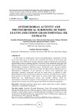
Although Zn is used as an inhibitor of cell death via caspase activation, direct evidence of Zn-mediated cell death has been obtained in a transient global ischemia [46]. There are complex systems for the regulation of intracellular zinc level such as the zinc transporters, Zn-1, -2, -3 or -4 [47–51]. Therefore, we used a zinc ionophore (Py) to investigate the cytotoxicity induced by elevation of intracellular Zn level. The addition of extracellular Zn (at concentrations of up to 200 lM) to HL-60 cells did not cause cytotoxicity. However, cytotoxicity of Zn was observed in the presence of Py (Fig. 1A). To determine the characteristics of cell death induced by Py/Zn treatment, we counted the number of necrotic and apoptotic cells by trypan blue staining and TUNEL methods, respectively. As shown in Fig. 1B,
Data were analyzed by analysis of variance, followed by Bonferroni multiple comparison test, or where applicable, by Student’s t-test. The acceptable level of significance was set at P < 0.05.
Fig. 1. Characterization of the features of cell death induced by treatment with Zn plus a Zn ionophore. (A) Cytotoxicity of Zn in the presence or absence of a Zn ionophore, pyrithione (Py). HL-60 cells (5 · 105 cellsÆmL)1) were treated with Zn at the indicated concentrations in the presence or absence of Py (1 lM) for 6 h. Then the viability was assessed by MTT assay. Data are means ± SD (n ¼ 4). *Significantly different from the Zn-treated cells without Py (P < 0.05). Data represent two independent experiments. (B) Induction of necrosis and apoptosis by Py/Zn treatment. After 6 h of treatment of Zn at the indicated concentration with or without Py, the number of necrotic and apoptotic cells were determined by trypan blue staining and TUNEL assay, respectively. Data are means ± SD (n ¼ 4). *Significantly different from the vehicle-treated cells (P < 0.05). ND, not determined. The results are representative of two independent experiments. (C) DNA fragmentation assay. After 6 h of treatment with Zn at the indicated concentration with or without Py, extracted DNA was subjected to electrophoresis on a 1.8% agarose and then stained with ethidium bromide. (D) Attenuation of Py/Zn-induced apoptosis by Zn chelator. After 2 h of treatment with TPEN (2 lM), cells were treated with 25 lM Zn plus 1 lM Py for 6 h, and then the appearance of DNA ladder was assayed. The results are representative of three independent experiments.
Zn and apoptosis (Eur. J. Biochem. 269) 6207
(cid:2) FEBS 2002
apoptotic cells appeared at 25 lM Zn in the presence of 1 lM Py, while necrotic cells appeared Zn concentrations of more than 50 lM in the presence of 1 lM Py. DNA ladder formation, a characteristic feature of apoptotic cells, was also observed at 25 lM Zn in the presence of 1 lM Py (Fig. 1C). Thus, the features of cell death changed depending on the intracellular Zn levels.
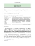
proteolysis of PARP (Fig. 3B) by Py/Zn. Induction of apoptosis and activation of caspases were observed at 6 h of treatment. Moreover, Z-Asp-CH2-DCB, a broad-spectrum inhibitor of caspases, attenuated the induction of apoptosis [41,42] (Fig. 3C) and also inhibited the proteolysis of PARP (Fig. 3D) induced by Py/Zn. These data suggest that Py/Zn-induced apoptosis is dependent on the activation of caspases.
Involvement of p38MAPK in apoptosis induced by Zn
Induction of apoptosis by the elevation of intracellular Zn levels at a low concentration but not a high concentration
Apoptotic signals are transferred to the apoptotic apparatus via various signal transduction pathways [53,54]. Recent studies have suggested that apoptotic stimuli are transferred to caspases via a MAPK cascade such as p38MAPK and JNK [59–62], and treatment with Zn has been shown to activate p38MAPK [37–39]. Therefore, we investigated the requirement of p38MAPK in the activation of caspases in Py/ Zn-induced apoptosis. Activation of p38MAPK was deter- mined by Western blot analysis using an antibody for the activated form of p38MAPK. As shown in Fig. 4A, activation of p38MAPK, determined by the phosphorylation of p38MAPK, occurred at 3 h prior to the caspase activation (6 h) and induction of apoptosis (6 h) (Figs 3A,B and 4A). Moreover, SB203580, a specific inhibitor of p38MAPK, inhibited the activation of p38MAPK and the induction of apoptosis (Fig. 4B,C). SB203580 also inhibited the activa- tion of caspases (Fig. 4D). Z-Asp-CH2-DCB did not inhibit the activation of p38MAPK (Fig. 4E). These data indicate that activation of p38MAPK is required for the caspases activation in Py/Zn-induced apoptosis.
D I S C U S S I O N
Next, we investigated the involvement of intracellular Zn levels in the induction of apoptosis by cotreatment of 1 lM Py and 25 lM Zn (Py/Zn treatment). Determination of trace elements in Py/Zn-treated cells by ICP-MS was performed. As described in Table 1, treatment of Py or Zn alone did not change intracellular Zn levels, but Py/Zn elevated intracel- lular Zn levels to a level about twofold greater than that of the vehicle-treated control. The intracellular Cu level was also increased by Py/Zn treatment. Although Py treatment alone, but not Zn treatment, enhanced intracellular Cu levels, Py did not induce apoptosis, indicating that the enhanced level of Cu is not sufficient to induce apoptosis (Fig. 1B,C). Therefore, elevation of the intracellular Zn levels was probably an essential event in the induction of apoptosis by Py/Zn. Indeed, TPEN, a specific Zn2+ chelator [52], abolished DNA ladder induced by Py/Zn treatment (Fig. 1D). To determine the relationship between intracellular Zn levels and induction of apoptosis, we investigate the appearance of a DNA ladder induced by Zn at concentrations in a narrow range in the presence of 1 lM Py. Although intracellular Zn levels increased with Zn concentration in a Zn dose-dependent manner, Zn-induced DNA ladder formation at concentrations of 20–30 lM with maximal induction at 25 lM (Fig. 2A,B). These findings indicated that the intracellular Zn level is an important factor in the determination of type of cell death, necrosis or apoptosis.
Involvement of caspase in apoptosis induced by Zn
Apoptosis, an endogenous suicide program, plays a central role in the maintenance of homeostasis, and there is an endogenous mechanism by which induction of apoptosis is controlled [53,54]. Several trace elements such as Zn, calcium and magnesium are known to be endogenous regulators of the induction of apoptosis. Calcium and magnesium are components of endonuclease, which plays a role in fragmentation of chromosomes into nucleosome fragments in apoptosis [63,64]. Interestingly, Zn functions as a inhibitor of apoptosis dependent on the inhibition of activation of caspases, and also as an inducer of apoptosis, dependent on the activation of caspases [23,65]. However, the mode of action of Zn in induction of apoptosis remains to be unclear. In this study, we investigated the molecular mechanism underlying the mode of action of the induc- tion of apoptosis by elevation of intracellular Zn levels.
There are various components of cell death machinery that induce apoptosis [53,54]. Caspases, a family of cysteine proteases, play a pivotal role in the execution of apoptosis [26]. We therefore examined the involvement of caspases in the apoptosis induced by Py/Zn. PARP is a substrate of caspases, and proteolysis of PARP is an index of activation of caspases [55–58]. Therefore, we first examined changes in the time courses of induction of apoptosis (Fig. 3A) and
Table 1. Changes in levels of trace elements by Py/Zn treatment. After 2 h of treatment of cells, the intracellular levels of trace elements were determined as described in Materials and methods. Data are means ± SD (n ¼ 4). Groups without a common superscript letter are significantly different at P < 0.05.
Treatment (lM) Trace elements (ng mg)1 protein)
Py Zn Mg Ca Mn Fe Cu Zn
0 1 0 1 0 0 25 25 1219.86 ± 30.66a 1194.45 ± 13.30a 1167.09 ± 13.16a 1376.34 ± 57.47b 42.86 ± 11.89a 25.44 ± 1.70b 23.42 ± 4.96b 20.82 ± 6.60b 2.32 ± 0.03a 1.81 ± 0.02b 1.06 ± 0.04c 1.02 ± 0.07c 20.59 ± 6.61a 14.49 ± 1.92a 11.27 ± 1.06b 13.68 ± 3.09a 13.42 ± 0.28a 26.68 ± 0.60b 11.99 ± 0.42a 31.66 ± 1.21c 99.46 ± 3.91a 95.03 ± 1.15a 90.48 ± 1.87a 200.24 ± 8.51b
6208 M. Kondoh et al. (Eur. J. Biochem. 269)
(cid:2) FEBS 2002
decreased
Fig. 3. Involvement of activation of caspases in the induction of apop- tosis by Py/Zn. (A,B) Time-course study. Cells were treated with 25 lM Zn plus 1 lM Py. DNA fragmentation (A) and proteolysis of PARP (B) were investigated at the indicated periods. (C,D) Effects of Z-Asp- CH2-DCB. Cells, after pretreatment with Z-Asp-CH2-DCB (100 lM) for 1 h, were incubated with 25 lM Zn plus 1 lM Py for 6 h. DNA fragmentation (C) and proteolysis of PARP (D) were investigated. The results are representative of three independent experiments.
211 ng ZnÆmg protein)1, above formation indicating that the ability of Zn to induce apoptosis is dependent on the intracellular Zn level. In fact, it has been shown that treatment with Zn (80–200 lM) induced apop- tosis in mouse thymocytes [22], and it was found in another study that Zn (0.5–5 mM) inhibited apoptosis in glucocor- ticoid-treated mouse thymocytes [21]. It is well known that Zn is an inhibitor of apoptosis mediated by inhibition of caspases at a millimolar concentration [65]. Indeed, although treatment with 25 lM Zn caused proteolysis of PARP in the presence of Py (1 lM), 50 lM Zn did not induce cleavage of PARP, indicating that a low level of intracellular Zn triggers activation of caspases (data not shown). Schrantz et al. [23] reported that caspase activation is required for Zn-induced apoptosis in human Burkitt lymphoma B cells. An increase in labile Zn in the cytoplasm may suppress apoptosis by inhibition of the action of caspase, cytoplasmic apoptosis inducer, via direct associ- ation of Zn with caspases [40]. Although we did not examine the localization of Zn introduced by Py, Zalewski et al. [66] reported that labile Zn introduced by Py was localized in the cytosol. Taken together, the results suggest that labile Zn released from Zn pools is a potent regulator of apoptosis via regulation of the activities of caspases.
Organisms are equipped with systems for intracellular Zn levels, and it has been shown that exogenous addition of Zn at a physiological concentration did not elevate intracellular Zn level [47–51]. Therefore, in this study, we used a Zn ionophore for the purpose of specific elevation of intracel- lular Zn levels. As the intracellular Zn level increased, the cytotoxicity of Zn in HL-60 cells was enhanced. However, the mode of cell death was dependent on the concentration of intracellular Zn. DNA ladder formation, a typical feature of apoptotic cells, was observed at 141 ng ZnÆmg protein)1 (basal level, 119 ng ZnÆmg protein)1), and DNA ladder
It has been reported that execution of caspase activation is preceded by intracellular signal transduction such as the activation of p38MAPK triggered by apoptotic stimuli [45,62]. Influx of Zn into the cytosol had already occurred at 2 h after the addition of Py/Zn in the present study (data not shown). Therefore, we turned our attention to the MAPK cascade as an apoptotic signal transduction machinery, which is involved in the early events of induc- tion of apoptosis [37,59–62,67]. Especially, p38MAPK was activated by Zn [37–39]. In this study, we found that
Fig. 2. Relationship between intracellular Zn levels and induction of apoptosis. Cells were incubated with Zn at the indicated concentration in the absence or presence of Py (1 lM). (A) Induction of apoptosis. After 6 h of treatment, induction of apoptosis was assessed by DNA ladder formation. The results are representative of three independent experiments. (B) Intracellular Zn level. Intracellular Zn levels were measured using an atomic absorption spectrophotometer after 2 h of treatment. Data are means ± SD (n ¼ 4). Groups without a common superscript letter are significantly different at P < 0.05. The results are representative of two independent experiments.
Zn and apoptosis (Eur. J. Biochem. 269) 6209
(cid:2) FEBS 2002
caspase, Zn might play a major role as an endogenous regulator of apoptosis in physiological conditions. More- over, activation of p38MAPK followed by caspase activation was found to be required for induction of apoptosis by Zn. This is the first study to show the involvement of a p38MAPK/caspase-dependent pathway in the induction of apoptosis by a low level of intracellular labile Zn.
A C K N O W L E D G M E N T S
This work was supported in part by a Grant-in-Aid for General Scientific Research from the Ministry of Education, Sciences, Sports and Culture of Japan.
R E F E R E N C E S
1. Eguchi, Y., Shimizu, S. & Tsujimoto, Y. (1997) Intracellular ATP levels determine cell death fate by apoptosis or necrosis. Cancer Res. 57, 1835–1840.
2. Leist, M., Single, B., Castoldi, A.F., Kuhnle, S. & Nicotera, P. (1997) Intracellular adenosine triphosphate (ATP) concentration: a switch in the decision between apoptosis and necrosis. J. Exp. Med. 185, 1481–1486.
3. Wyllie, A.H. (1980) Glucocorticoid-induced thymocyte apoptosis is associated with endogenous endonuclease activation. Nature 284, 555–556.
4. Liu, X., Kim, C.N., Yang, J., Jemmerson, R. & Wang, X. (1996) Induction of apoptosis program in cell-free extracts: requirement for dATP and cytochrome c. Cell 86, 147–157.
5. Nagata, S. (1997) Apoptosis by death factor. Cell 88, 355–365. 6. Radford, I.R. (1986) Evidence for a general relationship between the induced level of DNA double-strand breakage and cell-killing after X-irradiation of mammalian cells. Int. J. Radiat. Biol. Relat. Stud. Phys. Chem. Med. 49, 611–620. 7. Wood, K.A. & Youle, R.J. (1994) Apoptosis and free radicals. Ann. N.Y. Acad. Sci. 738, 400–407.
8. O’Brien, T., Babcock, G., Cornelius, J., Dingeldein, M., Talaska, G., Warshawsky, D. & Mitchell, K. (2000) A comparison of apoptosis and necrosis induced by hepatotoxins in HepG2 cells. Toxicol. Appl. Pharmacol. 164, 280–290.
9. Bonfoco, E., Krainc, D., Ankarcrona, M., Nicotera, P. & Lipton, S.A. (1995) Apoptosis and necrosis: two distinct events induced, respectively, by mild and intense insults with N-methyl-D-aspartate or nitric oxide/superoxide in cortical cell cultures. Proc. Natl Acad. Sci. USA 92, 7162–7166. Fig. 4. Involvement of activation of p38MAPK in the induction of apop- tosis by Py/Zn. (A) Time-course study of activation of p38MAPK. Cells were treated with 25 lM Zn plus 1 lM Py for the indicated periods. Activation of p38MAPK was assessed by the phosphorylation of p38MAPK by Western blotting analysis using an antibody for the activated p38MAPK. (B,C) Involvement of p38MAPK in the Py/Zn- treated cells. Cells, after pretreatment with SB203580 (5 lM) for 1 h, were incubated with 25 lM Zn plus 1 lM Py for 6 h. Activation of p38MAPK (B) and formation of DNA fragmentation (C) were inves- tigated. (D) Involvement of p38MAPK in the activation of caspases in Py/Zn-treated cells. Cells were treated with SB203580 for 1 h and then treated with 25 lM Zn plus 1 lM Py for 6 h. Activation of p38MAPK was then examined. (E) Involvement of caspases in the activation of p38MAPK in Py/Zn-treated cells. Cells were treated with Z-Asp-CH2- DCB for 1 h and then treated with 25 lM Zn plus 1 lM Py for 6 h. Proteolysis of PARP was then examined.
10. Dive, C., Gregory, C.D., Phipps, D.J., Evans, D.L., Milner, A.E. & Wyllie, A.H. (1992) Analysis and discrimination of necrosis and apoptosis (programmed cell death) by multiparameter flow cyto- metry. Biochim. Biophys. Acta 1133, 275–285.
(1994)
11. Huschtscha, L.I., Jeitner, T.M., Andersson, C.E., Bartier, W.A. & Tattersall, M.H. Identification of apoptotic and necrotic human leukemic cells by flow cytometry. Exp. Cell Res. 212, 161–165.
12. Danscher, G., Howell, G., Perez-Clausell, J. & Hertel, N. (1985) The dithizone, Timm’s sulphide silver and the selenium methods demonstrate a chelatable pool of zinc in CNS. A proton activation (PIXE) analysis of carbon tetrachloride extracts from rat brains and spinal cords intravitally treated with dithizone. Histochemistry 83, 419–422.
Py/Zn-induced apoptosis was mediated by activation of caspases followed by rapid activation of p38MAPK. Previous studies have demonstrated induction of apoptosis by dopamine or nitric oxide dependent on p38MAPK activation followed by activation of caspase-3 [61,62]. These findings support our results. The rapid phosphorylation of p38MAPK suggests that MAPK plays a role in the early stage of induction of Zn-induced apoptosis. The precise mechanism of activation of p38MAPK remains to be elucidated. One possible explanation is the involvement of production of reactive oxygen species induced by Zn. In fact, Kim et al. [68] showed that Zn-induced cytotoxicity was caused by the production of reactive oxygen species. p38MAPK was also reported to be activated by the production of reactive oxygen species in Cd-treated cells [45].
13. Frederickson, C.J., Manton, W.I., Frederickson, M.H., Howell, G.A. & Mallory, M.A. (1982) Stable-isotope dilution measure- ment of zinc and lead in rat hippocampus and spinal cord. Brain Res. 246, 338–341.
In summary, we demonstrated in this study that optimal intracellular Zn levels can be an initiator of apoptosis. Considering the ability of Zn to inhibit the activation of
14. Frederickson, C.J., Rampy, B.A., Reamy-Rampy, S. & Howell, G.A. (1992) Distribution of histochemically reactive zinc in the forebrain of the rat. J. Chem. Neuroanat. 5, 521–530.
6210 M. Kondoh et al. (Eur. J. Biochem. 269)
(cid:2) FEBS 2002
15. Smart, T.G., Xie, X. & Krishek, B.J. (1994) Modulation of inhibitory and excitatory amino acid receptor ion channels by zinc. Prog. Neurobiol. 42, 393–341. 16. Assaf, S.Y. & Chung, S.H. (1984) Release of endogenous Zn2+ 35. Wang, X., Martindale, J.L., Liu, Y. & Holbrook, N.J. (1998) The cellular response to oxidative stress: Influences of mitogen- activated protein kinase signalling pathways on cell survival. Biochem. J. 333, 291–300. from brain tissue during activity. Nature 308, 734–736.
36. Callsen, D. & Brune, B. (1999) Role of mitogen-activated protein kinases in S-nitrosoglutathione-induced macrophage apoptosis. Biochemistry 38, 2279–2286.
17. Charton, G., Rovira, C., Ben-Ari, Y. & Leviel, V. (1985) Spontaneous and evoked release of endogenous Zn2+ in the hip- pocampal mossy fiber zone of the rat in situ. Exp. Brain Res. 58, 202–205. 18. Choi, D.W., Yokoyama, M. & Koh, J.Y. (1988) Zinc neurotoxi- city in cortical cell culture. Neuroscience 24, 67–79. 37. McLaughlin, B., Pal, S., Tran, M.P., Parsons, A.A., Barone, F.C., Erhardt, J.A. & Aizenman, E. (2001) p38 activation is required upstream of potassium current enhancement and caspase cleavage in thiol oxidant-induced neuronal apoptosis. J. Neurosci. 21, 3303–3311.
19. Weiss, J.H., Hartley, D.M., Koh, J.Y. & Choi, D.W. (1993) AMPA receptor activation potentiates zinc neurotoxicity. Neuron 10, 43–49.
involvement 20. Koh, J.Y. & Choi, D.W. (1994) Zinc toxicity on cultured cortical receptors. of N-methyl-D-aspartate 38. Canesi, L., Betti, M., Ciacci, C. & Gallo, G. (2001) Insulin-like effect of zinc in mytilus digestive gland cells: modulation tyrosine kinase-mediated cell signaling. Gen. Comp. Endocrinol. 122, 60–66. neurons: Neuroscience 60, 1049–1057.
39. Samet, J.M., Graves, L.M., Quay, J., Dailey, L.A., Devlin, R.B., Ghio, A.J., Wu, W., Bromberg, P.A. & Reed, W. (1998) Activa- tion of MAPKs in human bronchial epithelial cells exposed to metals. Am. J. Physiol. 275, L551–L558. 21. Cohen, J.J. & Duke, R.C. (1984) Glucocorticoid activation of a calcium-dependent endonuclease in thymocyte nuclei leads to cell death. J. Immunol. 132, 38–42. 40. Truong-Tran, A.Q., Ruffin, R.E. & Zalewski, P.D.
22. Telford, W.G. & Fraker, P.J. (1995) Preferential induction od apoptosis in mouse CD4+CD8+ alpha beta TCRloCD3epsilonlo thymocytes by zinc. J. Cell Physiol. 164, 259–270. (2000) Visualization of labile zinc and its role in apoptosis of primary airway epithelial cells and cell lines. Am. J. Physiol. 279, L1172– L1183.
inhibitors of 23. Schrantz, N., Auffredou, M.T., Bourgeade, M.F., Besnault, L., Leca, G. & Vazquez, A. (2001) Zinc-mediated regulation of cas- pases activity: dose-dependent inhibition or activation of caspase-3 in the human Burkitt lymphoma B cells (Ramos). Cell Death Differ. 8, 152–161. 41. Dolle, R.E., Hoyer, D., Prasad, C.V., Schmidt, S.J., Helaszek, C.T., Miller, R.E. & Ator, M.A. (1994) P1 aspartate-based peptide alpha-(2,6-dichlorobenzoyl)oxy) methyl ketones as potent time- dependent interleukin-1beta-converting enzyme. J. Med. Chem. 37, 563–564.
42. Mashima, T., Naito, M., Kataoka, S., Kawai, H. & Tsuruo, T. (1995) Aspartate-based inhibitor of interleukin-1beta-converting enzyme prevents antitumor agent-induced apoptosis in human myeloid leukemia U937 cells. Biochem. Biophys. Res. Commun. 209, 907–915. 24. Nicholson, D.W., Ali, A., Thornberry, N.A., Vaillancourt, J.P., Ding, C.K., Gallant, M., Gareau, Y., Griffin, P.R., Labelle, M., Lazebnik, Y.A., Munday, N.A., Raju, S.M., Smulson, M.E. & Yamin, T.T., Yu, V.L. & Miller, D.K. (1995) Identification and inhibition of the ICE/CED-3 protease necessary for mammalian apoptosis. Nature 376, 37–43. 25. Cohen, G.M. (1997) Caspases: the executioners of apoptosis. Biochem. J. 326, 1–16. 26. Thornberry, N.A. & Lazebnik, Y. (1998) Caspases: enemies 43. Mosmann, T. (1983) Rapid colorimetric assay for cellular growth and survival: application to proliferation and cytotoxicity assays. J. Immunol. Methods 65, 55–63. within. Science 281, 1312–1316. 27. Tibbles, L.A. & Woodgett, J.R. (1999) The stress-activated protein 44. Watabe, M., Machida, K. & Osada, H. (2000) MT-21 is a syn- thetic apoptosis inducer that directly induces cytochrome c release from mitochondria. Cancer Res. 60, 5214–5222. kinase pathways. Cell Mol. Life Sci. 55, 1230–1254. 28. Seger, R. & Krebs, E.G. (1995) The MAPK signaling cascade. FASEB J. 9, 726–735.
45. Galan, A., Garcia-Bermejo, M.L., Troyano, A., Vilaboa, N.E., de Blas, E., Kazanietz, M.G. & Aller, P. (2000) Stimulation of p38 mitogen-activated protein kinase is an early regulatory event for the cadmium-induced apoptosis in human promonocytic cells. J. Biol. Chem. 275, 11418–11424. 29. Xia, Z., Dickens, M., Reingeaud, J., Davis, R.J. & Greenberg, M.E. (1995) Opposing effects of ERK and JNK-p38 MAP kinases on apoptosis. Science 270, 1326–1331.
30. Raingeaud, J., Gupta, S., Rogers, J.S., Dickens, M., Han, J., Ulevitch, R.J. & Davis, R.J. (1995) Pro-inflammatory cytokines and environmental stress cause p38 mitogen-activated protein kinase activation by dual phosphorylation on tyrosine and threonine. J. Biol. Chem. 270, 7420–7426. 46. Koh, J.Y., Suh, S.W., Gwag, B.J., He, Y.Y., Hsu, C.Y. & Choi, D.W. (1996) The role of zinc in selective neuronal death after transient global cerebral ischemia. Science 272, 1013–1016. 47. Palmiter, R.D. & Findley, S.D. (1995) Cloning and functional characterization of a mammlian zinc transporter that confers resistance to zinc. EMBO J. 14, 639–649.
48. Palmiter, R.D., Cole, T.B., Quaife, C.J. & Findley, S.D. (1996) ZnT-3, a putative transporter of zinc into synaptic vesicles. Proc. Natl Acad. Sci. USA 93, 14934–14939. 31. Chen, Y.R., Wang, X., Templeton, D., Davis, R.J. & Tan, T.H. (1996) The role of c-Jun N-terminal kinase (JNK) in apoptosis induced by ultraviolet C and gamma radiation. Duration of JNK activation may determine cell death and proliferation. J. Biol. Chem. 271, 31929–31936.
49. Palmiter, R.D., Cole, T.B. & Findley, S.D. (1996) ZnT-2, a mammalian protein that confers resistance to zinc by facilitating vesicular sequestration. EMBO J. 15, 1784–1791.
50. Huang, L. & Gitschier, J. (1997) A novel gene involved in zinc transport is deficient in the lethal milk mouse. Nat. Genet. 17, 292– 297. 51. McMahon, R.J. & Cousins, R.J. (1998) Mammalian zinc trans- porters. J. Nutr. 128, 667–670. 32. Juo, P., Kuo, C.J., Reynolds, S.E., Konz, R.F., Raingeaud, J., Davis, R.J., Biemann, H.P. & Blenis, J. (1997) Fas activation of the p38 mitogen-activated protein kinase signalling pathway requires ICE/CED-3 family proteases. Mol. Cell Biol. 17, 24–35. 33. Seimiya, H., Mashima, T., Toho, M. & Tsuruo, T. (1997) c-Jun NH2-terminal kinase-mediated activation of interleukin-1beta converting enzyme/CED-3-like protease during anticancer drug- induced apoptosis. J. Biol. Chem. 272, 4631–4636.
52. McCabe, M.J. Jr, Jiang, S.A. & Orrenius, S. (1993) Chelation of intracellular zinc triggers apoptosis in mature thymocytes. Laboratory Invest. 69, 101–110. 53. Hengartner, M.O. (2000) The biochemistry of apoptosis. Nature 407, 770–776. 34. Franklin, C.C., Srikanth, S. & Kraft, A.S. (1998) Conditional expression of mitogen-activated protein kinase phosphatase-1, MKP-1, is cytoprotective against UV-induced apoptosis. Proc. Natl Acad. Sci. USA 95, 3014–3019.
Zn and apoptosis (Eur. J. Biochem. 269) 6211
(cid:2) FEBS 2002
62. Junn, E. & Mouradian, M.M. (2001) Apoptotic signal 54. Ferri, K.F. & Kroemer, G. (2001) Organelle-specific initiation of cell death pathways. Nat. Cell Biol. 3, E255–E263.
in dopamine-induced cell death: the role of oxidative stress, p38 mitogen-activated protein kinase, cytochorome c and caspases. J. Neurochem. 78, 374–383.
55. Gu, Y., Sarnecki, C., Aldape, R.A., Livingston, D.J. & Su, M.S. (1995) Cleavage of poly (ADP-ribose) polymerase by interleukin- 1beta converting enzyme and its homologs TX and Nedd-2. J. Biol. Chem. 270, 18715–18718.
63. Kawabata, H., Anzai, N., Musutani, H., Hirama, T., Yoshida, Y. & Okuma, M. (1993) Detection of Mg(2+)-dependent endonuc- lease activity in myeloid leukemia cell nuclei capable of producing internucleosomal DNA cleavage. Biochem. Biophys. Res. Com- mun. 191, 247–254. 56. Lippke, J.A., Gu, Y., Sarnecki, C., Caron, P.R. & Su, M.S. (1996) Identification and characterization of CPP32/Mch2 homolog 1, a novel cysteine protease similar to CPP32. J. Biol. Chem. 271, 1825–1828.
64. Zhivotovsky, B., Cedervall, B., Jiang, S., Nicotera, P. & Orrenius, S. (1994) Involvement of Ca2+ in the formation of high molecular weight DNA fragments in thymocyte apoptosis. Biochem. Biophys. Res. Commun. 202, 120–127.
57. Tewari, M., Quan, L.T., O’Rourke, K., Desnoyers, S., Zeng, Z., Beidler, D.R., Poirier, G.G., Salvesen, G.S. & Dixit, V.M. (1995) Yama/CPP32beta, a mammalian homolog of CED-3, is a CrmA- inhibitable protease that cleaves the death substrate poly (ADP-ribose) polymerase. Cell 81, 801–809.
65. Perry, D.K., Smyth, M.J., Stennicke, H.R., Salvesen, G.S., Duriez, P., Poirier, G.G. & Hannun, Y.A. (1997) Zinc is a potent inhibitor of the apoptotic protease, caspase-3. A novel target for zinc in the inhibition of apoptosis. J. Biol. Chem. 272, 18530– 18533. 58. Lazebnik, Y.A., Kaufmann, S.H., Desnoyers, S., Poirier, G.G. & Earnshaw, W.C. (1994) Cleavage of poly(ADP-ribose) poly- merase by a proteinase with properties like ICE. Nature 371, 346– 347.
66. Zalewski, P.D., Forbes, I.J. & Betts, W.H. (1993) Correlation of apoptosis with change in intracellular labile Zn (II) using Zinquin [(2-methyl-8-toluenesulphonamido-6-quinolyloxy) acetic acid], a new specific fluorescent probe for Zn (II). Biochem. J. 296, 403– 408. 59. Troy, C.M., Rabacchi, S.A., Xu, Z., Maroney, A.C., Connors, T.J., Shelanski, M.L. & Greene, L.A. (2001) Beta-amyloid- induced neuronal apoptosis requires c-Jun N-terminal kinase activation. J. Neurochem. 77, 157–164.
60. Ura, S., Masuyama, N., Graves, J.D. & Gotoh, Y. (2001) MST1- JNK promotes apoptosis via caspase-dependent and independent pathways. Genes Cells 6, 519–530. 67. Iwama, K., Nakajo, S., Aiuchi, T. & Nakaya, K. (2001) Apoptosis induced by arsenic trioxide in leukemia U937 cells is dependent on activation of p38, inactivation of ERK and the Ca2+-dependent production of superoxide. Int. J. Cancer 92, 518–526.
61. Cheng, A., Chan, S.L., Milhavet, O., Wang, S. & Mattson, M.P. (2001) p38 MAP kinase mediates nitric oxide-induced apoptosis of neural progenitor cells. J. Biol. Chem. 276, 43320– 43327. 68. Kim, E.Y., Koh, J.Y., Kim, Y.H., Sohn, S., Joe, E. & Gwag, B.J. (1999) Zn2+ entry produces oxidative neuronal necrosis in cortical cell cultures. Eur. J. Neurosci. 11, 327–334.