HUE JOURNAL OF MEDICINE AND PHARMACY ISSN 3030-4318; eISSN: 3030-4326 31
Hue Journal of Medicine and Pharmacy, Volume 14, No.4/2024
DNA-PK inhibitor reduces cell viability on colorectal cancer
Bui Khac Cuong1,2, Tran Phuong Linh1, *Nguyen Thi Mai Ly3*
( 1) Laboratory Animal Research Center, Vietnam Military Medical University
(2) Department of Pathophysiology, Vietnam Military Medical University
(3) Department of Biochemistry, Military Hospital 103, Vietnam Military Medical University
Abstract
Background and Objectives: Colorectal cancer (CRC) is one of the most common malignant cancers of
the colon worldwide. Several novel approaches to cancer treatment have emerged, including gene therapy,
targeted therapy, and adjuvant therapies. One of the most dangerous effects of chemotherapy is the induction
of double-strand DNA breaks, which can lead to cell cycle arrest and cell death. Inhibition of DNA-PK can
increase cellular sensitivity to radiotherapy and DNA-damaging agents. The present study aimed to evaluate
the anti-cancer effect of the DNA-PK inhibitor (DNA-PKi) NU7441 on the HCT116 cell line. Method: HCT116
colorectal cancer cells were cultured under standard conditions. The effects of NU7441, a DNA-PKi, on cell
viability and cell cycle were evaluated. WST-1 and FACS cell cycle assays were used to assess cell proliferation
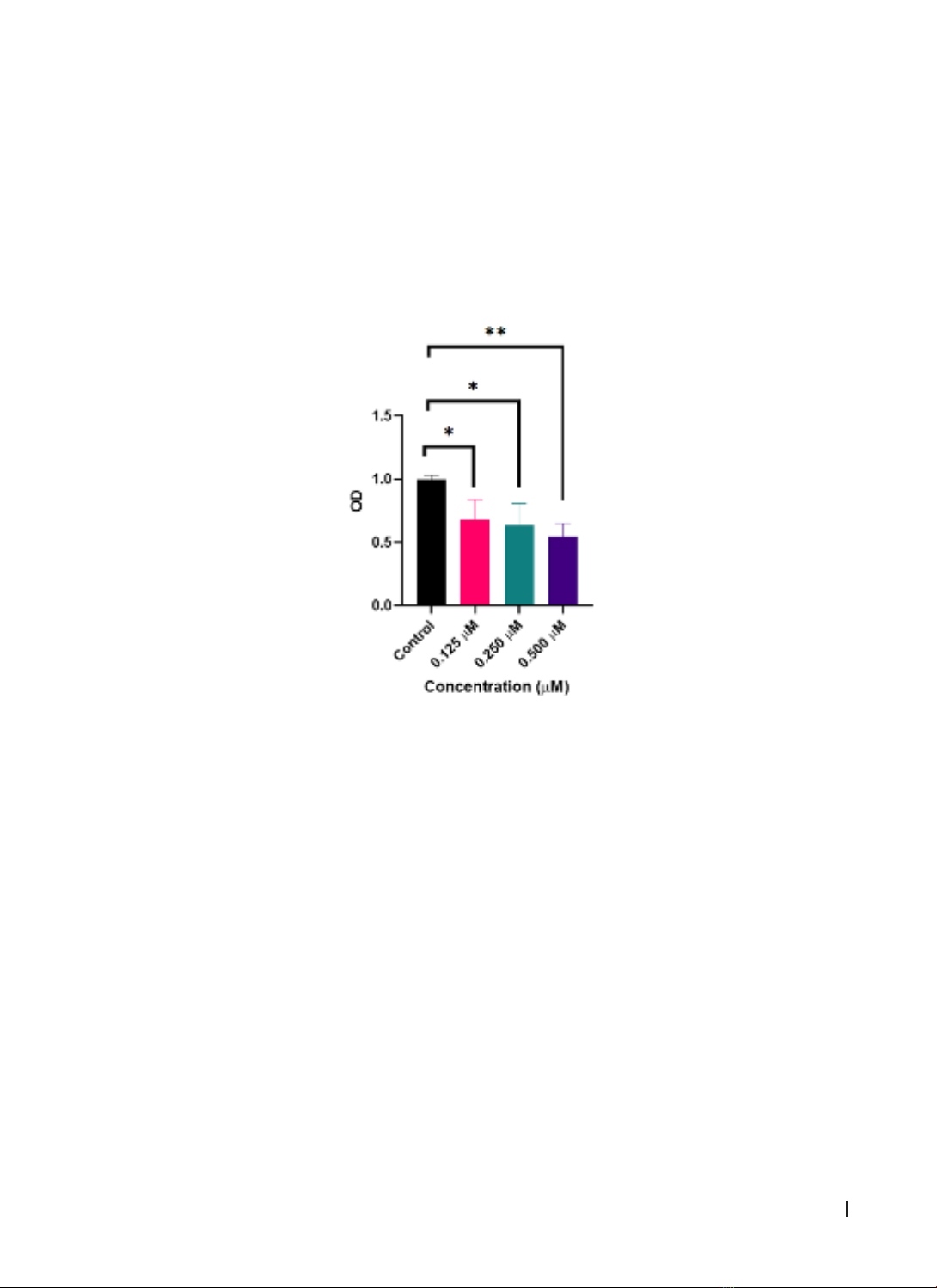
and cell cycle. Data were analyzed using GraphPad Prism 8.4. Results: DNA-PKi showed dose-dependent
effects on cell death and proliferation inhibition in HCT116 cells in vitro. The groups treated with DNA-PKi at
concentrations of 0.125, 0.250, and 0.500 μM all had significantly lower cell survival rates than the control
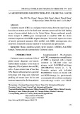
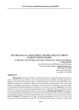
group (p < 0.05). However, DNA-PKi did not affect cell cycle distribution in HCT116 colorectal cancer cells.
Conclusion: DNA-PKi NU7441 exerted a dose-dependent effect on cell death and proliferation inhibition in
HCT116 cells in vitro.
Keywords: colorectal cancer, DNA-PK, DNA repair, Double-strand DNA breaks, target therapy, non-
homologous end joining.
1. INTRODUCTION
Colorectal cancer (CRC) remains a formidable
global health challenge and ranks among the leading
causes of cancer-related morbidity and mortality.
Colorectal cancer (CRC) is a significant health burden.
Globally, it is the third most common cancer in males
and the second most common in females, ranking as
the third leading cause of cancer-related deaths [1].
Colorectal cancer is trending toward younger ages,
with cases as young as 15 to 18 years [1]. Data from
2018 in Vietnam showed that colorectal cancer ranked
fifth among the top 10 most common cancers in both
genders, with 14,733 new cases and 7,856 deaths. By
the year 2020, in Vietnam, the incidence of new cases
of colorectal cancer had risen to the fourth position
in males and the third position in females, with a new
incidence rate of 9%, totaling 16,426 cases [2].
The conventional treatment methods often
applied in the comprehensive management of
colorectal cancer include surgery, chemotherapy,
radiation therapy, or a combination of these
approaches. However, the prognosis of colorectal
cancer remains limited, with a 5-year survival rate of
less than 20% [3]. Depending on the condition and
progression of the disease, treatment methods can
be used in combination. For localized malignancies,
surgical removal of the entire colon tumor is often
an option, and any tumor location requires therapy.
However, not all cancer cells can be eradicated
entirely. Approximately 66% of patients with colon
cancer undergo additional adjuvant treatment
with chemotherapy and/or radiotherapy [4]. These
treatments have many side effects because they are
nonspecific and toxic to healthy cells. Additionally,
even after receiving adjuvant therapy, up to 54% of
patients relapsed [4]. Therefore, the development of
more effective alternative therapies to treat patients
with CRC is urgently needed.
Targeted therapy is a type of cancer treatment
that focuses on specific molecules involved in
the growth and survival of cancer cells. The goal
of targeted therapy is to block the growth and
spread of cancer cells while minimizing damage
to normal cells. DNA double-strand breaks (DSBs)
are considered the most deleterious form of DNA
damage. The DNA damage response (DDR) pathway
encompasses a collection of intricate mechanisms,
including DNA damage repair, DNA damage tolerance
mechanisms, and cell-cycle checkpoint control. This
intricate system governs the accurate execution of
DNA replication and proliferation and, subsequently,
cell viability. The DDR pathway plays a pivotal role in
Corresponding author: Nguyen Thi Mai Ly; Email: dr.nguyenmaily@gmail.com
Received: 20/4/2024; Accepted:18/6/2024; Published: 25/6/2024
DOI: 10.34071/jmp.2024.4.4
HUE JOURNAL OF MEDICINE AND PHARMACY ISSN 3030-4318; eISSN: 3030-4326
32
Hue Journal of Medicine and Pharmacy, Volume 14, No.4/2024
preserving genomic integrity and stability through
DNA damage repair. Strand breakage resulting from
base alterations, single-strand breaks (SSBs), or
double-strand breaks (DSBs) can lead to chromosome
breakage, resulting in gene loss. It is mostly caused
by altered DNA replication forks, ROS, ionizing
radiation, and physical or mechanical stress [5]. DNA-
dependent protein kinase (DNA-PK) is an important
enzyme in DDR pathways that plays a crucial role in
the repair of DSBs via non-homologous end joining
(NHEJ) [6]. NU7441 represents a new generation of
selective DNA-PK inhibitors. The aim of the present
study was to investigate the anti-colorectal cancer
effect of the DNA-PK inhibitor NU7441, which inhibits
DNA-DSB repair by blocking the NHEJ pathway, in the
HCT116 cell line.
2. MATERIALS AND METHODS
2.1. Cell lines and culture conditions
The antitumor efficacy of DNA-PKi was assessed
using HCT116 cells as an experimental model. The
HCT116 cells, sourced from the American Type
Culture, USA, were cultured in Dulbecco’s Modified
Eagle Medium (DMEM) obtained from Cytiva, USA.
The culture medium was supplemented with 10%
fetal bovine serum (FBS) provided by Cytiva and 1%
penicillin-streptomycin (Sigma-Aldrich, USA). Cells
were incubated in an atmosphere of 5% CO2 and
95% air at 37°C.
2.2. Reagent:
DNA-PKi NU7441 (KU-57788, Catalog No.S2638,
Selleckchem) was stored at -20°C for subsequent
experiments. The dosage was determined during
the experiments.
2.3. WST-1 assay:
Cells were seeded at a density of 1000 cells/well
in a 96-well plate and cultured at 37 °C overnight.
Cells were treated with DNA-PKi for 72h at increasing
concentrations of 0.000, 0.125, 0.250, and 0.500 µM.
Next, 100 µL of 2% WST-1 solution (Roche, Switzerland)
was added to each well, and the plate was incubated at
37oC for 2h. A microplate reader (Tecan, Switzerland)
was used to measure the absorbance at 450 nm and
620 nm was used as the reference wavelength. Cell
viability was assessed with Excel ver. 2016 (Microsoft,
USA) using the following formula:
2.4. FACS cell cycle assay:
HCT116 cells were cultured in 6-well plates of
500,000 cells each for 24 h. After the cells reached
the appropriate amount, they were treated with
DNA-PKi at different temperatures (0.125 µM/ml;
0.25 µM/ml) and the control group was the normal
cultured without treatment. Cells were imaged at
0, 24, and 48 h after drug testing. After 48 h, cells
were scraped off the plate, cycled with EtOH, and
incubated overnight at -30°C. The cells were stained
with Propidium Iodide (PI, 10 µg/ml), incubated for
30 min, and analyzed using a flow cytometer. PI is a
common fluorescent red nuclear pigment antagonist.
Because propidium iodide does not permeate living
cells, it is commonly used to detect dead cells in
populations. Stain PI provides information about cell
cycle and amount of DNA of population. The flow
cytometer will analyze the data and charts from the
obtained cells.
2.5. Statistical analysis:
GraphPad Prism version 8.4 (GraphPad Software,
Inc., California, USA) was used for statistical analysis.
The study results are presented as mean ± standard
error (SD). One-way ANOVA was used to test the
difference in the mean when comparing more than
two groups. Differences were considered statistically
significant when the p-value was less than 0.05.
3. RESULTS
3.1. Cell culture and proliferation assay
Figure 1. Morphological characteristics of HCT116 cell line in culture: (A) HCT116 cells on day 1; (B) HCT116
cells on day 2; (C) HCT116 cells on day 7. After 24h, cells adhered to the bottom 40 - 50% of the culture
area. After seven days, the cultured cells covered 80% of the culture area.
HUE JOURNAL OF MEDICINE AND PHARMACY ISSN 3030-4318; eISSN: 3030-4326 33
Hue Journal of Medicine and Pharmacy, Volume 14, No.4/2024
The images in Figure 1 show the status, form, and
density of HCT116 cells cultured after 1, 2, and 7 days.
HCT116 cells appeared as basal epithelial cells, and
monolayer growth, having many different shapes from
oval to polygonal, was approximately 20 - 25 µm in size,
although this size could vary depending on the stage of
the cell cycle. In addition, HCT116 cells had large nuclei
that occupied most of their cytoplasm. After 24h of
culturing, the HCT116 cells adhered to the bottom of
the culture dish and showed robust proliferation. The
proliferation rate of HCT116 was about 40 - 50% within
24 hours. After seven days, the cultured cells covered
80% of the flask area. The cell proliferation rate was
monitored, and when it reached approximately 80%
of the plate area, the cells were transferred to a new
culture plate.
3.2. Evaluation of the impact of DNA-PKi on the viability of colorectal cancer cells using the WST-1 assay
Figure 2. Percentage of viable cells 72 h after DNA-PKi treatment at concentrations of 0.125, 0.250,
and 0.500 µM. Data were analyzed by one-way ANOVA and post-hoc Tukeys test; results are shown as
mean ± SD. *P < 0.05, **P < 0.01
Based on the WST-1 results, the number of living
cells was proportional to the OD index displayed on
the spectrophotometer. The OD index after reading
was calibrated by the index at 650 nm wavelength
minus the index at 450 nm wavelength. The
higher the OD index, the higher is the percentage
of living cells.
To investigate the effect of DNA-PKi on the
viability of colorectal cancer cells, HCT116 cells
were treated with DNA-PKi at concentrations: 0.125
µM, 0.250 µM and 0.500 µM and the control group
was not treated with DNA-PKi. Employing one-way
ANOVA and post hoc Tukey’s pairwise comparison
tests, we observed that at the 72-hour time
point after DNA-PKi treatment, the control group
exhibited the highest cell survival rate. In contrast,
the groups treated with DNA-PKi at concentrations
of 0.125 μM, 0.250 μM, and 0.500 μM all had
significantly lower cell survival rates compared to
the control group (p < 0.05, Figure 2). This indicates
that DNA-PKi at concentrations of 0.125, 0.250, and
0.500 μM can induce cell death and inhibit in vitro
proliferation in HCT116 colorectal cancer cell lines.
Additionally, DNA-PKi showed a dose-dependent
effect on cell death and proliferation inhibition in
HCT116 cells in vitro. Therefore, we concluded that
the inhibitory effect of DNA-PKi on HCT116 cells was
dose-dependent.
HUE JOURNAL OF MEDICINE AND PHARMACY ISSN 3030-4318; eISSN: 3030-4326
34
Hue Journal of Medicine and Pharmacy, Volume 14, No.4/2024
3.3. Evaluation of the effect of DNA-PKi on the cell cycle was performed on colorectal cancer cells
Figure 3. HCT116 colorectal cancer cells in the control group and DNA-PKI treatment groups at
concentrations of 0.125 μM and 0.250 μM at 0h, 24h and 48h after treatment. Cells in the control group
and the 0.125 µM treatment group grew in large patches and spread evenly on the surface while the
0.25µM treatment group grew in small discrete clusters.
Figure 4. Flow cytometry results of cell cycle assay between the control group and DNA-PKi treatment
groups at concentrations of 0.125 μM and 0.250 μM on HCT116 cell line after 48 h of treatment. Nuclear
DNA was stained with Propidium Iodide and analyzed by Flow cytometry. If PI penetrates the cell
membrane, it can cause damage.
Figure 5. Comparison of cell cycle test results between the control group and the DNA-PKi treatment
groups at concentrations of 0.125 μM and 0.250 μM on the human colorectal cancer cell line HCT116 after
48 h of treatment. Data were analyzed by one-way ANOVA test and post-hoc Tukey test, and the results are
presented as mean ± SD.
To analyze the effect of DNA-PKi on the phases
of the division cycle of HCT116 cells, cells were
treated with different concentrations (0.125 µM and
0.25 µM), and then nuclear DNA was stained with
Propidium Iodide and analyzed by Flow cytometry.
Cells in the control group and the 0.125 µM
treatment group grew in large patches and spread
evenly on the surface while the 0.25 µM treatment
HUE JOURNAL OF MEDICINE AND PHARMACY ISSN 3030-4318; eISSN: 3030-4326 35
Hue Journal of Medicine and Pharmacy, Volume 14, No.4/2024
group grew in small discrete clusters (Figure 3).
Two-way ANOVA and Dunnetts post-test were
used to compare the proportion of cells in each
stage between the groups. We did not observe a
difference in the cell cycle distribution after treatment
with DNA-PKi compared to that in the control group
in all groups. Specifically, the average percentage of
cells in S phase in all groups ranged between 17 - 18%.
Similarly, the ratio of cells in the G1 and G2/M phases
was the same in all groups (Figure 4-5). Therefore,
DNA-PKi did not affect the cell cycle.
4. DISCUSSION
Colorectal cancer is one of the leading causes
of cancer in the world, so there is increasing
appreciation for the development of novel methods
for the treatment of colorectal cancer. DNA-PK is
an essential component of the NHEJ pathway, and
its activation is necessary for DNA repair. Inhibiting
the phosphorylation process of DNA-PK prevents
the repair of DNA damage, leading to programmed
cell death. NU7441, a potent and specific DNA-PKcs
inhibitor, is predicted to enhance the effectiveness
of disease treatment. A previous study has shown a
combination of pharmacological agents with specific
chemotherapeutic agents against lung cancer.
Inhibition of DNA-PKcs counteracts cell proliferation
in A549 lung cancer cells when topoisomerase
inhibitors are applied . NU7441 inhibits the repair
of DNA damage caused by topoisomerase inhibitors
in non-small cell lung carcinoma (NCSLC) cells,
subsequently leading to programmed cell death.
Therefore, the results indicate that the combination
of traditional therapy inhibitors and NU7441 is a
potential treatment option for NCSLC [7].
Targeting DNA is a promising approach for cancer
treatment, but it has some limitations. Despite
promising findings in vitro, DNA-PK inhibitors have
failed to induce changes in animal studies. One
reason for these findings may be related to the limited
solubility and poor pharmacokinetic properties
of DNA-PK inhibitors. NU7441 is poorly absorbed
and rapidly metabolized in mice, which hinders its
clinical application [8]. In addition, its poor water
solubility hampers the possibility of higher dosing.
Based on promising pharmacokinetics, studies on
the effectiveness of combined therapies have been
conducted, showing that NU7441 doubles the delay
in tumor development caused by etoposide without
increasing toxicity to unacceptable levels.
Targeting DNA-PK as a therapeutic intervention
in human malignancy began entering clinical trials,
especially in combination with chemotherapy
or radiotherapy. CC-122, an inhibitor of DNA-
PK, currently undergoing phase I clinical trial
for solid tumors, non-Hodgkin lymphoma, and
multiple myeloma. Similarly, CC-115, designed as
a dual inhibitor of DNA-PK and mTOR, is in phase
I trial for advanced solid tumors and hematologic
malignancies. Notably, phase I studies have
revealed that oral formulations of CC-122 and CC-
115 demonstrate favorable tolerability profiles, with
no unexpected toxicities or adverse effects reported
[9], [10]. The utilization of CC-122 in brain cancer
has yielded promising outcomes[10]. Additionally,
ZSTK474, primarily a PI3K inhibitor with DNA-PK
inhibitory activity, is undergoing phase I trials for
advanced solid malignancies [11]; the DNA-PKi
MSC2490484A, is being investigated in combination
with radiotherapy for the treatment of advanced
solid tumors or chronic lymphocytic leukemia [12].
These clinically significant DNA-PKi hold promise for
potentially offering therapeutic benefits in the fight
against cancer. Peposertib is a powerful and selective
small molecule DNA-PK inhibitor that is taken orally
[13]. It has been shown to have radio sensitizing
and anticancer action in xenograft models and
to be well-tolerated when used in monotherapy.
However, enrollment was discontinued because of
insufficient exposure at that dose, and the RP2D was
not formally declared [14].
DNA-dependent protein kinase inhibitors (DNA-
PKis) have significant effects on the cell cycle. In
response to DNA damage, cells often undergo cell
cycle arrest to allow time for DNA repair. DNA-PKi-
induced inhibition of DNA-PK can lead to G1 phase
arrest [15]. In this phase, the cell checks for DNA
damage and either repairs it or initiates apoptosis
(programmed cell death) if the damage is too
severe to be repaired. DNA-PK also plays a role
in regulating cell progression through the S phase
of the cell cycle, during which DNA replication
occurs. Inhibition of DNA-PK can slow down or
delay progression through the S phase as the cell
attempts to repair DNA lesions before replicating
the damaged DNA [16].
Jarah showed that DNA-PKI can induce cell
cycle arrest at G2/M phase in glioblastoma cells.
DNA demethylating agents such as 5-Aza-CdR and
Zebularine induce G2/M arrest in various cancer
cell types [17], and it has been shown that the
release from Zebularine-induced G2/M arrest
and DNA repair efficiency correlates well with
phosphorylation of ATM, p53, and Chk1 proteins. If
cells with persistent DNA damage undergo mitosis
due to a G2/M checkpoint deficiency, mitotic