doi:10.1046/j.1432-1033.2003.03776.x
Eur. J. Biochem. 270, 3822–3828 (2003) (cid:1) FEBS 2003
Dolastatin 15 binds in the vinca domain of tubulin as demonstrated by Hummel–Dreyer chromatography
Zobeida Cruz-Monserrate1, Jeffrey T. Mullaney2, Patrick G. Harran3, George R. Pettit2 and Ernest Hamel1 1Screening Technologies Branch, Developmental Therapeutics Program, Division of Cancer Treatment and Diagnosis, National Cancer Institute at Frederick, National Institutes of Health, Frederick, Maryland, USA; 2Cancer Research Institute and Department of Chemistry and Biochemistry, Arizona State University, Tempe, Arizona, USA; 3Department of Biochemistry, University of Texas Southwestern Medical Center at Dallas, Dallas, Texas, USA
and a second, nonradiolabeled drug. No inhibition was detected with either the colchicine site agent combretastatin A-4 or with an analog of the antimitotic marine peptide diazonamide A (both the analog and diazonamide A are potent inhibitors of tubulin assembly). Weak inhibition was observed with cemadotin, a structural analog of dolasta- tin 15, and with the depsipeptide cryptophycin 1. Moderate inhibition occurred with vinblastine and vincristine, and strong inhibition with maytansine, halichondrin B, and the peptides dolastatin 10 and phomopsin A. These observa- tions suggest that the binding site(s) for peptide and depsi- peptide antimitotic drugs may consist of a series of overlapping domains rather than a well-defined locus on the surface of b-tubulin.
Keywords: cemadotin; dolastatin 10; cryptophycin 1; dola- statin 15; tubulin.
The antimitotic depsipeptide dolastatin 15 was radiolabeled with tritium in its amino-terminal dolavaline residue. Dolastatin 15, although potently cytotoxic, is a relatively weak inhibitor of tubulin assembly and does not inhibit the binding of any other ligand to tubulin. The only methodo- logy found to demonstrate an interaction between the dep- sipeptide and tubulin was Hummel–Dreyer equilibrium chromatography on Sephadex G-50 superfine. The average apparent Kd value obtained in these studies was about 30 lM, with no difference observed when column size or tubulin concentration was varied. This relatively high dis- sociation constant is consistent with the apparent weak interaction of dolastatin 15 with tubulin demonstrated indirectly in the assembly assay. We attempted to gain insight into the binding site for dolastatin 15 on tubulin by studying inhibitory effects of other drugs when the gel fil- tration column was equilibrated with both [3H]dolastatin 15
of radiolabeled cemadotin to tubulin [10]. Therefore, it seemed possible that dolastatin 15 and cemadotin might bind in a unique site on tubulin. To evaluate this idea, we synthesized radiolabeled dolastatin 15 and sought a method to study its interaction with tubulin, both in the presence and absence of potential competitors. Our primary goal was to gain new insights into the potential relationships between binding site(s) for different antimitotic peptides and between binding sites for the peptides and for other compounds that inhibit binding of vinca alkaloids to tubulin.
is the the subunit protein of microtubules, Tubulin, intracellular target of an increasing number of antimitotic peptides and depsipeptides. These compounds are all natural products containing unusual, frequently unique, amino acids, and they invariably inhibit microtubule assembly (recently reviewed [1]). Most of these peptides are highly cytotoxic, inhibiting the growth of cultured cells with picomolar or low nanomolar IC50 values, and, as is typical with drugs that target tubulin, treated cells arrest in the mitotic phase of the cell cycle. Most of these peptides and synthetic analogs inhibit the binding of vinca alkaloids to tubulin in a noncompetitive manner [2–6].
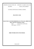
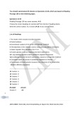
A notable exception to this generalization has been the depsipeptide dolastatin 15, originally extracted from the sea hare Dolabella auricularia [7], and its synthetic analog cemadotin [8] (structures in Fig. 1). Neither compound inhibited the binding of vinblastine to tubulin [8,9], and it was also reported that vinblastine did not inhibit the binding
Despite many efforts to detect a tubulinÆdolastatin 15 interaction, we only succeeded when we employed the cumbersome Hummel–Dreyer chromatographic method [11]. Utilizing progressively smaller columns, we were able to adapt this technique to obtain qualitative information about relative inhibitory effects on the binding of dolasta- tin 15 by a number of vinca domain drugs. We were unable, however, to determine which compounds might be com- petitive or noncompetitive inhibitors of dolastatin 15 bind- ing, because we did not have adequate amounts of either [3H]dolastatin 15 or most of the potential inhibitors to perform the detailed studies at multiple concentrations required for such an evaluation.
Most of the vinca domain drugs that we examined inhibited dolastatin 15 binding in parallel with their
Correspondence to E. Hamel, Building 469 Room 104, National Cancer Institute at Frederick, Frederick, Maryland 21702, USA. Fax: +1 301 846 6014, Tel.: +1 301 846 1678, E-mail: hamele@mail.nih.gov (Received 4 June 2003, revised 24 July 2003, accepted 31 July 2003)
Binding of dolastatin 15 to tubulin (Eur. J. Biochem. 270) 3823
(cid:1) FEBS 2003
Sephadex LH-20 column. The column was developed with methanol, and fractions containing [3H]Dov were pooled and the solvent removed under vacuum. A mixture containing 6 mg [3H]Dov, 30 mg des-Dov-dolastatin 15, 12 lL triethylamine, 12 lL diethyl phosphocyanidate, and 1.5 mL CH2Cl2 was stirred for 3 h at room temperature. The solvent was evaporated under a stream of N2 and the residue dissolved in a 1 : 1 mixture of hexane and acetone. The solution was applied to a column of silica gel (Merck), and the column was developed with the same solvent mixture. Fractions containing [3H]dolastatin 15 were pooled, and the solvent was evaporated under vacuum. The [3H]dolastatin 15 was stored dry in liquid nitrogen until used.
inhibitory effects on dolastatin 10 binding and vinca alkaloid binding to tubulin. The major exception was cryptophycin 1. Although a strong inhibitor of dolastatin 10 and vinblastine binding [3], this depsipeptide had minimal inhibitory effect on dolastatin 15 binding. This suggests to us that the vinca domain is most likely a relatively large binding pocket on the surface of b-tubulin that is able to accommodate different complex natural product ligands by diverse binding mech- anisms, presumably involving different, possibly adjacent and/or overlapping, subsites.
Fig. 1. Molecular structures of dolastatin 15 and cemadotin.
Materials and methods
Materials
With Sephadex G-50 (superfine) columns we observed a classic Hummel–Dreyer [11] pattern for the binding of dolastatin 15 to tubulin. Over the course of the studies presented here, all performed at room temperature (21–23 (cid:2)C), we progressively reduced the size of the columns to conserve both [3H]dolastatin 15 and potential inhibitory drugs. We saw no significant differences in apparent Kd values obtained for dolastatin 15 as a function either of column size or the amount of tubulin applied to the column. The column sizes used in these studies, with the average flow rates and fraction sizes, were: (a) 48 · 1.5 cm at 0.4 mLÆmin)1 and 1.3 mL fractions; (b) 24 · 1.5 cm at 0.5 mLÆmin)1 and 1.4 mL fractions; and (c) 30 · 0.7 cm at 0.2 mLÆmin)1 and 0.6 mL fractions. The specific activity of the [3H]dolastatin 15 was 2.8 c.p.m.Æpmol)1 in most of the experiments presented here. Protein in fractions was quantitated by the Lowry procedure, with BSA as standard. The columns were equilibrated with the [3H]dolastatin 15 at 8–10 lM in 0.1 M 4-morpholineethanesulfonate (pH 6.9 with NaOH in 1 M stock solutions)/0.5 mM MgCl2/1% (v/v) dimethyl sulfoxide. The Sephadex G-50 (superfine) from which these columns were made was swollen either in buffer containing only [3H]dolastatin 15 or both [3H]do- the lastatin 15 and the desired potential inhibitor at indicated concentration. After column equilibration, the desired amount of tubulin was diluted to 1 mL with the column buffer and applied to the column. The stock tubulin solution was 76 mgÆmL)1, and 3 or 10 mg was applied to the 48 · 1.5-cm columns, 10 mg was applied to the 24 · 1.5-cm columns, and 5 mg was applied to the 30 · 0.7-cm columns. Data were analyzed in all cases by assuming a simple
Bovine brain tubulin was purified as described previously [12], including gel filtration chromatography on Sephadex G-50 (superfine) [13]. Nonradiolabeled dolastatin 15, dolast- atin 10, and combretastatin A-4 were synthesized as described previously [14–16]. Cemadotin, maytansine, vin- blastine, vincristine, and halichondrin B were generously provided by the Drug Synthesis and Chemistry Branch, National Cancer Institute. Cryptophycin 1 [17] was a gener- ous gift from Merck, Sharp and Dohme Research Labor- atories (Rahway, NJ, USA), and compound 1, an analog of diazonamide A, was synthesized as described elsewhere [18]. Phomopsin A was obtained from Calbiochem.
tubulin-drug equilibrium:
tubulin þ dolastatin 15 (cid:2)(cid:2)*)(cid:2)(cid:2) tubulin (cid:3) dolastatin 15
and
Kd ¼ ½tubulin(cid:6)½dolastatin 15(cid:6)=½tubulin (cid:3) dolastatin 15(cid:6)
Methods The [3H]dolastatin 15 was synthesized by coupling des- Dov-dolastatin 15, the carboxy-terminal segment of the depsipeptide (synthesized as described in [15]), to [3H]Dov, its amino-terminal residue. The [3H]Dov was prepared by reductive alylation of [3H]valine (Moravek Biochemicals) with H2 over a platinum catalyst. The [3H]valine (5 mCi; 10 CiÆmol)1) in 1.5 mL water was mixed with 10 mg nonradiolabeled valine, 5 mg 10% platinum on carbon, and 1.5 mL 37% formaldehyde. The mixture was stirred under a slight positive (balloon) pressure of H2 for 8 h at room temperature (21–23 (cid:2)C) and filtered through celite. The filtrate was concentrated under vacuum and the residue dissolved in 1.5 mL methanol and applied to a
The equilibrated column radiolabel was used as the concentration of unbound dolastatin 15, the radiolabel above this level in a protein-containing fraction was taken as the tubulinÆdolastatin 15 concentration, and the free tubulin concentration was obtained by subtraction (total tubulin concentration, from the protein assay, minus the tubulinÆdolastatin 15 concentration). Two apparent Kd values were obtained in each experiment by using the two fractions containing the highest amounts of protein for these calculations.
3824 Z. Cruz-Monserrate et al. (Eur. J. Biochem. 270)
(cid:1) FEBS 2003
Results and discussion
Interaction of dolastatin 15 with tubulin
manifest itself as reduced radiolabel associated with the protein peak, resulting in an increase in the calculated apparent Kd value for dolastatin 15. Alternatively, if no inhibition occurred, the apparent Kd value should be unaltered.
Initially we examined nonequilibrium methods (DEAE- filters, centrifugal gel filtration) for evaluating the binding of [3H]dolastatin 15 to tubulin, but no interaction of drug with protein was detectable. Singer and Himes [19,20] described a Hummel–Dreyer [11] procedure using HPLC gel filtration columns for measuring the interactions of various vinca alkaloids with tubulin, and we attempted to use a similar column to measure the binding of dolastatin 15 to the protein. Although the column was readily equilibrated with the [3H]dolastatin 15 preparation, when tubulin was applied to the column there again appeared to be no interaction of the protein with dolastatin 15.
This was tested with cemadotin, on the one hand, and with combretastatin A-4, a potent colchicine site drug [21], on the other (Table 1). With the highest feasible cemadotin concentration (determined by the amount of the drug we could obtain), 100 lM, there was > 50% reduction in the amount of [3H]dolastatin 15 bound to tubulin, with a concomitant rise in the calculated apparent Kd value to 85 lM (Table 1). In contrast, two concentrations of com- bretastatin A-4, which is at least 10-fold more active than both dolastatin 15 and cemadotin as an inhibitor of tubulin assembly and a powerful competitive inhibitor of the binding of colchicine to tubulin [21], did not alter the apparent Kd value for the binding of [3H]dolastatin 15 to tubulin, nor greatly affect the stoichiometry of binding of dolastatin 15 to tubulin (Table 1).
Drugs that bind in the ‘vinca domain’
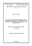
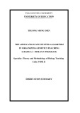
Despite this failure, the Hummel–Dreyer procedure was undertaken again with standard gel filtration on Sephadex G-50 (superfine). This method proved to be successful (Fig. 2), and multiple runs with different size columns and different amounts of tubulin yielded apparent Kd values in the range of 5–49 lM, with an average of 28 lM [± 14 lM (SD); ± 2.7 lM (SEM); n ¼ 26]. Most (19) of the values obtained were > 20 lM, and, if only these are considered, the average apparent Kd value is 35 ± 8.5 (SD) lM. For comparison, Jordan et al. [10] obtained an apparent Kd value of 19 lM for [14C]cemadotin by equilibrium dialysis.
Binding of dolastatin 15 to tubulin is inhibited by its synthetic analog cemadotin but not by a colchicine site drug
We reasoned that if the column were equilibrated with both dolastatin 15 and another drug, an inhibitory effect would
In studies with radiolabeled vinca alkaloids and radiolabeled dolastatin 10, a potent antimitotic peptide also originally obtained from D. auricularia, various compounds yielded either competitive or noncompetitive patterns of inhibition (relative potencies of most of these inhibitory effects are summarized in Table 2). Thus, maytansine, rhizoxin, and vinblastine competitively inhibit the binding of [3H]vincris- tine to tubulin, while the peptides dolastatin 10 and phomopsin A noncompetitively inhibit vincristine binding [2]. The depsipeptide cryptophycin 1 [3] and the peptide hemiasterlin [4] are noncompetitive inhibitors of [3H]vin- blastine binding, as are a synthetic analog of dolastatin 10 [5], the complex peptide vitilevuamide [6], and the macro- cyclic polyethers halichondrin B [22] and spongistatin 1 [23]. With [3H]dolastatin 10 as the test ligand, competitive inhibition occurs with the peptides 19aR-isodolastatin 10 [3], which is essentially identical to dolastatin 10 in its interactions with tubulin, hemiasterlin [4], and phomop- sin A (unpublished data) and with the depsipeptide crypto- phycin 1 [3], while spongistatin 1 is a noncompetitive inhibitor [23] of the binding of [3H]dolastatin 10 to tubulin. With the strongest inhibitors, whether a competitive or noncompetitive pattern is obtained, near total inhibition occurs if a sufficiently high concentration of inhibitor is used. In contrast to these inhibitory effects, the complex bicyclic peptide diazonamide A and its analog compound 1 (structures in Fig. 3) resemble dolastatin 15 in that they do not inhibit the binding of either [3H]vinblastine or [3H]dolastatin 10 to tubulin. Unlike dolastatin 15, however, these two peptides are potent inhibitors of tubulin assembly and its associated GTP hydrolysis, equivalent in activity to dolastatin 10 [24].
Such findings led to the concept of the (cid:2)vinca domain(cid:3), which was envisaged as distinct sites for the vinca alkaloids, for the inhibitory peptides and depsipeptides, and for spongistatin 1, with the sites impinging on each other sterically when occupied by their ligands [2]. This would explain both the near total inhibition and noncompetitive inhibitory pattern obtained with some of these drugs.
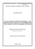
Fig. 2. Hummel–Dreyer chromatographic demonstration of the binding of [3H]dolastatin 15 to tubulin. The Sephadex G-50 (superfine) column (48 · 1.5 cm) was equilibrated and developed with a solution con- taining 0.1 M 4-morpholineethanesulfonate pH 6.9, 0.5 mM MgCl2, and a mixture of [3H]dolastatin 15 and nonradiolabeled dolastatin 15 at (cid:7) 8 lM (2.8 c.p.m.Æpmol)1). Tubulin (10 mg) was applied to the column and fractions were collected. Aliquots of the fractions were counted in a scintillation counter or assayed for protein by the Lowry procedure. The data obtained from the fractions with the two highest protein concentrations were used to calculate Kd values for the binding of dolastatin 15 to tubulin. The values obtained from this column were 33 and 44 lM dolastatin 15. Dolastatin 15 (radiolabel), s. Tubulin (protein), n.
Binding of dolastatin 15 to tubulin (Eur. J. Biochem. 270) 3825
(cid:1) FEBS 2003
Table 1. Average stoichiometries of binding and apparent Kd values obtained for [3H]dolastatin 15 with Hummel–Dreyer columns equilibrated with both dolastatin 15 and a potential inhibitory drug. Only positive values were used in calculations of average stoichiometry. The stoichiometry of binding of the two fractions from each Sephadex G-50 (superfine) column containing the highest amounts of tubulin were determined and the means calculated. The number of Sephadex G-50 (superfine) columns used for each reaction condition is shown in parentheses in the last column of the table. Apparent Kd values were calculated, as described in the text, for each of the two fractions from each Sephadex G-50 (superfine) column containing the highest amounts of tubulin and averaged.
Apparent Kd value obtained [lM ± SEM (number of columns)] Second drug in column (lM) Average stoichiometry ± SEM (% inhibition) (mol dolastatin 15 bound per mol tubulin)
a Radiolabel in the second protein-containing fraction from this column was not above baseline. b A second column was run in which there was no increase of radiolabel in the tubulin-containing fractions. c Nominal concentration, for addition of 50 lM cryptophycin 1 to an aqueous solution caused the solution to become turbid, presumably due to limited solubility of the depsipeptide.
None Combretastatin A-4 (20 lM) Combretastatin A-4 (50 lM) Cemadotin (50 lM) Cemadotin (100 lM) Vinblastine (50 lM) Vincristine (50 lM) Maytansine (50 lM) Halichondrin B (50 lM) Phomopsin A (20 lM) Dolastatin 10 (20 lM) Dolastatin 10 (50 lM) Cryptophycin 1 (10 lM) c) Cryptophycin 1 (50 lM Diazonamide A analog (50 lM) 0.29 ± 0.03 0.27 ± 0.04 (7) 0.27 ± 0.05 (7) 0.15 ± 0.005 (48) 0.095 ± 0.003 (67) 0.11 ± 0.04 (62) 0.058 ± 0.006 (80) 0.025 ± 0.01 (91) 0.013a (96) 0.010 ± 0.001 (97) 0.0046 ± 0.0001 (98) 0.0071 ± 0.002 (98) 0.31 ± 0.009 (0) 0.096 ± 0.00004 (67) 0.32 ± 0.07 (0) 28 ± 3 (13) 33 ± 7 (1) 25 ± 7 (1) 51 ± 2 (1) 85 ± 3 (1) 170 ± 40 (4) 190 ± 30 (2) 720 ± 200 (3) 840a (1b) 820 ± 80 (3) 2200 ± 3 (1b) 1500 ± 500 (1) 23 ± 1 (2) 81 ± 0.1 (1) 19 ± 6 (2)
Table 2. Comparison of drug inhibitory effects on the binding of radiolabeled dolastatins 10 and 15 and vinblastine/vincristine to tubulin. Qualitative terms are used to describe relative inhibitory effects. Published lM Ki values are shown in parentheses.
[3H]Vinblastine/vincristine binding [3H]Dolastatin 10 binding Inhibitor [3H]Dolastatin 15 binding
Weak Noninhibitory [25] Noninhibitory (unpublished) Strong (0.7–0.8) [4,23,25]
Dolastatin 15 Cemadotin 19aR-isodolastatin 10 Dolastatin 10 Cryptophycin 1 Phomopsin A Diazonamide A analog Vinblastine Vincristine Maytansine Strong Weak Strong Noninhibitory Moderate Moderate Strong Strong (2.1) [3] Strong [23,25] Noninhibitory [24] Weak [23] Moderate [23] Moderate [23]
Inhibition of dolastatin 15 binding by vinca domain drugs
obtained for dolastatin 15 is consistent with this possibility, being substantially higher than the apparent Kd values of 26 nM and 0.1–0.45 lM obtained, respectively, for dolasta- tin 10 [25] and cryptophycin 52 [26], which is a synthetic analog of cryptophycin 1 with activity equivalent to that of the natural product [27].
site on tubulin different
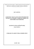
Alternatively, it is possible that dolastatin 15 binds at from that an independent where dolastatin 10, phomopsin A, cryptophycin 1, and hemiasterlin bind. Examination of this possibility became more important with the recent discovery that diazona- mide A and its close analog compound 1, like dolastatin 15, do not significantly inhibit the binding of [3H]vinblastine
As noted above, dolastatin 15 differs from most other antimitotic peptides and depsipeptides in not inhibiting vinca alkaloid binding to tubulin, nor does it inhibit dolastatin 10 binding to tubulin. Moreover, despite its potent cytotoxic properties, dolastatin 15 is about 5 to 20-fold less active as an inhibitor of tubulin assembly than dolastatin 10, cryptophycin 1, hemiasterlin, and phomop- sin A, depending on precise reaction conditions. Thus, it is conceivable that dolastatin 15 binds with lower affinity in the same site as the other peptides. The apparent Kd we
Halichondrin B Strong Moderate [23] Noninhibitory [9] Noninhibitory [8] Strong [25] Strong (1.4 vs. vincristine) [2,28] Strong (3.9 vs. vinblastine) [3] Strong (2.8 vs. vincristine) [2,28] Noninhibitory [24] Moderate (6.6 vs. vincristine) [2,28] Strong (1.3 vs. vinblastine) [23] Strong (3.1 vs. vincristine) [2,28]; (0.9 vs. vinblastine) [22] Moderate-strong (5.0 vs. vinblastine) [22]
3826 Z. Cruz-Monserrate et al. (Eur. J. Biochem. 270)
(cid:1) FEBS 2003
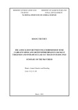
Fig. 3. Structures of diazonamide A and its analog, compound 1, which was used in the Hummel–Dreyer chromatographic analysis of [3H]do- lastatin 15 binding. The arrow indicates the position of the difference between the two compounds.
and [3H]dolastatin 10 to tubulin. An additional similarity between diazonamide A/compound 1 and dolastatin 15 is that these compounds do not interfere significantly with nucleotide exchange on b-tubulin. In contrast to dolasta- tin 15, however, diazonamide A and compound 1 are as potent as dolastatin 10 as inhibitors of tubulin assembly [24].
the equilibrium level was (cid:7) 1% of the average value. Combining these small increases in radiolabel with the protein concentrations in the two fractions yielded apparent Kd values of (cid:7) 1300 and (cid:7) 800 lM. Thus, for purposes of this discussion, an increase in the apparent Kd value to 50–100 lM will be considered weak inhibition, to 100–500 lM as moderate inhibition, and to > 500 lM as strong inhibition.
Examination of various vinca domain drugs and com- pound 1 for inhibitory effects on the tubulinÆdolastatin 15 interaction might provide information about the binding site of the depsipeptide on the protein relative to the other compounds. The selection of agents was determined only by availability of sufficient compound to equilibrate and develop the Hummel–Dreyer columns. The results obtained are summarized quantitatively in Table 1, in terms of the effects of specific concentration(s) of inhibitors on the stoichiometry of binding of [3H]dolastatin 15 to tubulin and on the apparent Kd value obtained for dolastatin 15. Our Hummel–Dreyer results are also summarized qualitatively in Table 2 for comparison with available data on drug inhibitory effects on the binding of vinca alkaloids and dolastatin 10 to tubulin.
Every compound examined, with the exceptions of compound 1 and the colchicine site drug combretasta- tin A-4, inhibited binding of dolastatin 15 to tubulin (Table 1). Weak inhibition was observed with saturating (nominally 50 lM) cryptophycin 1, moderate inhibition with vinblastine and vincristine, and strong inhibition with dolastatin 10, phomopsin A, maytansine, and halichond- rin B. A complicating factor in interpretation of these inhibitory data is that dolastatin 10 [2,28], phomopsin A [2,29], cryptophycin 1 [3,30,31], cemadotin [8], and vin- blastine and vincristine [32], besides inhibiting microtubule assembly, all induce formation of complex, morphologi- cally distinct, structurally aberrant oligomers and/or polymers of tubulin. It is possible that a binding site for dolastatin 15 could be inaccessible in these aberrant structures. However, while difficult to exclude, such a phenomenon has not yet been demonstrated as occurring between two classes of drug that inhibit microtubule assembly. In fact, it has often been observed that different drug classes tend to enhance each other’s binding, an observation usually explained in terms of ligand stabiliza- tion of the overall conformation of tubulin.
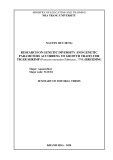
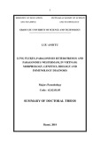
While there appears to be a wide range of effects on the apparent Kd values, a critical examination of the actual data used to generate the values shown in Table 1 revealed that values over about 500 lM should be considered equivalent and indicative of a strong inhibitory effect. Values > 500 lM were all observed when inhibition of dolasta- tin 15 binding was > 90%, with differences in the calculated apparent Kd values therefore of limited reliability. As a specific example, the experimental data obtained from one of the columns, in which the effect of 50 lM maytansine was examined, are shown in Fig. 4, with details summarized in the legend. The radiolabel associated with the two fractions containing the highest amounts of protein was only (cid:7) 2% above the column equilibrium level, and the standard deviation of the radiolabel in the fractions used to establish
When the inhibitory effects of the compounds examined with [3H]dolastatin 15 are compared qualitatively with those previously observed with radiolabeled dolastatin 10 and radiolabeled vinca alkaloids (Table 2), the results for
Fig. 4. Effect of coequilibrating the Hummel–Dreyer column with [3H]dolastatin 15 and 50 lM maytansine. The procedure was as described in the legend of Fig. 2, except the column equilibration/ development buffer also contained 50 lM maytansine. The same scales are used as in Fig. 2 to plot the [3H]dolastatin 15 and tubulin con- centrations of the appropriate fractions. The fractions used to calculate the equilibrated [3H]dolastatin 15 concentration had an average of 2137 c.p.m., with a standard deviation of 26 c.p.m. The two fractions with the two highest protein concentrations contained 38 and 49 c.p.m. above the background average; and these numbers, when combined with the tubulin concentrations, yielded Kd values of 1300 and 800 lM, respectively. Dolastatin 15 (radiolabel), s. Tubulin (protein), n.
Binding of dolastatin 15 to tubulin (Eur. J. Biochem. 270) 3827
(cid:1) FEBS 2003
The peptide site was originally proposed to explain findings with dolastatin 10 and with chiral isomers and segments of dolastatin 10 [2]. The concept seemed to be validated when hemiasterlin, phomopsin A, and crypto- phycin 1 were all found to be competitive inhibitors of the binding of [3H]dolastatin 10 to tubulin. The marked differ- ence shown here between the effects on [3H]dolastatin 15 binding of, on the one hand dolastatin 10 and phomo- psin A, and, on the other, of cryptophycin 1 suggests to us that the (cid:2)vinca domain(cid:3), and particularly the (cid:2)peptide site(cid:3), is most likely a relatively large binding pocket on the surface of b-tubulin. Presumably this pocket is able to accommo- date different complex natural product ligands by diverse binding mechanisms, involving different, possibly adjacent and/or overlapping, subsites.
the most part are reasonably parallel. They do not, however, seem to be related to whether competitive or noncompeti- tive inhibition patterns were observed. Thus, dolasta- tin 10/19aR-isodolastatin 10 and phomopsin A are strong inhibitors of the binding of all three ligands, while dolast- atin 15/cemadotin and compound 1 are weak inhibitors or noninhibitory. Maytansine and halichondrin B were mod- erate inhibitors of dolastatin 10 binding and strongly inhibited vinca alkaloid binding and dolastatin 15 binding. In cross-inhibition studies with the two vinca alkaloids, vincristine seemed to bind to tubulin more tightly than vinblastine, and these relative activities were reflected in greater inhibition by vincristine than vinblastine of dolast- atin 10 binding. In the present studies, however, we found little difference in their inhibitory effects on dolastatin 15 binding. Our observation of moderate inhibition of dolast- atin 15 binding by vinblastine contrasts with the failure of Jordan et al. [10] to observe inhibition by vinblastine of the binding of cemadotin to tubulin, using the centrifugal gel filtration methodology.
We wish to note that, in principle, it should be possible to adapt the Hummel–Dreyer method to obtain more specific data about drug binding sites (e.g. whether binding is competitive or noncompetitive). This would require accu- mulation and analysis of data obtained at multiple drug concentrations, but we did not have adequate amounts of either [3H]dolastatin 15 or most of the potential inhibitors to undertake such an evaluation.
The most distinctive pattern was obtained with crypto- phycin 1. Despite the depsipeptide’s potent inhibition of [3H]vinblastine and of [3H]dolastatin 10 binding to tubulin, it had only a weak inhibitory effect on [3H]dolastatin 15 binding. This would suggest a significant difference in the topography of binding of cryptophycin 1 to tubulin vs. the binding of dolastatin 10/19aR-isodolastatin 10 and pho- mopsin A, despite the kinetic evidence that the dolastatin 10 peptides, phomopsin A, and cryptophycin 1 bind at the same site on tubulin.
to inhibit
In addition, the failure of compound 1, and presumably [3H]dolastatin 15 binding diazonamide A, re-emphasizes the unique properties of these new antimitotic inhibition of assembly, peptides. Despite their potent suggesting a relatively strong interaction with tubulin, these peptides seem unable to inhibit the binding of vinblastine, dolastatin 10, dolastatin 15, or colchicine to tubulin. This may indicate an exceptionally weak interaction with unpoly- merized tubulin (the ab-tubulin heterodimer), presumably in the vinca domain, a truly unique binding site, or, perhaps, a specific affinity for microtubule ends and not for the heterodimer.
Finally, the relatively low affinity of both dolastatin 15 and cemadotin for tubulin contrasts with their potent cytotoxicity. Although this apparent discrepancy could indicate that the tubulin–microtubule system is not the primary or only target for these drugs, this seems unlikely. Dolastatin 15 and cemadotin cause classic mitotic arrest with disappearance of intracellular microtubules [8,9], and cemadotin dramatically suppresses microtubule dynamics [10]. These are cellular effects always observed with drugs that are believed to exert their cytotoxicity through an inhibitory interaction with the microtubule system. One possible explanation for the potent cytotoxicity of dolast- atin 15 and cemadotin is that these agents are metabolized intracellularly to a compound with increased affinity for tubulin, perhaps involving hydrolysis of the ester bond of dolastatin 15 or removal of the benzamide group in cemadotin. Pettit et al. [33] described the synthesis of a large series of dolastatin 15 analogs, all possessing the amino-terminal pentapeptide and different groups at the carboxy-side of the ester bond. None was as active as dolastatin 15 in a tubulin-based assay, but they all had antimitotic activity equivalent to that of dolastatin 15, leading to the proposal that a common intracellular metabolite might be responsible for the activity of the compounds. A similar proposal has been made to account for the activity of cemadotin [8], but an active intracellular metabolite with enhanced affinity for tubulin has yet to be documented.
References
1. Hamel, E. & Covell, D.G. (2002) Antimitotic peptides and depsipeptides. Curr. Med. Chem. – Anti-Cancer Agents 2, 19–53. 2. Bai, R., Pettit, G.R. & Hamel, E. (1990) Binding of dolastatin 10 to tubulin at a distinct site for peptide antimitotic agents near the exchangeable nucleotide and vinca alkaloid sites. J. Biol. Chem. 265, 17141–17149.
In conclusion, we have been able to demonstrate by Hummel–Dreyer chromatography that dolastatin 15 has a relatively low binding affinity (apparent Kd of (cid:7) 30 lM) for the ab-tubulin heterodimer and that the Hummel–Dreyer method may be modified to document inhibitory effects on ligand binding. Because of the large number of compounds that interfered with the tubulin–dolastatin 15 interaction, we were unable to definitively characterize the binding site for dolastatin 15 on tubulin. This binding site does appear to be within the (cid:2)vinca domain(cid:3), even though both compet- itive and noncompetitive inhibitors of vinca alkaloid binding to tubulin had similar inhibitory effects on the binding of dolastatin 15 to tubulin. The most unexpected findings were the weak inhibition of dolastatin 15 binding by cryptophycin 1 and the complete failure of the diazon- amide A analog compound 1 to inhibit dolastatin 15 binding. While our results here and in earlier studies [24] have provided no insight into the binding site for diazon- amide A on tubulin, the cryptophycin 1 result suggests that our concept of a (cid:2)peptide site(cid:3) should be modified.
3. Bai, R., Schwartz, R.E., Kepler, J.A., Pettit, G.R. & Hamel, E. (1996) Characterization of the interaction of cryptophycin 1 with
3828 Z. Cruz-Monserrate et al. (Eur. J. Biochem. 270)
(cid:1) FEBS 2003
true structure and origin of the natural isolates. Angew. Chem. Int. Ed. Engl. 40, 4770–4773. tubulin: binding in the Vinca domain, competitive inhibition of dolastatin 10 binding, and an unusual aggregation reaction. Cancer Res. 56, 4398–4406.
19. Singer, W.D., Hersh, R.T. & Himes, R.H. (1988) Effect of solution variables on the binding of vinblastine to tubulin. Biochem. Pharmacol. 37, 2691–2696.
4. Bai, R., Durso, N.A., Sackett, D.L. & Hamel, E. (1999) Inter- actions of the sponge-derived antimitotic tripeptide hemiasterlin with tubulin: comparison with dolastatin 10 and cryptophycin 1. Biochemistry 43, 14302–14310. 20. Singer, W.D. & Himes, R.H. (1992) Cellular uptake and tubulin binding properties of four vinca alkaloids. Biochem. Pharmacol. 43, 545–551.
5. Natsume, T., Watanabe, J., Tamoki, S., Fujio, N., Miyasaka, K. & Kobayashi, M. (2000) Characterization of the interaction of TZT-1027, a potent antitumor agent, with tubulin. Jpn. J. Cancer Res. 91, 737–747. 21. Lin, C.M., Ho, H.H., Pettit, G.R. & Hamel, E. (1989) Antimitotic natural products combretastatin A-4 and combretastatin A-2: studies on the mechanism of their inhibition of the binding of colchicine to tubulin. Biochemistry 28, 6984–6991.
6. Edler, M.C., Fernandez, A.M., Lassota, P., Ireland, C.M. & Barrows, L.R. (2002) Inhibition of tubulin polymerization by vitilevuamide, a bicyclic marine peptide, at a site distinct from colchicine, the vinca alkaloids and dolastatin 10. Biochem. Phar- macol. 63, 707–715.
22. Bai, R., Paull, K.D., Herald, C.L., Malspeis, L., Pettit, G.R. & Hamel, E. (1991) Halichondrin B and homohalichondrin B, marine natural products binding in the vinca domain of tubulin: discovery of tubulin-based mechanism of action by analysis of differential cytotoxicity data. J. Biol. Chem. 266, 15882–15889. 23. Bai, R., Taylor, G.F., Cichacz, Z.A., Herald, C.L., Kepler, J.A., Pettit, G.R. & Hamel, E. (1995) The spongistatins, potently cytotoxic inhibitors of tubulin polymerization, bind in a distinct region of the vinca domain. Biochemistry 34, 9714–9719.
7. Pettit, G.R., Kamano, Y., Dufresne, C., Cerny, R.L., Herald, C.L. & Schmidt, J.M. (1989) Isolation and structure of the cytostatic linear depsipeptide dolastatin 15. J. Org. Chem. 54, 6005–6006. 8. de Aruda, M., Cocchiaro, C.A., Nelson, C.M., Grinnell, C.M., Janssen, B., Haupt, A. & Barlozzari, T. (1995) LU103793 (NSC D-669356): a synthetic peptide that interacts with microtubules and inhibits mitosis. Cancer Res. 55, 3085–3092.
24. Cruz-Monserrate, Z., Vervoort, H.C., Bai, R., Newman, D.J., Howell, S.B., Los, G., Mullaney, J.T., Williams, M.D., Pettit, G.R., Fenical, W. & Hamel, E. (2003) Diazonamide A and a synthetic structural analog: disruptive effects on mitosis and cel- lular microtubules and analysis of their interactions with tubulin. Mol. Pharmacol. 63, 1273–1280. 9. Bai, R., Friedman, S.J., Pettit, G.R. & Hamel, E. (1992) Dolas- tatin 15, a potent antimitotic depsipeptide derived from Dolabella auricularia: interaction with tubulin and effects on cellular microtubules. Biochem. Pharmacol. 43, 2637–2645.
25. Bai, R., Taylor, G.F., Schmidt, J.M., Williams, M.D., Kepler, J.A., Pettit, G.R. & Hamel, E. (1995) Interaction of dolastatin 10 with tubulin: induction of aggregation and binding and dissocia- tion reactions. Mol. Pharmacol. 47, 965–976. 10. Jordan, M.A., Walker, D., de Arruda, M., Barlozzari, T. & Panda, D. (1998) Suppression of microtubule dynamics by binding of cemadotin to tubulin: possible mechanism of its antitumor action. Biochemistry 37, 17571–17578.
11. Hummel, J.P. & Dreyer, W.J. (1962) Measurement of protein- binding phenomena by gel filtration. Biochim. Biophys. Acta 63, 530–532.
12. Hamel, E. & Lin, C.M. (1984) Separation of active tubulin and microtubule-associated proteins by ultracentrifugation and isola- tion of a component causing the formation of microtubule bun- dles. Biochemistry 23, 4173–4184. 13. Grover, S. & Hamel, E. (1994) The magnesium–GTP interaction 26. Panda, D., Ananthnarayan, V., Larson, G., Shih, C., Jordan, M.A. & Wilson, L. (2000) Interaction of the antitumor compound cryptophycin-52 with tubulin. Biochemistry 39, 14121–14127. 27. Moore, R.E. (1997) Presentation at the Fourth Anticancer Drug Discovery and Development Symposium, Annapolis, Maryland. 28. Bai, R., Pettit, G.R. & Hamel, E. (1990) Dolastatin 10, a powerful cytostatic peptide derived from a marine animal: inhibition of tubulin polymerization mediated through the vinca alkaloid binding domain. Biochem. Pharmacol. 39, 1941–1949. in microtubule assembly. Eur. J. Biochem. 222, 163–172.
14. Pettit, G.R., Singh, S.B., Hogan, F., Lloyd-Williams, P., Herald, D.L., Burkett, D.D. & Clewlow, P.J. (1989) The absolute config- uration and synthesis of natural (–)-dolastatin 10. J. Am. Chem. Soc. 111, 5463–5465.
29. To¨ nsing, E.M., Steyn, P.S., Osborn, M. & Weber, K. (1984) Phomopsin A, the causative agent of lupinosis, interacts with microtubules in vivo and in vitro. Eur. J. Cell Biol. 35, 156–164. 30. Kerksiek, K., Mejillano, M.R., Schwartz, R.E., Georg, G.I. & Himes, R.H. (1995) Interaction of cryptophycin 1 with tubulin and microtubules. FEBS Lett. 377, 59–61. 15. Pettit, G.R., Thornton, T.J., Mullaney, J.T., Boyd, M.R., Herald, D.L., Singh, S.-B. & Flahive, E.J. (1994) A convenient synthetic route to dolastatin 15. Tetrahedron 50, 12097–12108.
31. Smith, C.D. & Zhang, X. (1996) Mechanism of action of cryp- tophycin: interaction with the Vinca alkaloid domain of tubulin. J. Biol. Chem. 271, 6192–6198. 16. Pettit, G.R., Singh, S.B., Hamel, E., Lin, C.M. & Alberts, D.S. (1989) Isolation and structure of the strong cell growth and tubulin inhibitor combretastatin A-4. Experientia 45, 209–211.
32. Himes, R.H. (1991) Interactions of the Catharanthus (Vinca) alkaloids with tubulin and microtubules. Pharmac. Ther. 51, 257–267.
17. Schwartz, R.E., Hirsch, C.F., Sesin, D.F., Flor, J.E., Chartrain, M., Fromtling, R.E., Harris, G.H., Salvatore, M.J., Liesch, J.M. & Yudin, K. (1990) Pharmaceuticals from cultured algae. J. Indust. Microbiol. 5, 113–124.
18. Li, J., Burgett, A.W.G., Esser, L., Amezcua, C. & Harran, P.G. (2001) Total synthesis of nominal diazonamides – part 2: on the 33. Pettit, G.R., Flahive, E.J., Boyd, M.R., Bai, R., Hamel, E., Pettit, R.K. & Schmidt, J.M. (1998) Synthesis and cancer cell growth inhibitory studies of dolastatin 15 structural modifications. Anti- Cancer Drug Design 13, 47–66.