Enzymes of mannitol metabolism in the human pathogenic fungus Aspergillus fumigatus – kinetic properties of mannitol-1-phosphate 5-dehydrogenase and mannitol 2-dehydrogenase, and their physiological implications Stefan Krahulec, Guilliano Cem Armao, Mario Klimacek and Bernd Nidetzky
Institute of Biotechnology and Biochemical Engineering, Graz University of Technology, Austria
Keywords Aspergillus fumigatus; mannitol; mannitol metabolism; mannitol-1-phosphate 5- dehydrogenase; mannitol 2-dehydrogenase
Correspondence B. Nidetzky, Institute of Biotechnology and Biochemical Engineering, Graz University of Technology, Petersgasse 12 ⁄ I, A-8010 Graz, Austria Fax: +43 316 873 8434 Tel: +43 316 873 8400 E-mail: bernd.nidetzky@TUGraz.at
(Received 17 December 2010, revised 1 February 2011, accepted 4 February 2011)
doi:10.1111/j.1742-4658.2011.08047.x
Abbreviations AbM2DH, mannitol 2-dehydrogenase from Agaricus bisporus; AfM1PDH, mannitol-1-phosphate 5-dehydrogenase from Aspergillus fumigatus; AfM2DH, mannitol 2-dehydrogenase from Aspergillus fumigatus; EcM1PDH, mannitol-1-phosphate 5-dehydrogenase from Escherichia coli; Fru, D-fructose; Fru6P, D-fructose 6-phosphate; KIE, kinetic isotope effect; Man-ol, D-mannitol; Man-ol1P, D-mannitol 1-phosphate; M1PDH, mannitol-1-phosphate 5-dehydrogenase; M2DH, mannitol 2-dehydrogenase; NADD, (4S)-[2H]-NADH; PSLDR, polyol- specific long-chain dehydrogenase ⁄ reductase; PsM2DH, mannitol 2-dehydrogenase from Pseudomonas fluorescens.
FEBS Journal 278 (2011) 1264–1276 ª 2011 The Authors Journal compilation ª 2011 FEBS
1264
The human pathogenic fungus Aspergillus fumigatus accumulates large amounts of intracellular mannitol to enhance its resistance against defense strategies of the infected host. To explore their currently unknown roles in studied A. fumigatus mannitol-1-phosphate mannitol metabolism, we 5-dehydrogenase (AfM1PDH) and mannitol 2-dehydrogenase (AfM2DH), each recombinantly produced in Escherichia coli, and performed a detailed steady-state kinetic characterization of the two enzymes at 25 (cid:2)C and pH 7.1. Primary kinetic isotope effects resulting from deuteration of alco- hol substrate or NADH showed that, for AfM1PDH, binding of D-manni- tol 1-phosphate and NAD+ is random, whereas D-fructose 6-phosphate binds only after NADH has bound to the enzyme. Binding of substrate and NAD(H) by AfM2DH is random for both D-mannitol oxidation and D-fructose reduction. Hydride transfer is rate-determining for D-mannitol 1-phosphate oxidation by AfM1PDH (kcat = 10.6 s)1) as well as D-fructose reduction by AfM2DH (kcat = 94 s)1). Product release steps control the maximum rates in the other direction of the two enzymatic reactions. Free energy profiles for the enzymatic reaction under physiological boundary conditions suggest that AfM1PDH primarily functions as a D-fructose-6- phosphate reductase, whereas AfM2DH acts in D-mannitol oxidation, thus establishing distinct routes for production and mobilization of mannitol in A. fumigatus. ATP, ADP and AMP do not affect the activity of AfM1PDH, suggesting the absence of flux control by cellular energy charge at the level of D-fructose 6-phosphate reduction. AfM1PDH is remarkably resistant to inactivation by heat (half-life at 40 (cid:2)C of 20 h), consistent with the idea that formation of mannitol is an essential component of the tem- perature stress response of A. fumigatus. Inhibition of AfM1PDH might be a useful target for therapy of A. fumigatus infections.
S. Krahulec et al.
Enzymes of mannitol metabolism in A. fumigatus
Introduction
dependent M2DH produces Fru, which is then phos- phorylated from ATP by hexokinase (EC 2.7.1.1) to regenerate Fru6P [12]. It is currently not clear whether stockpiled mannitol could also be utilized by going through the steps of path 1 in reverse [13–16], whereby Man-ol would have to become phosphorylated by a suitable kinase. [1]. stress fungi, In parasitic
The six-carbon polyol d-mannitol (Man-ol) is ubiqui- tous throughout the fungal kingdom, and one of the most abundant sugar alcohols in nature. Aside from other physiological functions that have been ascribed to it, intracellular accumulation of Man-ol is a wide- spread mechanism by which fungi cope with different including high temperature forms of external stress, and oxidative the improved stress resistance resulting from accumulated Man-ol appears to confer a substantially enhanced ability to deal with defense strategies of the infected host [2–6]. The human pathogen Aspergillus fumigatus, which is the most common agent causing invasive aspergillosis in immunosuppressed patients, produces enough Man-ol to raise the serum Man-ol level of the infected animal [7,8]. High thermotolerance is a major component of A. fumigatus pathogenicity, and involves Man-ol indirectly [9]. Decreased susceptibility to reac- tive oxygen species produced by human phagocytes in response to the microbial infection is yet another viru- lence factor of A. fumigatus, and includes direct partic- ipation of Man-ol as a free radical scavenger [10,11]. The inhibition of fungal Man-ol production therefore presents a potential target in advanced therapies for A. fumigatus infections. Unfortunately, little is cur- rently known about the enzyme system of Man-ol metabolism in A. fumigatus.
M1PDH and NAD+-dependent M2DH have recently been characterized from A. fumigatus [17,18]. On the basis of sequence similarity, both enzymes were classified as members of the polyol-specific long-chain family [12]. The dehydrogenase ⁄ reductase (PSLDR) PSLDRs constitute a diverse group of NAD(P)- dependent oxidoreductases that are widespread among microorganisms but are lacking in humans. Their acti- vity is not dependent on a metal cofactor and is exclu- sively targeted towards polyol ⁄ ketose substrates [12,19]. In a family-wide categorization of the PSLDRs, M1PDH and M2DH were clearly separated from each other, showing only a distant evolutionary relationship [12,19]. Structurally, PSLDRs are composed of two separate domains. The N-terminal domain adopts an expanded Rossmann fold, and provides the residues for binding the coenzyme. The C-terminal domain has a lar- gely a-helical structure, and serves mainly in substrate binding. The active site is located in the interdomain cleft, and contains a highly conserved tetrad of residues (Lys ⁄ Asn ⁄ Asn ⁄ His), whereby Lys is the catalytic acid– base of the enzymatic reaction [20,21]. A recent study has demonstrated, at the level of both gene transcript that A. fumigatus M1PDH and translated protein, (AfM1PDH) becomes strongly upregulated during heat shock [9]. Enhanced biosynthesis of Man-ol via AfM1PDH-catalyzed conversion of Fru6P might contribute extra robustness to A. fumigatus under high- temperature conditions.
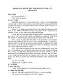
A number of studies suggest that two different meta- bolic paths contribute to the biosynthesis of Man-ol in fungi (Scheme 1; reviewed in [1]). Reduction of d-fruc- tose 6-phosphate (Fru6P) by an NADH-dependent mannitol-1-phosphate 5-dehydrogenase (M1PDH; EC 1.1.1.17) gives d-mannitol 1-phosphate (Man-ol1P), which, upon hydrolysis of phosphate ester by a phos- phatase (EC 3.1.3.22), yields Man-ol (path 1). In the alternative route (path 2), Fru6P is converted to d-fructose (Fru), which, in turn, is reduced to Man-ol by NADPH-dependent or NADH-dependent mannitol 2-dehydrogenase (M2DH; EC 1.1.1.138 or 1.1.1.67). Mobilization of Man-ol occurs through quasireversal of path 2, whereby an NADP+-dependent or NAD+-
Scheme 1. Metabolic pathways for the biosynthesis of mannitol in fungi. HXK, hexokinase; M1Pase, mannitol-1-phosphatase.
FEBS Journal 278 (2011) 1264–1276 ª 2011 The Authors Journal compilation ª 2011 FEBS
1265
In this study, an enzymological approach was chosen to examine the roles of AfM1PDH and A. fumigatus M2DH (AfM2DH) in the metabolism of Man-ol in A. fumigatus. A detailed steady-state kinetic character- ization was performed with each enzyme, providing the basis for the construction of free energy profiles for the enzymatic reactions under physiological boundary con- ditions as defined from the literature. The results pro- vide clear assignment of a biosynthetic function to AfM1PDH (path 1), which behaves kinetically as a Fru6P reductase, whereas AfM2DH is essentially a Man-ol-oxidizing enzyme (path 2, backwards). The results of inhibition studies show that the activity of AfM1PDH would not be affected by changes in the lev- els of ATP, ADP and AMP, suggesting that flux
S. Krahulec et al.
Enzymes of mannitol metabolism in A. fumigatus
conclude that the phosphate moiety in Man-ol1P and Fru6P is essential for substrate recognition and ⁄ or catalysis by AfM1PDH.
through Fru6P reduction is not under direct control of the cellular energy charge. Considering the results of studies on the human pathogenic fungus Cryptococ- cus neoformans, showing that a low Man-ol-producing mutant strain was a 5000-fold less potent agent than the wild type [2,3], we propose that inhibition of AfM1PDH might be exploitable in the development of novel thera- peutic strategies against A. fumigatus infection.
Results
Substrate specificity of Af M1PDH and Af M2DH
Purified preparations of recombinant AfM1PDH and AfM2DH were assayed for activity in the directions of alcohol oxidation by NAD+ (pH 10.0) and carbonyl group reduction by NADH (pH 7.1), with a range of possible alternative substrates. Both enzymes showed only trace activity for utilization of NADP+. Catalytic efficiencies (in terms of kcat ⁄ KNADP) were more than three orders of magnitude below those obtained with NAD+ [18]. AfM1PDH and AfM2DH contain a con- served Asp (Asp33 and Asp77, respectively) in their coenzyme-binding pockets that is known from previous studies of a related PSLDR, M2DH from Pseudomo- nas fluorescens (PsM2DH), to prevent accommodation of the 2¢-phosphate group of NADP+ [20,22]. Reactions of A. fumigatus enzymes that are dependent on NADP+ or NADPH were therefore not further investigated.
d-mannose, l-arabinose,
Alcohol oxidation by AfM2DH was assayed across substrates utilized above with the same series of AfM1PDH. l-Arabinitol, d-arabinitol, d-ribose, 2-deoxy- d-galactose and 2-deoxy-d-glucose were additionally tested as alcohol substrates. In the reduction direction, l-sorbose, d-xylulose and dihydroxyacetone were exam- ined. Above the 1% level of activity with Man-ol and Fru, two polyols (d-arabinitol and d-sorbitol) and two ketoses (d-xylulose and l-sorbose) gave significant con- version rates. The known regioselectivity of M2DH in the oxidation of polyols [23], indicated in Table 1, allows assignment of d-xylulose and l-sorbose as the products of oxidation of d-arabinitol and d-sorbitol, respectively. Table 1 summarizes the results of kinetic parameter determination for polyol–ketose substrate pairs of AfM2DH. Structural comparison of polyols that are reactive for NAD+-dependent oxidation by AfM2DH with those that are not substrates of the enzyme (Fig. S1) reveals that a d-arabo configuration is required for a polyol to become reactive. The C2 (R) configuration (Man-ol) is preferred over the C2 (S) con- figuration (d-sorbitol). A model of AfM2DH was gener- ated with the crystal structure of PsM2DH as the template (Fig. S2) [20]. Residues contributing to the substrate-binding site of PsM2DH are fully conserved in AfM2DH, explaining the observed substrate specific- ity of the A. fumigatus enzyme [12,20]. It can be assumed from the way in which Man-ol interacts with the sub- strate-binding site of PsM2DH in the crystal structure that ketose substrates must bind in their open-chain free-carbonyl form [20]. The values of Km and kcat ⁄ Km in Table 1 are appropriately corrected for the available proportion of reactive ketose substrate present.
Kinetic characterization
Table 1. Kinetic constants of AfM2DH for reactions with different polyol and ketose substrates. Km and kcat ⁄ Km data for carbonyl substrates are corrected for the available proportion of open-chain free-carbonyl forms present in aqueous solution (Fru, (cid:2) 1% [42]; D-xylulose, (cid:2) 20% [43]; and L-sorbose, 0.2% [44]). Numbers in parentheses show values as measured. Ketose reductions, pH 7.1; polyol oxidations, pH 10.0. ND, not detectable (enzyme could not be saturated with L-sorbose).
Substrate
)1)
kcat (s)1)
Km (mM)
kcat ⁄ Km (s)1ÆmM
0.60 ± 0.07 (60 ± 7)
1.7 ± 0.1 (8.3 ± 0.4)
ND
D-Fructose D-Xylulose L-Sorbose D-Mannitol D-Arabinitol D-Sorbitol
86 ± 5 64 ± 1 ND 212 ± 3 162 ± 5 60 ± 1
13 ± 1 163 ± 13 680 ± 30
140 ± 20 (1.4 ± 0.2) 39 ± 2 (7.7 ± 0.4) 1.1 ± 0.1 (0.0022 ± 0.0001) 17 ± 1 0.99 ± 0.08 0.088 ± 0.004
FEBS Journal 278 (2011) 1264–1276 ª 2011 The Authors Journal compilation ª 2011 FEBS
1266
A full steady-state kinetic characterization of AfM1PDH and AfM2DH was carried out under physiological pH Above a level of 1% activity with Man-ol1P (1.0 mm), AfM1PDH did not catalyze the oxidation of Man-ol, d-sorbitol, d-ribitol, xylitol, d-xylose, l-xylose, d-glucose, d-arabinose, d-galactose, l-fucose, and d-lyxose. The enzyme was also inactive above a level of 1% activity with Fru6P (100 mm) for reduction of Fru, l-sorbose, d-xylulose, d-fructose 1,6-bisphosphate, d-glucose 6-phosphate (150 mm), and d-glucose 1-phosphate (150 mm). There- fore, AfM1PDH appears to be fairly specific for its natu- ral pair of substrates, Man-ol1P and Fru6P. From the highly truncated activity with Man-ol and Fru, we
S. Krahulec et al.
Enzymes of mannitol metabolism in A. fumigatus
the regulation of
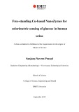
of (cid:2) 800 lm, AMP binds one order of magnitude less strongly to EcM1PDH than does ATP [25]. To exam- two A. fumigatus ine possible enzymes by components of the cellular energy charge (ATP, ADP, and AMP), we measured inhibition of AfM1PDH and AfM2DH by each of these adenine nucleotides. Figure S3 shows double-reciprocal plots of initial-rate data recorded at different concentrations of inhibitor. The observed inhibition was, in each case, best described by competitive binding of coenzyme and EI) were adenine nucleotide. Inhibition constants (Ki obtained from nonlinear fits of Eqn (5) to the data. They are summarized in Table 3. Both AfM1PDH and AfM2DH were inhibited weakly by adenine nucleo- tides as compared with the inhibition of EcM1PDH by ATP. conditions. Lineweaver–Burk plots of initial-rate data (Fig. 1) gave a pattern of intersecting lines, for each enzyme and in both reaction directions, consistent with a kinetic mechanism in which substrate and coenzyme must bind to the enzyme to form a ternary complex prior to the release of the first product. Kinetic param- eters were obtained from nonlinear least-squares fits of Eqn (3) or Eqn (4) to the experimental data. They are summarized in Table 2, and their internal consistency was verified with Haldane relationship analysis, com- paring the kinetically determined reaction equilibrium constant (Keq) from Eqn (7) with those reported in the literature. Table 2 shows the useful agreement between previously published and calculated Keq values [23,24]. It is interesting that, in terms of Km, AfM1PDH binds Man-ol1P two orders of magnitude more tightly than AfM2DH binds Man-ol.
Kinetic isotope effects (KIEs)
Inhibition of Af M1PDH and Af M2DH by components of the cellular energy charge
(polyol oxidation) or coenzyme
Primary KIEs resulting from deuterium substitution of the hydrogen atom undergoing hydride transfer from (ketose substrate reduction) were determined for AfM1PDH and AfM2DH at pH 7.1. Initial-rate data recorded with protio and deuterio substrate or coenzyme were fitted with Eqn (6), and KIEs on kinetic parameters are M1PDH from Escherichia coli (EcM1PDH) is evolu- tionary related to AfM1PDH by common membership of the family of PSLDRs [12,19]. It is strongly inhib- ited by ATP, which acts as a competitive inhibitor against NADH with a Ki of (cid:2) 60 lm [25]. With a Ki
A
B
C
D
Fig. 1. Double reciprocal plots of initial-rate data obtained for AfM1PDH (A, B) and AfM2DH (C, D) at pH 7.1 and 25 (cid:2)C.
FEBS Journal 278 (2011) 1264–1276 ª 2011 The Authors Journal compilation ª 2011 FEBS
1267
S. Krahulec et al.
Enzymes of mannitol metabolism in A. fumigatus
Table 2. Kinetic parameters of AfM1PDH and AfM2DH at 25 (cid:2)C and pH 7.1. NA, not applicable.
Parameter
AfM1PDH
AfM2DH
10.6 ± 0.7
0.13 ± 0.05
14.2 ± 0.3 11 ± 2
80 ± 30
)1Æs)1)
0.8 ± 0.3 1.6 ± 0.8
1.3 ± 0.2 0.11 ± 0.02 2.0 ± 0.4
132 ± 3
3.2 ± 0.2
)1Æs)1)
41 ± 3 14 ± 1 2 ± 1
94 ± 4 40 ± 20 2 ± 1 15 ± 8 NA
1.4 ± 0.9
NA 300
b (M) c (M)
3 · 10)10 5 · 10)10 [24]
20 4 · 10)9 5 · 10)9 [23]
kox (s)1) KMan-ol(1P ) (mM) kox ⁄ KMan-ol(1P ) (mM KNAD (mM) aox kred (s)1) KFru(6P ) (mM) kred ⁄ KFru(6P ) (mM KNADH (lM) KiNADH (lM) ared 1 ⁄ app Keq (pH 7.1)a Keq Keq
0.3) represents a mean value and standard deviation for three independent experiments evaluated in either of the ways described above. The KIE on kcat ⁄ Km for Fru was calculated from the kcat and Km values obtained with NADH and NADD. The KIE on kcat ⁄ Km for coenzyme was calculated as the Dkcat ⁄ DKm ratio, where the value of DKm was obtained by dividing Km data for reactions with NADH and NADD. Note that Km (NADH) was invariant (± 15%) across a wide range of Fru concen- trations (40–800 mm), and so the choice of the constant level of Fru was not critical for determination of DKm (1.23 ± 0.15). The KIE data in Table 4 are instrumen- tal in delineating the kinetic mechanism of AfM1PDH and AfM2DH, as described in the Discussion.
a The dimensionless app Keq at pH 7.1 was calculated with the Hal- dane relationship (Eqn 7), using kinetic parameters from this Table. b The given Keq is pH-independent, and was obtained from the dimensionless app Keq at pH 7.1. c Experimentally determined, pH- independent equilibrium constant.
Free energy profile analysis for reactions catalyzed by Af M1PDH and Af M2DH
catalyzed
EI is a constant describing competitive inhibition
Table 3. Inhibition of AfM1PDH and AfM2DH by adenine nucleotides at pH 7.1 and 25 (cid:2)C. Ki of AMP, ADP or ATP against NAD+ or NADH. ND, not detectable.
AfM1PDH
AfM2DH
Fru6P reduction
Man-ol1P oxidation
Fru reduction
Man-ol oxidation
EI
EI
EI
4.6 ± 0.6 5.6 ± 1.9 6.5 ± 1.4
1.5 ± 0.1 2.0 ± 0.2 1.4 ± 0.1
5.6 ± 0.5 5.7 ± 0.4 ND
2.9 ± 0.7 4.8 ± 1.3 ND
Ki Ki Ki
AMP (mM) ADP (mM) ATP (mM)
FEBS Journal 278 (2011) 1264–1276 ª 2011 The Authors Journal compilation ª 2011 FEBS
1268
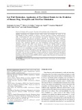
Interpretation of the kinetic properties of an enzyme in the context of its role in cellular metabolism relies on knowledge of the physiological concentrations of sub- strates, products, coenzymes and relevant effectors. We are not aware of a study reporting intracellular metab- olite levels in A. fumigatus (except for an early study commenting on the cellular Man-ol content). However, data for the closely related fungus Aspergillus niger are available. Table 5 shows a list of metabolite concentra- tions calculated from values in the literature, thus defining plausible reaction conditions for AfM1PDH and AfM2DH in vivo. Because values for the intracel- lular concentration of Fru are not available for asper- gilli, we approximated the level of Fru with the known intracellular Fru concentration of Rhizobium legumin- osarum [26]. With the assumption of the conditions in Table 5, kinetic constants from Table 2 (including the EI values for ATP, ADP and AMP from Table 3) Ki were used to construct free energy profiles for the by AfM1PDH and transformations AfM2DH. These free energy profiles (Fig. 2) show that both enzymes operate under nonequilibrium reaction conditions that involve a substantial thermodynamic driving force for reduction of Fru6P and oxidation of shown in Table 4. A nomenclature is used whereby superscript D designates the KIE (e.g. Dkcat). A KIE greater than unity means that hydrogen fi deuterium replacement caused slowing down of the enzymatic reac- tion analyzed. NADH-dependent reduction of Fru cata- lyzed by AfM2DH was characterized by substrate inhibition at high Fru concentrations (KiS = 2 ± 1 m). Substrate inhibition was observed irrespective of whether the NADH concentration used was saturating or limiting in the sub-Km range. With the use of (4S)- [2H]-NADH (NADD) instead of NADH, this substrate inhibition disappeared (Fig. S4), precluding the use of a single equation for calculation of the KIEs. We there- fore obtained Dkcat from a direct comparison of kcat data derived from nonlinear fits of Eqn (2) and Eqn (1) with initial rates recorded with NADH and NADD, respec- tively and additionally by fitting Eqn (6) to the experi- mental data obtained below the occurrence of substrate inhibition. Dkcat for Fru reduction in Table 4 (2.0 ±
S. Krahulec et al.
Enzymes of mannitol metabolism in A. fumigatus
Table 4. KIEs for reactions of AfM1PDH and AfM2DH at pH 7.1.
AfM1PDH
2.9 ± 0.2 2.4 ± 0.5 2.4 ± 0.4
1.5 ± 0.1 3.1 ± 0.4 0.8 ± 0.2
Man-ol1P oxidation Dkcat Dkcat ⁄ KMan-ol1P Dkcat ⁄ KNAD Fru6P reduction Dkcat Dkcat ⁄ KFru6P Dkcat ⁄ KNADH
AfM2DH
1.0 ± 0.1 1.2 ± 0.2 1.6 ± 0.2
2.0 ± 0.3 1.2 ± 0.1 1.6 ± 0.3
Man-ol oxidation Dkcat Dkcat ⁄ KMan-ol Dkcat ⁄ KNAD Fru reduction Dkcat Dkcat ⁄ KFru Dkcat ⁄ KNADH
Table 5. Internal metabolite concentrations from the literature.
Metabolite
mM
Species
Ref.
Man-ol. Considering the uncertainty in the in vivo levels of NADH, Fru and Man-ol1P used, we per- formed a sensitivity analysis in which the effects of changes in intracellular reactant concentrations on the thermodynamic boundary conditions for the action of AfM1PDH and AfM2DH were examined. The allowed concentration ranges were comprehensive: 5–150 lm NADH; 0.01–10 mm Fru; and 10–400 lm Man-ol1P. The results (see the shaded area in Fig. 2) indicate that the overall conclusion of this work, that the physiolog- ical direction of the reaction of AfM1PDH is Fru6P reduction, whereas that of AfM2DH is Man-ol oxida- tion, was not affected by the assumed variation in the reactant concentrations. Equations (10) and (11) are (simplified) kinetic expressions that can be used to esti- mate the net direction of the enzymatic reaction (knet = oxidation – reduction) with the given substrate and product concentrations. Applying the concentra- tions from Table 5 to Eqns (10, 11), we find, for AfM1PDH, that the direction parameter knet is nega- tive or, in other words, Fru6P reduction is preferred. With AfM2DH, by contrast, knet is positive, indicating that the reaction proceeds in the direction of Man-ol oxidation.
High-temperature stability of Af M1PDH
Man-ol1P Fru6P Man-ol Fru ATP ADP AMP NAD+ NADH
0.05a 0.23 50a 0.4 2.6 0.47 0.09 0.83 0.01
Aspergillus niger A. niger Aspergillus fumigatus Rhizobium leguminosarum A. niger A. niger A. niger A. niger A. niger
[30] [45] [46] [26] [45] [45] [45] [45] [45]
a Calculated from lmolÆg)1 dry mycelium with application of an intracellular volume of 1.2 mLÆg)1 dry weight as determined for the mycelium of A. niger [45].
involves production upregulated
The literature suggests that the heat stress response of A. fumigatus of M1PDH [9]. Because the function of AfM1PDH at ele- vated temperatures might require pronounced resis- tance of the enzyme to inactivation by heat, we recorded time courses of irreversible loss of activity at temperatures, and use half-life times (sH), different (Fig. S5), as calculated from these measurements
A
B
Fig. 2. Free energy profiles for reactions of AfM1PDH (A) and AfM2DH (B) under in vivo boundary conditions. The reaction coordinate, from left to right, shows reduction of ketose substrate by NADH. Therefore, E, A, B, P and Q are enzyme, NADH, Fru(6P ), Man-ol(1P ) and NAD+, respectively. E-A-B and E-Q-P are ternary complexes. TS is the transition state. DG values were obtained with Eqns (13)–(19), using kinetic constants from Table 3 and 4 and reactant concentrations from Table 5 (solid line). The shaded areas between the dashed lines depict the results of a sensitivity analysis in which the reactant levels were assumed to vary between upper and lower boundaries: NADH, 5–150 lM; Man-ol1P, 10–400 lM; and Fru, 0.01–10 mM.
FEBS Journal 278 (2011) 1264–1276 ª 2011 The Authors Journal compilation ª 2011 FEBS
1269
S. Krahulec et al.
Enzymes of mannitol metabolism in A. fumigatus
Table 6. Thermal stability of AfM1PDH and AfM2DH; sH is the half-life of the enzyme under the indicated conditions.
AfM1PDH
AfM2DH
sH (h) (0.23 mgÆmL)1)a
sH (h) (6 lgÆmL)1)a
sH (h) (0.5 mgÆmL)1)a
sH (h) (3 lgÆmL)1)a
Temperature ((cid:2)C)
Temperature ((cid:2)C)
30 40 50
43 ± 4 23 ± 1 0.26 ± 0.03
31 ± 1 18 ± 2 0.16 ± 0.02
0 25 30
93 ± 7 3.6 ± 0.2 0.42 ± 0.04
15 ± 1 0.64 ± 0.03 0.06 ± 0.01
a Protein concentration used.
of that random binding
also randomness in the binding of substrate and coen- zyme in each direction of the reaction catalyzed by AfM2DH. By way of comparison, an earlier study on M1PDH from A. niger also reported random binding of NAD+ and Man-ol1P [29]. It is reasonable to by reactants assume AfM1PDH and AfM2DH occurs in rapid equilibrium, and the absence of curvature in Lineweaver–Burk plots (Fig. 1) supports this notion. The parameter a in Table 2 is therefore interpreted as a substrate– for which a value coenzyme interaction coefficient, greater than unity indicates that binding of one reac- tant weakens the affinity of the enzyme for binding of the other reactant. Kcoenzyme and Ksubstrate are dissocia- tion constants for binary enzyme complexes with coen- zyme and substrate, respectively. KiNADH in Table 2 is the dissociation constant of AfM1PDH–NADH, whereas KmNADH represents an apparent binding
parameters for stability. The stability of AfM2DH was analyzed in the same way, and the results for both enzymes are summarized in Table 6. AfM1PDH is much more stable than AfM2DH, about two orders of magnitude in terms of sH at 30 (cid:2)C. The stability of AfM1PDH was hardly affected by increasing the enzyme concentration in the assay from 0.006 mgÆmL)1 to 0.23 mgÆmL)1, whereas a comparable change in concentration for AfM2DH (0.003 mgÆmL)1 to 0.5 mgÆmL)1) resulted in a substantial (approximately six- fold) increase in sH. Further mechanistic analysis of AfM1PDH and AfM2DH inactivation was beyond the scope of this work. AfM1PDH displays remarkable sta- bility at 40 (cid:2)C and even at 50 (cid:2)C, so it can be considered to be a thermotolerant enzyme, fitting with the thermo- tolerance of the organism. Interestingly, M1PDHs from aspergilli (A. niger and Aspergillus parasiticus [27]) that are less resistant to high temperature than A. fumigatus have stabilities (at 30 (cid:2)C) about one order of magnitude below the sH of AfM1PDH.
Discussion
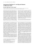
Scheme 2. Steady-state reaction mechanisms for (A) AfM1PDH and (B) AfM2DH at pH 7.1. The thick lines in (B) show the pre- ferred paths for addition of substrate and release of product. The dashed line shows the formation of an abortive ternary complex (see Discussion for details).
Kinetic mechanism of Af M1PDH and Af M2DH
FEBS Journal 278 (2011) 1264–1276 ª 2011 The Authors Journal compilation ª 2011 FEBS
1270
The theory developed by Cook and Cleland is used to deduce the kinetic mechanism of AfM1PDH and AfM2DH from KIE data in Table 4 [28]. The pattern of KIEs observed for AfM1PDH, in which Dkcat ⁄ KmNADH was not different from unity within the limits of error, whereas Dkcat ⁄ KmFru6P had a large value of (cid:2) 3, indi- cates that NADH binds to the enzyme prior to binding of Fru6P. The absence of a KIE on kcat ⁄ Km for the substrate binding first is a clear requirement of the strictly ordered kinetic mechanism, because, at satu- rating concentrations of the substrate which is added second, the commitment to catalysis becomes infinite, and so the KIE is completely suppressed. It follows from the additional sets of KIE data in Table 4, where Dkcat ⁄ Km values for substrate and coenzyme are both that binding of Man-ol1P and greater than unity, NAD+ by AfM1PDH is not ordered, and that there is
S. Krahulec et al.
Enzymes of mannitol metabolism in A. fumigatus
constant. Scheme 2 summarizes the proposed kinetic mechanisms of AfM1PDH and AfM2DH. more those expected from a true dehydrogenase acting in the direction of NAD+-dependent alcohol oxidation (see later).
involving
Inhibition by ketose substrate during NADH-depen- dent reduction of Fru and its absence under conditions in which NADD is used (Fig. S4) is plausibly explained by an expanded random kinetic mechanism of AfM2DH (Scheme 2), an abortive enzyme–NAD+–Fru complex, which releases NAD+ at a rate slow enough to partially limit the overall reaction rate. KIE data indicating that hydride transfer is rate-determining for reduction of Fru by AfM2DH suggest that the relative amount of enzyme–NAD+ available for binding of Fru at the steady state can- not be very high. However, slowing the reaction by using NADD in place of NADH will further restrict the availability of enzyme–NAD+, explaining the complete lack of substrate inhibition under these con- ditions.
reaction step not a
Proposed function of Af M1PDH and Af M2DH in mannitol metabolism
indicates function of implying a physiological
A value of Dkcat ⁄ Km well above unity implies that the isotope-sensitive step of hydride transfer contrib- utes significantly to rate limitation for the sequence of reaction steps included in the kcat ⁄ Km analyzed. For example, kcat ⁄ KmFru6P involves all steps from binding of Fru6P to AfM1PDH–NADH up to release of the first product, Man-ol1P or NAD+. In random bireac- tant systems, kcat ⁄ Km stands for reaction of the vari- able reactant with the corresponding binary enzyme– substrate complex. Inspection of the KIE data in Table 4 reveals that hydride transfer is partly rate- determining in either direction of each of the two enzy- matic reactions. Comparison of KIEs on kcat and kcat ⁄ Km distinguishes datasets in which Dkcat is smaller than Dkcat ⁄ Km from others in which Dkcat roughly equals Dkcat ⁄ Km. The first case (Dkcat < Dkcat ⁄ Km) indicates that, under kcat conditions where the concen- trations of coenzyme and substrate are both saturat- included in kcat ⁄ Km, ing, is partly presumably release of the second product, (Dkcat > 1) or completely (Dkcat = 1) rate-determining overall. This applies to Fru6P reduction by AfM1PDH as well as Man-ol oxidation by AfM2DH. The second case (Dkcat (cid:3) Dkcat ⁄ Km > 1) that hydride transfer is rate-determining for the overall enzymatic to Man-ol1P oxidation by reaction and applies AfM1PDH as well as Fru reduction by AfM2DH.
it
turnover Fru the for
requires product
chemical
FEBS Journal 278 (2011) 1264–1276 ª 2011 The Authors Journal compilation ª 2011 FEBS
1271
Now, considering a kinetic scenario for AfM1PDH in which kcat for Man-ol1P oxidation is governed by hydride transfer, whereas kcat for Fru6P reduction is partly limited by product dissociation, is worth remarking that the reduction kcat exceeds the oxidation kcat by a factor of 12 (Table 2). These kcat conditions imply that chemical reaction of AfM1PDH in the reduction direction proceeds much faster than the cor- responding chemical reaction in the oxidation direc- tion. Under physiological pH conditions, therefore, AfM1PDH shows a clear preference for catalysis in the reduction direction, so the enzyme may be con- sidered to be a Fru6P reductase. In AfM2DH, by reduction contrast, (kcat = 94 s)1) is limited by chemical transformation, whereas the kcat of 14.2 s)1 for Man-ol oxidation reflects slow product release. With the reasonable assumption that complete suppression of the KIE on the oxidation kcat release to be minimally about 10 times slower than the hydride transfer, transformation in oxidation by AfM2DH should occur with a rate constant of 142 s)1 or higher. In comparison with AfM1PDH, therefore, the kinetic properties of AfM2DH at pH 7.1 resemble much The results of free energy profile analysis strongly sup- port the suggestion that Fru6P reduction is the pre- ferred direction of catalytic action of AfM1PDH in vivo, the enzyme in Man-ol biosynthesis via path 1 of Scheme 1. The proposed role of AfM1PDH is in good agreement with evidence from M1PDH gene disruption studies in other fungi, showing that Dm1pdh mutants accumulate no Man-ol (A. niger mycelium) [30] or have a five-fold to 10-fold decreased Man-ol content (Alternaria alter- nata and Phaeosphaeria nodorum) in the mycelium [15,31]. Furthermore, A. niger undergoing sporulation displayed 4.5-fold enhanced production of Man-ol as compared with nonsporulating mycelium, and this change was correlated with a similar, about six-fold, increase in the level of M1PDH activity [30]. We also show in this work that AfM1PDH activity is not sensi- tive to submillimolar alterations in the levels of ATP, ADP or AMP, indicating that, in contrast to E. coli, where inhibition by ATP is a probable mechanism of downregulation of M1PDH activity for Fru6P reduc- in A. fumigatus the cellular energy charge tion [25], exercises no control at the protein level over the rate of Fru6P conversion into Man-ol1P. The results of transcriptomic and proteomic analysis of the heat shock response in A. fumigatus resulting from a shift in growth temperature from 30 (cid:2)C to 48 (cid:2)C suggest that regulation of AfM1PDH activity is achieved at the level of enzyme synthesis [9]. The marked resis- tance of isolated AfM1PDH to inactivation by temper-
S. Krahulec et al.
Enzymes of mannitol metabolism in A. fumigatus
atures promoting the heat shock (40 (cid:2)C and 50 (cid:2)C) is consistent with a possible role of the enzyme in confer- ring thermotolerance to the fungus via enhanced Man- ol production under temperature stress conditions. for Man-ol biosynthesis
saturated with both alcohol in vivo levels of Man-ol and Fru from Table 5, we reach the immediate conclusion that reduction of Fru by NADPH is not thermodynamically feasible under these conditions. In other words, AfM1PDH is the key in A. fumigatus, enzyme irrespective of a possible multiplicity of NADP+ ⁄ NAD+-dependent M2DH activities in the organism. A side remark in this respect is that gene expression and gene disruption studies in P. nodorum and A. niger could possibly have overlooked the existence of the NAD+-dependent M2DH (P. nodorum, Q0UEB6; A. niger, A2QGA1) [15,33].
Features of Af M1PDH structure and function that might be exploited for inhibition Considering the reactant concentrations in Table 5, NAD+-dependent oxidation of Man-ol proceeds ther- modynamically downhill. From its kinetic properties (Tables 1 and 2), AfM2DH appears to be well primed for catalysis for mobilization of Man-ol (not its syn- thesis) under these conditions. The enzyme would be almost substrate and NAD+, whereas the assumed intracellular concentra- tion of Fru is about two orders of magnitude below the apparent Km of 40 mm. A physiological role of AfM2DH in the utilization of intracellular Man-ol via reversal of path 2 in Scheme 1 is therefore proposed. the In light of
target
that compounds good chance
suggested interplay between AfM1PDH and AfM2DH, it is interesting that deletion of the m1pdh gene in some fungi resulted in poor growth (A. alternata) the absence thereof [31] or (P. nodorum) [15] on Man-ol as carbon source. The corresponding Dm2dh mutants, however, showed sub- stantial growth under these conditions [15,31]. It is possible, therefore, that M1PDH has an additional physiological function in the catabolism of external Man-ol once the substrate has become phosphorylated during or after uptake into the cell. However, a clear requirement for AfM1PDH to change directional pref- erence in catalytic action would be that levels of intra- cellular metabolites undergo a large shift from the situation portrayed in Table 5 to another one that favors Man-ol1P, NAD+ or both. Are oxidoreductases other
reductase sorbitol ⁄ xylulose
is a Inhibition of the fungal biosynthesis of Man-ol promising strategy for development of new therapies against infection by A. fumigatus. The results of this work show that antagonism of AfM1PDH presents a clear for achieving the desired inhibition. Moreover, because the human genome does not encode proteins homologous to AfM1PDH or any other protein of the current PSLDR family, there is a raised against AfM1PDH will not show substantial cross-reactivity with respect the human to binding of proteins of host. The high specificity of AfM1PDH for reactions with phosphorylated substrates could be a feature of recognition to be exploited in the design of selective substrate and ⁄ or transition state analogs. The N-ter- minal NAD+-binding domain of AfM1PDH could be another target for selective inhibition, given that, in this domain adopts a somewhat unusual PSLDRs, Rossmann fold that is distinct in many details from isofunctional domains in enzymes of other dehydroge- nase ⁄ reductase families. Precedents from the litera- ture, on lactate dehydrogenase [34] for example, are highly encouraging in showing that, by using inhibi- tors targeted towards the coenzyme-binding site, it may be possible to achieve inhibition that is not only selective for a particular enzyme type, as would be necessary for inhibition of AfM1PDH, but can even discriminate between the same enzyme from parasite and host.
existence of tentative the
Experimental procedures
Recombinant AfM1PDH and AfM2DH were produced in E. coli and purified as described recently [17,18]. Unless otherwise indicated, highly purified preparations of recom-
Materials
FEBS Journal 278 (2011) 1264–1276 ª 2011 The Authors Journal compilation ª 2011 FEBS
1272
than AfM1PDH and AfM2DH involved in the metabolism of mannitol by A. fumigatus? We searched the genome of the organ- ism, and identified an ORF whose translated product is 35.2% identical in amino acid sequence to the M2DH from Agaricus bisporus (AbM2DH) [12]. This putative protein of A. fumigatus is currently annotated as Sou1-like (UniProt ⁄ TrEMBL entry Q4WZX5). In contrast to AfM2DH, AbM2DH is an NADP+-dependent enzyme. From its sequence and three-dimensional structure, AbM2DH is classified as member of the short-chain dehydroge- nase ⁄ reductase superfamily of proteins and enzymes, and shows no significant evolutionary relationship with AfM2DH. Now, an NADP+-dependent M2DH in A. fumigatus made it necessary to examine the possibility that Man-ol is produced from Fru by reduction with NADPH. How- ever, assuming a value of 0.62 for the ratio of intracel- lular concentrations of NADPH and NADP+ (data from A. niger [32]) and applying the values for the
S. Krahulec et al.
Enzymes of mannitol metabolism in A. fumigatus
several constant concentrations of
Measurements performed with AfM2DH involved various concentrations of Fru (8.5–430 mm) and Man-ol (3–110 mm) at several constant concentrations of NADH (0.014–0.20 mm) and NAD+ (0.085–1.4 mm), respectively. Inhibition by adenine nucleotides (AMP, ADP and ATP) was analyzed by measuring initial rates under conditions in which the concen- tration of NADH (0.008–0.25 mm) or NAD+ (0.05–2.5 mm) was varied at the respective adenine nucleotide in the range 0.63–5.0 mm. The concentration of carbonyl or polyol substrates was constant and saturating.
binant AfM1PDH and AfM2DH were used in all experiments. Enzymatic production of Man-ol1P and 5-[2H]-Man-ol1P was carried out with previously reported methods [17]. d-Xylulose was synthesized by microbial oxidation of d-arabinitol, employing a previously described protocol [35]. NADD was prepared with an enzymatic procedure that used glucose dehydrogenase from Baci- llus megaterium and 1-[2H]-d-glucose. Deutero-NADH was purified by MonoQ anion exchange chromatography, as previously described [23,36,37]. The optical purity of NADD and its degree of deuteration (95 ± 1%) were ana- lyzed by 1H-NMR and MS, respectively. 2-[2H]-Man-ol was prepared by enzymatic conversion of Fru, and was purified as described recently [38,39]. The isotopic purity of 2-[2H]- Man-ol was determined by MS (> 99%). No residual Fru, NAD+ or NADH was detected by 13C-NMR. Man-ol, Fru, Fru6P, b-nicotinamide adenine dinucleotides (NAD+ and NADH) and adenine nucleotides (ATP, ADP and AMP) at a purity ‡ 95% were obtained from commercial sources.
Primary deuterium KIEs on apparent kinetic parameters of AfM1PDH and AfM2DH were obtained from a compari- son of initial rates recorded with unlabeled or deuterium- labeled substrates or coenzymes. Oxidation of Man-ol1P and 5-[2H]-Man-ol1P was measured under conditions in which the concentration of NAD+ (0.08–8 mm) or Man- ol1P ⁄ 5-[2H]-Man-ol1P (0.04–6.2 mm) was varied at a con- stant and saturating concentration of the respective other substrate (NAD+, 5.7 mm; Man-ol1P ⁄ 5-[2H]-Man-ol1P, 1.0 mm). Fru6P reduction was measured under conditions in which the concentration of NADH ⁄ NADD (0.012–0.2 mm) or Fru6P (0.45–45 mm) was varied at a constant and saturat- ing concentration of the respective other substrate (NADH ⁄ NADD, 0.2 mm; Fru6P, 45 mm). Likewise, the conditions used for determination of KIEs on kinetic parameters for AfM2DH were as follows. Oxidation: Man-ol ⁄ 2-[2H]-Man-ol, 0.9–180 mm, and NAD+, 4 mm; NAD+, 0.08–4 mm, and Man-ol ⁄ 2-[2H]-Man-ol, 260 mm. Reduction: Fru, 4–840 mm, and NADH ⁄ NADD, 0.25 mm; NADH ⁄ NADD, 0.002–0.2 mm, and Fru, 800 mm.
concentration of
Assays
regression
analysis with
Protein concentrations were determined with the Bio-Rad protein assay (Bio-Rad Laboratories, Hercules, CA, USA) referenced against known concentrations of BSA. Initial- rate data were collected with a DU800 spectrophotometer (Beckman Coulter, Fullerton, CA, USA) at 25 (cid:2)C, and are based on the measurement of formation or depletion of NAD(P)H at 340 nm (eNADH = 6.22 cm)1Æmm)1). Unless otherwise indicated, substrate screening was performed at a 300 mm. Tris ⁄ HCl buffer constant (100 mm; pH 7.1) was used for ketose reduction. Gly- cine ⁄ NaOH buffer (100 mm; pH 10.0) was used for polyol oxidation. Different pH conditions were chosen because alcohol oxidation by NAD+ generally proceeds best at high pH, whereas a lower pH value is normally suitable for car- bonyl group reduction by NADH. The concentrations of NADH and NAD+ used in these assays were 0.2 and 2.0 mm, respectively. Apparent kinetic parameters (kcat and Km) were obtained for those compounds that had shown significant conversion rates in screening assays, using a threshold of 1% of the activity with the respective native substrate (Man-ol and Fru; Man-ol1P and Fru6P). By application of this criterion, the following substrates were selected for reaction with AfM2DH in the given concentra- tion range: Fru (2–1000 mm), d-xylulose (0.5–450 mm), l- sorbose (20–1000 mm), Man-ol (1–400 mm), d-arabinitol (3–1300 mm) and d-sorbitol (30–1600 mm).
Kinetic parameters were obtained from a nonlinear fit of the appropriate equation to the data. Unweighted nonlinear sigma plot 9.0 least-squares (SYSTAT Software; San Jose, CA, USA) was used. In Eqns (1)-(6), v is the initial rate, kcat is the kinetic turnover number, E and S are the molar concentrations of enzyme and substrate, Km is an apparent Michaelis constant, and KiS is a substrate inhibition constant. E was obtained from the protein concentration, using molecular masses of 44.2 kDa and 57.6 kDa for AfM1PDH and AfM2DH, respectively [17,18]. Equation (3) implies ordered binding of substrates A and B in a bisubstrate reaction where KiA is the dissociation constant for A. Equation (4) is used for random bisubstrate kinetics, where KA and KB are dissocia- tion constants for A and B, and a is a factor describing EI is a how bound A affects the binding of B. In Eqn (5), Ki competitive inhibition constant, and I is the molar inhibitor concentration. Unless mentioned, KIEs were obtained by using Eqn (6) [40], where EV and EV ⁄ K are isotope effects minus 1 on kcat and kcat ⁄ Km, respectively. Fi is the fraction of deuterium in the labeled substrate.
Tris ⁄ HCl buffer (100 mm; pH 7.1) was used in all further kinetic studies. Full kinetic characterization of AfM1PDH involved initial-rate measurements under conditions of vari- ous concentrations of Fru6P (0.17–8.7 mm) and Man-ol1P (0.02–2.3 mm) at several constant concentrations of NADH (0.0017–0.17 mm) and NAD+ (0.06–5.9 mm), respectively.
FEBS Journal 278 (2011) 1264–1276 ª 2011 The Authors Journal compilation ª 2011 FEBS
1273
Data processing
S. Krahulec et al.
Enzymes of mannitol metabolism in A. fumigatus
ð1Þ
v ¼ kcat (cid:4) E (cid:4) S=ðK m þ SÞ
(cid:6) ¼ K NADðHÞ (cid:4) ð1 þ AMP=K EI
i AMP
i ADP þ ATP=K EI
i ATPÞ
(cid:6) ¼ K iNADH (cid:4) ð1 þ AMP=K EI i ADP þ ATP=K EI
i AMP i ATPÞ
(cid:6)
K NADðHÞ ð8Þ þ ADP=K EI ð2Þ v ¼ kcat (cid:4) E (cid:4) S=½Km þ S (cid:4) ð1 þ S=KiSÞ(cid:5) K iNADH ð9Þ þ ADP=K EI v ¼ kcat (cid:4) E (cid:4) A (cid:4) B=ðKiA (cid:4) KmB þ KmA (cid:4) B þ KmB (cid:4) A þ A (cid:4) BÞ ð3Þ
(cid:6) (cid:4) KFruÞ
ð10Þ knet M2DH ¼ kox (cid:4) NADþ (cid:4) Man-ol=ðaox (cid:4) KNAD (cid:4) KMan-olÞ (cid:7) kred (cid:4) NADH (cid:4) Fru= v ¼ kcat (cid:4) E (cid:4) A (cid:4) B=ðA (cid:4) B þ a (cid:4) KA (cid:4) Bþa (cid:4) KB (cid:4) A þ a (cid:4) KA (cid:4) KBÞ ð4Þ ðared (cid:4) KNADH
i Þ þ S(cid:5)
(cid:6) (cid:4) KMan-ol1PÞ
(cid:6) (cid:4) KmFru6PÞ ð11Þ
ð5Þ v ¼ kcat (cid:4) E (cid:4) S=½K m (cid:4) ð1 þ I=K EI knet M1PDH ¼ kox (cid:4) NADþ (cid:4) Man-ol1P= ðaox (cid:4) KNAD v ¼ kcat (cid:4) E (cid:4) S=½K m (cid:4) ð1 þ Fi (cid:4) EV=KÞ þ S (cid:4) ð1 þ Fi (cid:4) EV Þ(cid:5) ð6Þ (cid:7) kred (cid:4) NADH (cid:4) Fru6P=ðKiNADH
K ¼ NADþ (cid:4) ROH=ðNADH (cid:4) ROÞ ð12Þ
ð13Þ DGKeq ¼ (cid:7) R (cid:4) T (cid:4) lnð1=KeqÞ
ð14Þ DGK ¼ (cid:7) R (cid:4) T (cid:4) lnðKÞ
(cid:6) (cid:4) KROH (cid:4) a=
ð15Þ DGKeff ¼ DGKeq(cid:7)DGK
DGE-NAD-ROH ¼ R (cid:4) T (cid:4) ln½KNAD ð16Þ ðNADþ (cid:4) ROHÞ(cid:5) þ DGKeff
(cid:6) (cid:4) KmRO=ðNADH (cid:4) ROÞ(cid:5) ð17Þ
DGE-NADH-RO ¼ R (cid:4) T (cid:4) ln½KiNADH
indicates a preference
(cid:6) (cid:4) KRO (cid:4) a=ðNADH (cid:4) ROÞ(cid:5) ð18Þ DGTS ¼ (cid:7) R (cid:4) T (cid:4) ln½kox (cid:4) NADþ (cid:4) ROH=ðKNAD (cid:4) KROH (cid:4) aÞ(cid:5) ð19Þ
DGE-NADH-RO ¼ R (cid:4) T (cid:4) ln½KNADH
þ R (cid:4) T (cid:4) lnðk (cid:4) T=hÞ þ DGKeff
Acknowledgements
is
V. Pacher and K. Longus are thanked for expert tech- nical assistance. We are grateful to M. Murkovic (Institute of Biochemistry, Graz University of Technol- ogy) and H. Weber (Institute of Organic Chemistry, Graz University of Technology) for MS and NMR measurements, respectively. Financial support from the Austrian Science Fund FWF (P18275-B09 to B. Nide- tzky) is gratefully acknowledged.
References
Planck
and
the
1 Solomon PS, Waters OD & Oliver RP (2007) Decoding
Equation (7) is the Haldane relationship for an ordered bi-bi kinetic mechanism, where app Keq is the (kinetically determined) equilibrium constant of the reaction; kox and kred are kcat values for alcohol oxidation and ketose reduc- tion; KiNADH and KiNAD are dissociation constants for NADH and NAD+; and KmRO and KmROH are Michaelis constants for ketose and polyol. Note: in the random bi- substrate mechanism, the expression aÆKAÆKB is used to replace KiAÆKmB in Eqn (7). The combined effect of com- petitive inhibition by adenine nucleotides is accounted for by Eqn (8) or Eqn (9), where an asterisk indicates the apparent binding constant. Equations (10) and (11) are simplified expressions of enzyme directional preference (knet), based solely on catalytic efficiencies. Formally, they are derived from the complete rate equation for a rapid equilibrium random bireactant kinetic mechanism [41], by leaving out all terms in the denominator. Strictly, these equations would be applicable only for limiting reactant concentrations. However, they are qualitatively useful because, at given reactant concentrations, a positive value of knet for alcohol oxidation, whereas a negative value signifies a preference for ketose reduction. Note that the term aÆKAÆKB applies to a ran- dom mechanism, as in Eqn (10). When the mechanism is ordered, the aÆKAÆKB term is replaced by KiAÆKmB, as in Eqn (11). Gibbs free energy (DG, kJÆmol)1) profiles for catalyzed by AfM2DH and AfM1PDH at reactions pH 7.1 were constructed with the use of Eqns (12)-(19). R is the gas constant (0.008314 kJÆmol)1ÆK)1), and T is the temperature (298.15 K). The parameter K in Eqn (12) is the mass action ratio calculated by applying intracellular reactants. DGKeff the difference concentrations of between DG at Keq and DG at a given value of K. Calcu- lation of DG values at ternary complexes (indicated by subscript) and the transition state (subscript TS) is shown in Eqns (16)–(19). k and h are the Boltzmann constant (1.38 · 10)26 kJÆK)1) constant (6.63 · 10)37 kJÆs), respectively.
ð7Þ
the mannitol enigma in filamentous fungi. Trends Microbiol 15, 257–262.
appK eq¼ kox (cid:4) K iNADH (cid:4) K mRO=ðkred (cid:4) K iNAD (cid:4) K mROHÞ
FEBS Journal 278 (2011) 1264–1276 ª 2011 The Authors Journal compilation ª 2011 FEBS
1274
S. Krahulec et al.
Enzymes of mannitol metabolism in A. fumigatus
2 Chaturvedi V, Flynn T, Niehaus WG & Wong B (1996) Stress tolerance and pathogenic potential of a mannitol mutant of Cryptococcus neoformans. Microbiology 142(Pt 4), 937–943.
15 Solomon PS, Waters OD, Jorgens CI, Lowe RG, Rechberger J, Trengove RD & Oliver RP (2006) Mannitol is required for asexual sporulation in the wheat pathogen Stagonospora nodorum (glume blotch). Biochem J 399, 231–239.
3 Chaturvedi V, Wong B & Newman SL (1996) Oxidative killing of Cryptococcus neoformans by human neutro- phils. Evidence that fungal mannitol protects by scavenging reactive oxygen intermediates. J Immunol 156, 3836–3840.
4 Ve´ l€ez H, Glassbrook NJ & Daub ME (2008) Mannitol biosynthesis is required for plant pathogenicity by Alternaria alternata. FEMS Microbiol Lett 285, 122–129.
16 Strandberg GW (1969) d-mannitol metabolism by Aspergillus candidus. J Bacteriol 97, 1305–1309. 17 Krahulec S, Armao GC, Weber H, Klimacek M & Nidetzky B (2008) Characterization of recombinant Aspergillus fumigatus mannitol-1-phosphate 5-dehydro- genase and its application for the stereoselective synthe- sis of protio and deuterio forms of d-mannitol 1- phosphate. Carbohydr Res 343, 1414–1423.
18 Krahulec S, Armao GC, Bubner P, Klimacek M &
5 Jennings DB, Ehrenshaft M, Pharr DM & Williamson JD (1998) Roles for mannitol and mannitol dehydroge- nase in active oxygen-mediated plant defense. Proc Natl Acad Sci USA 95, 15129–15133.
Nidetzky B (2009) Polyol-specific long-chain dehydrogenases ⁄ reductases of mannitol metabolism in Aspergillus fumigatus: biochemical characterization and pH studies of mannitol 2-dehydrogenase and mannitol- 1-phosphate 5-dehydrogenase. Chem Biol Interact 178, 274–282.
6 Voegele RT, Hahn M, Lohaus G, Link T, Heiser I & Mendgen K (2005) Possible roles for mannitol and mannitol dehydrogenase in the biotrophic plant patho- gen Uromyces fabae. Plant Physiol 137, 190–198.
7 Latge´ JP (1999) Aspergillus fumigatus and aspergillosis.
Clin Microbiol Rev 12, 310–350.
19 Klimacek M, Kavanagh KL, Wilson DK & Nidetzky B (2003) Pseudomonas fluorescens mannitol 2-dehydroge- nase and the family of polyol-specific long-chain dehy- drogenases ⁄ reductases: sequence-based classification and analysis of structure–function relationships. Chem Biol Interact 143–144, 559–582.
8 Wong B, Brauer KL, Tsai RR & Jayasimhulu K (1989) Increased amounts of the Aspergillus metabolite d-man- nitol in tissue and serum of rats with experimental aspergillosis. J Infect Dis 160, 95–103.
9 Albrecht D, Guthke R, Brakhage AA & Kniemeyer O (2010) Integrative analysis of the heat shock response in Aspergillus fumigatus. BMC Genomics 11: 32, doi:10.1186/1471-2164-11-32.
20 Kavanagh KL, Klimacek M, Nidetzky B & Wilson DK (2002) Crystal structure of Pseudomonas fluorescens mannitol 2-dehydrogenase binary and ternary com- plexes. Specificity and catalytic mechanism. J Biol Chem 277, 43433–43442.
21 Kavanagh KL, Klimacek M, Nidetzky B & Wilson DK (2003) Crystal structure of Pseudomonas fluorescens mannitol 2-dehydrogenase: evidence for a very divergent long-chain dehydrogenase family. Chem Biol Interact 143–144, 551–558.
10 Stevens DA, Moss RB, Kurup VP, Knutsen AP, Green- berger P, Judson MA, Denning DW, Crameri R, Brody AS, Light M et al. (2003) Allergic bronchopulmonary aspergillosis in cystic fibrosis – state of the art: Cystic Fibrosis Foundation Consensus Conference. Clin Infect Dis 37(Suppl. 3), 225–264.
11 Shen B, Jensen RG & Bohnert HJ (1997) Mannitol pro- tects against oxidation by hydroxyl radicals. Plant Phys- iol 115, 527–532.
22 Bubner P, Klimacek M & Nidetzky B (2008) Structure- guided engineering of the coenzyme specificity of Pseu- domonas fluorescens mannitol 2-dehydrogenase to enable efficient utilization of NAD(H) and NADP(H). FEBS Lett 582, 233–237.
23 Slatner M, Nidetzky B & Kulbe KD (1999) Kinetic
study of the catalytic mechanism of mannitol dehydro- genase from Pseudomonas fluorescens. Biochemistry 38, 10489–10498.
24 Wolff JB & Kaplan NO (1956) d-Mannitol 1-phosphate dehydrogenase from Escherichia coli. J Biol Chem 218, 849–869.
25 Klungsøyr L (1967) The binding of adenine nucleotides to the mannitol-I-phosphate dehydrogenase of Escheri- chia coli. Biochim Biophys Acta 146, 10–17.
12 Nidetzky B & Klimacek M (2007) Fungal mannitol 2-de- hydrogenases and mannitol-1-phosphate 5-dehydro- genases constitute novel branches in the protein family of polyol-specific long-chain dehydrogenases and reductases. In Enzymology and Molecular Biology of Carbonyl Meta- bolism (Weiner H, Maser E, Lindahl R & Plapp B eds), pp 305–322. Purdue University Press, West Lafayette. 13 Ueng ST, Hartanowicz P, Lewandoski C, Keller J, Holick M & McGuinness ET (1976) d-Mannitol dehydrogenase from Absidia glauca. Purification, metabolic role, and subunit interactions. Biochemistry 15, 1743–1749.
14 Lee WH (1967) Mannitol acetyl phosphate phospho- transferase of Aspergillus. Biochem Biophys Res Com- mun 29, 337–342.
26 Glenn AR, Arwas R, McKay IA & Dilworth MJ (1984) Fructose metabolism in wild-type, fructokinase-negative and revertant strains of Rhizobium leguminosarum. J Gen Microbiol 130, 231–237.
FEBS Journal 278 (2011) 1264–1276 ª 2011 The Authors Journal compilation ª 2011 FEBS
1275
S. Krahulec et al.
Enzymes of mannitol metabolism in A. fumigatus
40 Blanchard JS & Cleland WW (1980) Kinetic and
chemical mechanisms of yeast formate dehydrogenase. Biochemistry 19, 3543–3550.
41 Segel IH (1993) Enzyme Kinetics – Behavior and
27 Foreman JE and Niehaus WG Jr (1985) Zn2+-induced cooperativity of mannitol-1-phosphate dehydrogenase from Aspergillus parasiticus. J Biol Chem 260, 10019– 10022.
Analysis of Rapid Equilibrium and Steady-State Enzyme Systems. Wiley, New York.
28 Cook PF (1991) Kinetic and regulatory mechanisms of enzymes from isotope effects. In Enzyme Mechanism from Isotope Effects (Cook PF ed), pp 203–231. CRC Press, Boca Raton.
42 Collins P & Ferrier R (1995) Monosaccharides: Their Chemistry and Their Roles in Natural Products. Wiley, Chichester.
29 Kiser RC & Niehaus WG Jr (1981) Purification and
kinetic characterization of mannitol-1-phosphate dehy- drogenase from Aspergillus niger. Arch Biochem Biophys 211, 613–621.
30 Ruijter GJG, Bax M, Patel H, Flitter SJ, van de
43 Wu J, Serianni AS & Vuorinen T (1990) Furanose ring anomerization: kinetic and thermodynamic studies of the d-2-pentuloses by 13C-n.m.r. spectroscopy. Carbohydr Res 206, 1–12.
44 Hayward LD & Angyal SJ (1977) A symmetry rule for the circular dichroism of reducing sugars, and the pro- portion of carbonyl forms in aqueous solutions thereof. Carbohydr Res 53, 13–20.
Vondervoort PJI, de Vries RP, vanKuyk PA & Visser J (2003) Mannitol is required for stress tolerance in Asper- gillus niger conidiospores. Eukaryot Cell 2, 690–698. 31 Ve´ l€ez H, Glassbrook NJ & Daub ME (2007) Mannitol metabolism in the phytopathogenic fungus Alternaria alternata. Fungal Genet Biol 44, 258–268.
32 Poulsen BR, Nohr J, Douthwaite S, Hansen LV,
45 Ruijter GJG & Visser J (1996) Determination of inter- mediary metabolites in Aspergillus niger. J Microbiol Methods 25, 295–302.
Iversen JJL, Visser J & Ruijter GJG (2005) Increased NADPH concentration obtained by metabolic engineer- ing of the pentose phosphate pathway in Aspergillus niger. FEBS J 272, 1313–1325.
33 Aguilar-Osorio G, Vankuyk PA, Seiboth B, Blom D,
46 Yamamoto Y, Nitta K, Tango K, Saito T & Tsuchim- uro M (1965) Studies on the metabolic products of a strain of Aspergillus fumigatus (DH 413). I. Isolation and chemical structures of metabolites. Chem Pharm Bull (Tokyo) 13, 935–941.
Supporting information
Solomon PS, Vinck A, Kindt F, Wo¨ sten HA & de Vries RP (2010) Spatial and developmental differentiation of mannitol dehydrogenase and mannitol-1-phosphate dehydrogenase in Aspergillus niger. Eukaryot Cell 9, 1398–1402.
34 Vander Jagt DL, Deck LM & Royer RE (2000) Gossy- pol: prototype of inhibitors targeted to dinucleotide folds. Curr Med Chem 7, 479–498.
35 Mayer G, Kulbe KD & Nidetzky B (2002) Utilization
of xylitol dehydrogenase in a combined microbial ⁄ enzy- matic process for production of xylitol from d-glucose. Appl Biochem Biotechnol 98–100, 577–589.
36 Mostad SB & Glasfeld A (1993) Using high field NMR
to determine dehydrogenase stereospecificity with respect to NADH. J Chem Educ 70, 504–506. 37 Orr GA & Blanchard JS (1984) High-performance ion-exchange separation of oxidized and reduced nicotinamide adenine dinucleotides. Anal Biochem 142, 232–234.
38 Klimacek M & Nidetzky B (2002) Examining the rela-
tive timing of hydrogen abstraction steps during NAD+-dependent oxidation of secondary alcohols cata- lyzed by long-chain d-mannitol dehydrogenase from Pseudomonas fluorescens using pH and kinetic isotope effects. Biochemistry 41, 10158–10165.
The following supplementary material is available: Fig. S1. Structural comparison of polyol substrates of AfM2DH. Fig. S2. Comparison of the structural model of AfM2DH with the experimentally determined crystal structure of PsM2DH (1lj8). Fig. S3. Kinetic analysis of inhibition of AfM1PDH (A–F) and AfM2DH (G–L) by adenine nucleotides at pH 7.1. Fig. S4. Michaelis–Menten plot for Fru reduction by AfM2DH employing NADH or NADD. Fig. S5. Irreversible inactivation of AfM1PDH (A, B) and AfM2DH (C, D) at different temperatures, repre- sented as semilogarithmic plots. This supplementary material can be found in the online version of this article.
39 Klimacek M & Nidetzky B (2010) The oxyanion hole of Pseudomonas fluorescens mannitol 2-dehydrogenase: a novel structural motif for electrostatic stabilization in alcohol dehydrogenase active sites. Biochem J 425, 455– 463.
FEBS Journal 278 (2011) 1264–1276 ª 2011 The Authors Journal compilation ª 2011 FEBS
1276
Please note: As a service to our authors and readers, this journal provides supporting information supplied by the authors. Such materials are peer-reviewed and may be re-organized for online delivery, but are not copy-edited or typeset. Technical support issues arising from supporting information (other than missing files) should be addressed to the authors.