Journal of Medicine and Pharmacy - No.5
56
GROWTH INHIBITION BY A GREEN TEA
STANDARDIZED EXTRACT (POLYPHENON E)
IN PROSTATE CANCER CELLS
Phu Thi Hoa 1,Ngo Viet Quynh Tram1, Gianfranco Pintus2
(1) Hue University of Medicine and Pharmacy, Vietnam
(2) University of Sassari, Italy
Abstract
Objective: Green tea consumption has been shown to exhibit cancer-preventive activities in preclinical
studies. Polyphenon E (Poly E) is a green tea standardized extract. This study was undertaken to
examine the antiproliferative effect and pro-oxidant activity of Poly E on PC3 prostate cancer cells.
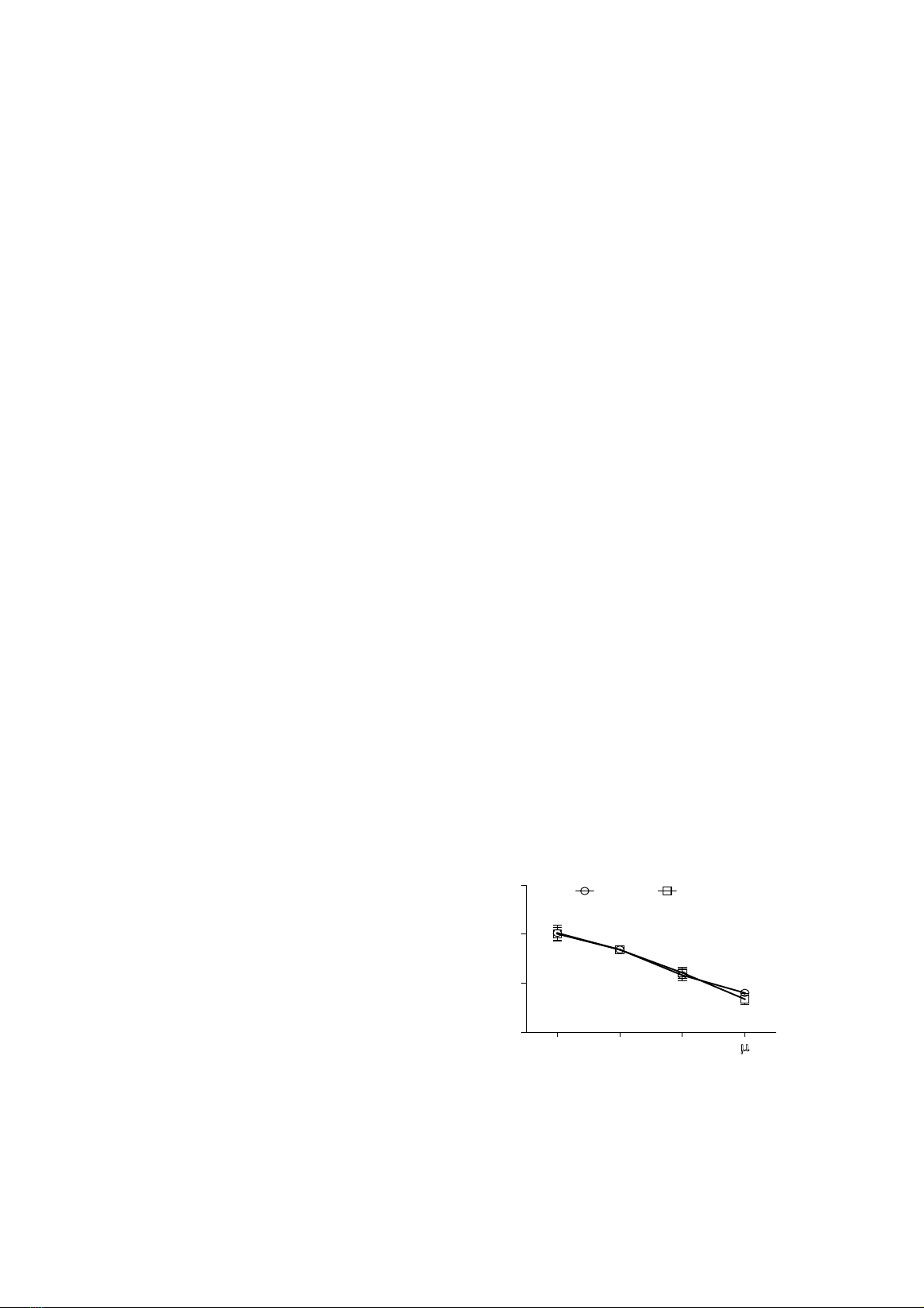
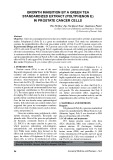
Experimental Design and results: - PC3 prostate cancer cells were used as model system. Treatment
of PC3 cells with 30 and 100 µg/ml Poly E significantly decreased cell viability and proliferation. At
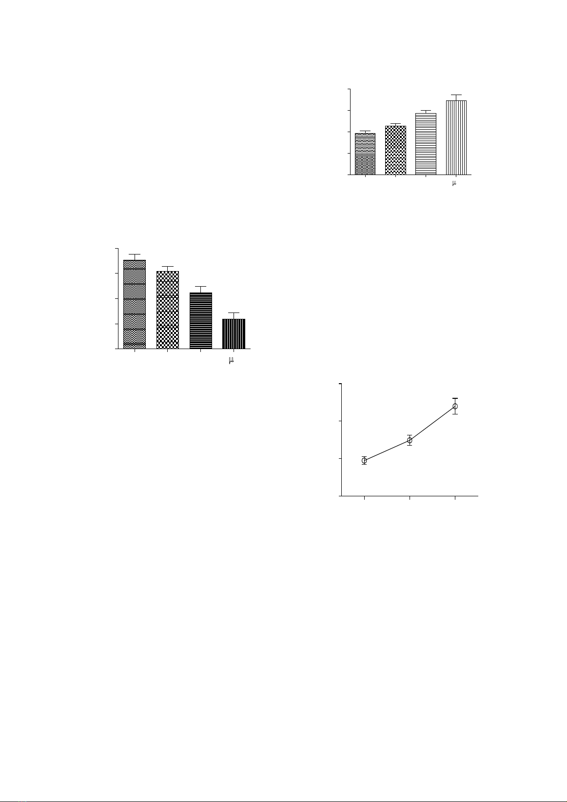
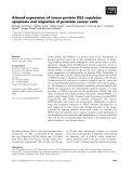
all tested concentrations, Poly E elicited pro-oxidant effect at 30 and 100 µg/ml. This effect of Poly E
is consistent with the observed cytotoxicity, thus establishing a correlation between pro-oxidant activity
and the antiproliferative effect of Poly E in PC3 cells. Conclusion: Our data showed the antiproliferative
effect of Poly E and suggest Poly E-induced pro-oxidant effect involved in this activity.
Key words: Polyphenon E, pro-oxidant effect, prostate cancer cells.
1. INTRODUCTION
Prostate cancer (PCa) is one of the most
frequently diagnosed male cancer in the Western
countries and continues to represent a major
cause of cancer-related mortality, despite medical
advances. Asian-Americans seem to be at the
lowest risk for PCa [9]. About less than 10% of
PCa has been shown to be inherited suggesting that
a variety of genetic and environmental factors may
be important contributions to PCa development
[17]. The Asians appear to have the lowest risk of
developing PCa which may be due to consuming
specific dietary constituents daily over many years.
Over the last two decades many epidemiological
studies, both cohort and case-control studies, have
suggested that green tea consumption correlates
with a lower risk of certain cancers such as breast,
colon, and prostate [7].
Green tea contains many polyphenols, which
include flavanols, flavandiols, flavonoids and
phenolic acids. Most of the green tea polyphenols
are flavanols, commonly known as catechins
[1]. EGCG is the major catechin in green tea,
which possesses antioxidant, anti-mutagenic,
anti-proteolytic and anti-proliferative activity
[14]. While many studies have focused on the
effects and mechanism of EGCG on various cell
types, the effects of Polyphenon E (Poly E) on
tumor cells, as well as its mechanism of action,
have to be elucidated yet. Polyphenon E is a
well-defined pharmaceutical-grade mixture of
polyphenols that contain about 50% EGCG and
30% other catechins [2]. Since the formulation is
highly reproducible and easily prepared, Poly E
is an attractive derivative of green tea for clinical
chemoprevention trials [16].
In the present study, we show that Poly E, a
green tea standardized extract can inhibit PC3
cell growth. We also demonstrate that Poly E
can induce pro-oxidant effect, suggesting a
correlation between pro-oxidant activity and the
antiproliferative effect of Poly E in PC3 cells.
2. MATERIALS AND METHODS
Reagents
Polyphenon E (Poly E), a green tea standardized
extract was manufactured by Mitsui Norin Co. Ltd.
(Shizuoka, Japan). Poly E was dissolved in PBS
plus or cell medium with 2.5% FBS.
2.1 Cell culture and treatments
PC3 human prostate cancer cells from
ATCC (Rockville, MD) were cultured in Fk12
nutrient mixture 1X (Invitrogen, Carlsbad,
CA) respectively, supplemented with 7% fetal
bovine serum (FBS) and penicillin G (100
U/ml), streptomycin (100 μg/ml) and 0,25 μg/ml
amphotericin B. Cells were maintained at 37°C
and 5% CO2 in a humid environment.
- Corresponding author: Phu Thi Hoa, email: phuthihoa2010@gmail.com
- Received: 28/4/2014 Revised: 16/6/2014 Accepted: 25/6/2014 DOI: 10.34071/jmp.2014.1e.9
Journal of Medicine and Pharmacy - No.5 57
The PC3 (70-80% confluent) were treated with
Poly E (10, 30, and 100 μg/ml) with different time
points depending on experiments. Cells used as
controls were incubated with the vehicle only.
2.2. Cell viability assay (ATP assay)
Cell viability assay was assessed by using the
CellTiter-Glo®Luminescent Cell Viability Assay.
This is a homogeneous method to determine the
number of viable cells in culture based on quantitation
of the produced ATP, which signals the presence of
metabolically active cells. Briefly, after treatment
for 24 h, equal volume of CellTiter-Glo reagent was
added directly to the wells. These contents were
mixed for 2 minutes to induce cell lysis. Plates were
incubated at room temperature for 10 minutes to
stabilize luminescent signal. The luminescence was
measured on microplate reader (Tecan). The results
are expressed as percent of control.
2.3. Cell metabolic assay (MTT assay)
Cell metabolic activity was assessed
in 96-well plates (BD Falcon) by using the
colorimetric 3-(4,5-dimethylthiazol-2-yl)-2,5-
diphenyltetrazolium bromide (MTT) assay
(Promega, Madison, WI). After treatments for
24 h, cells were added with 20 µl MTT solution
(5 mg/ml) in medium M199 and incubated at 37°C
in a cell incubator for 60 min. At the end of the
incubation period, the medium was removed and
cell monolayer was washed twice with HBSS.
The converted dye was solubilized with acidic
isopropanol and plates were analyzed at 570 nm
with background subtraction at 650 nm. Results
were expressed as a percent of control.
2.4. Cell proliferation assay (BrdU
incorporation Assay)
Cell proliferation was assessed by using
chemiluminescent immunoassay, which based on
the measurement of BrdU incorporation during
DNA synthesis. Confluent cells was treated and
cell proliferation was evaluated at 24 h. BrdU is
added to cells cultured in microplates, followed by
incubation for 10 hours. After the culture supernatant
is removed, cells are fixed by Fix-Denat solution
for 30 min. Fix-Denat was discarded and cells was
incubated with an anti-BrdU antibody (anti-BrdU-
POD) for 90 min. After rinsing three times with
washing buffer, substrate solution was added and
allowed to react for 6 min at room temperature.
Light emission was read by using a microplate
reader. Results were expressed as means ± SD.
2.5. Measurements of intracellular ROS
Intracellular ROS levels were determined
by using the ROS molecular probe 2’,7’-
dichlorodihydrofluorescein diacetate (H2DCF-
DA) [12]. After treatments, cells were incubated
for 30 min with PBS plus containing 1µM H2DCF-
DA, then washed twice with PBS and fluorescence
was measured with a plate reader (Tecan). Results
were corrected for background fluorescence and
protein concentration and expressed as a percent
of untreated cells.
2.6. Statistic
Data are expressed as mean±SD of four
different experiments. One-way analysis of variance
followed by a post hoc Newman-Keuls multiple
comparison test was used to detect differences of
means among treatments with significance defined
as P< 0.05 (GraphPad Prism version 5.00).
3. RESULTS
3.1. Dose-dependent effect of Poly E on cell
viability and metabolic activity of PC3
Cell viability was evaluated by using the
ATP assay. Cells were stimulated with increasing
concentrations (10, 30, 100 µg/ml) of Poly E for
24 h, while untreated cells were used as control
(CTRL). Although a reduction of cell viability
was observed at a concentration of 10 µg/ml
Poly E, it is not significant compared to the
control. In contrast, the treatment of cells with
the higher concentrations (30, 100 µg/ml) of Poly
E, significantly lowered the viability of cells in
compa rison to the untreated ones (Fig.1).
Similar to the observed cell death, a
significant decrease in cell metabolic activity was
induced by both 30 and 100 µg/ml of Poly E, as
depicted by the data reported in Fig.1 obtained
with the MTT assay. Moreover, a correlation was
evident between MTT and ATP data. Results are
expressed as percent of untreated controls.
CTRL 10 30 100µ/ml
0
50
100
150 ATP MTT
*
*
% of control
Fig. 1. Confluent PC3 cells were stimulated by
Poly E for 24 h with various concentrations. Cell
viability and cell metabolic activity were assessed
after treatments by ATP assay and MTT assay.
The results are expressed as percent of control.
*Significantly different from the control (p< 0.05).
Journal of Medicine and Pharmacy - No.5
58
3.2. Dose-dependent effect of Poly E on PC3
cell proliferation
Further investigation on the cytotoxicity
of Poly-E on PC3 was conducted by using the
BrdU assay. Cells were treated with different
concentrations of Poly E and cell proliferation
was assessed after 24 h. As reported in
Fig. 2, 24 h treatment of Poly E induced a dose-
dependent decrease in the DNA synthesis of PC3.
Consistent with data of previous experiments, this
result demonstrated that Poly E is inhibiting the
proliferation and inducing cell death in PC3 at
both concentrations 30 and 100 µg/ml. Results are
expressed as means ± SD.
BrdU incorporation
CTRL 10 30 100µg/ml
0
2000
4000
6000
8000
*
#
*
#
#
(RFUs)
Fig. 2. Confluents PC3 were treated with
different concentrations of Poly E for 24 h.
Cell proliferation was evaluated by using BrdU
assay. Quantification of cell proliferation in
cultured PC3 in the absence (CTRL) or presence
of the indicated treatments. Poly E caused
dose-dependent inhibition of proliferation of
PC3. *Significantly different from the control,
#significantly different from each other (p< 0.05).
3.3. Dose and time-dependent effects of Poly
E on PC3 ROS levels
To gain further mechanistic insight on the
effect Poly E upon PC3 cell we assessed the potential
variation of intracellular ROS levels. Intracellular
ROS generation was examined in PC3 in response
to Poly-E using 2’,7’-dichlorodihydrofluorescein
diacetate (H2DCF-DA). This probe enters the cells
and is oxidized in the presence of ROS, generating
the fluorescent compound, DCF. To determine
the effects of Poly E on PC3, cells were treated
with the previously indicated concentrations and
intracellular ROS levels were assessed after 2 h
of stimulation, by a fluorescence detector. Fig.
3A shown that ROS levels were significantly
increased by the Poly E treatment at dose of 30 and
100µg/ml. The observed pro-oxidant effect is
consistent with the previously reported cytotoxicity
results, suggesting the Poly E-induced pro-oxidant
effect as responsible for the reported PC3 death.
ROS levels (2 hrs)
CTRL 10 30 100µg/ml
0
50
100
150
200
*
*
#
#
#
(RFUs) % of control
Fig. 3A. Confluent PC3 cells were stimulated with
various concentrations of Poly E. ROS levels were
assessed 2 h after treatment. Quantification of
ROS levels in cultured PC3 in the absence (CTRL)
or presence of the indicated treatments. The results
are expressed as percent of control. *Significantly
different from the control, #significantly different
from each other (p< 0.05).
With dose of 30 µg/ml, we next assess ROS
generation with the intent to investigate a potential
time-dependent effect of Poly E at 2 h, 6 h,
12 h. Fig. 3B shown that ROS were increasingly
generated after 12 h of Poly E treatment in PC3.
ROS levels
2 h rs 6 h rs 12 h rs
0
2
4
6
(RFUs)
ratio treated/untreated
Fig. 3B. ROS levels were assessed after 2 h, 6 h
and 12 h of treatment with 30 µg/ml of Poly E. The
results are expressed as ratio treated and control.
4. DISCUSSION
Tea, next to water, is the most widely
consumed beverage in the world. The tea
plant Camellia sinensis has been cultivated
in Asia for thousands of years, and green tea
has been used for centuries in China, Japan,
and Thailand as a traditional medicine with a
variety of applications. Green tea possesses
anti-carcinogenic effects, such as inhibition of
growth proliferation, induction of apoptosis,
induction of phase II detoxifying enzymes, and
reduction of oxidative damage to DNA [11].
Several studies have more specifically shown
Journal of Medicine and Pharmacy - No.5 59
that consumption of green tea polyphenols is
associated with decreased risk and/or slower
progression of prostate cancer [7, 11]
EGCG, the most abundant catechin in green
tea, has been shown to be the main effector of
the anti-carcinogenic properties. For this reason,
EGCG is the most commonly studied green tea
catechins (GTCS). However, whole mixtures of
GTCs may more accurately reflect the human
consumption of green tea, possibly due to the
fact that tea constituents other than catechins may
also have anti-carcinogenic activity [3]. Poly E, a
decaffeinated pharmaceutical preparation of tea
catechins that contains approximately 50% EGCG
and 30% other catechins may be preferable to
EGCG. The catechins in this mixture may exert
synergistic effects [15]. In addition, the effects
of Poly E on tumor cells and its mechanisms
of action are poorly known [16]. In the present
study, we examine the effects of Poly E on PC3
prostate cancer cells. Cell viability, cell metabolic
activity and cell proliferation were evaluated
by ATP, MTT and BrdU assay, respectively.
The results demonstrate that Poly E is able
to induce loss of both viability and inhibition
of PC3 DNA synthesis in dose-dependent
manner (Fig.1-2). At concentration of 30 and
100µg/ml, a cytotoxic effect is shown as judged
by the low viability. Similar effects of this
compound were seen in studying the colon cancer,
where Poly E preferentially inhibited growth of
the Caco2, HCT116, HT29, SW480, and SW837
colon cancer cells compared to the FHC normal
human fetal colon cell line [15]. It has been also
demonstrated that Poly E inhibited proliferation
of immortalized Barrett’s cells as well as various
adenocarcinoma cells by suppressing cyclin D1
expression through both transcriptional and post-
translational mechanisms [16].
While tea and other plant polyphenols are
generally considered as antioxidants [10], it is
known that tea polyphenols also have pro-oxidant
properties [5]. The pro-oxidant effect of green
tea polyphenols has been described in vitro [6].
It has also been reported that EGCG may induce
the production of H2O2 in the culture media [21].
Inhibition of cancer cell viability and induction of
apoptosis by green tea polyphenols in vitro appear
to be, in part, due to the production of ROS.
Treatment of HL-60 cells with 50 µM EGCG
caused the generation of ROS and a concomitant
increase in apoptosis [4].
In our experiments, the pro-oxidant effect
of Poly E was assessed by ROS generation.
The results show that Poly E causes a rapid and
significant ROS generation in PC3 cells at 2 h
(Fig. 3A) and ROS reached a maximum level at 12
h (Fig. 3B). Interestingly, at cytotoxicity-inducing
concentrations, Poly E causes ROS formation.
ROS includes free radicals such as superoxide,
hydroxyl radical, and non radical derivatives of
oxygen such as hydrogen peroxide [8]. ROS are
essential for normal cell function where they play
key roles in regulating signal transduction events,
enzyme activity, and cytokine production [20].
Indeed, ROS have been shown to be involved in
regulation of both cell death and survival [18].
Cancer cells become more dependent on increased
ROS levels and a highly functional antioxidant
system than healthy cells, and as a consequence,
they are more sensitive to agents that deteriorate
antioxidant capacity or induce further oxidative
stress levels [13]. When ROS reach a toxic
threshold, they can trigger cancer cell death [19].
In our study, ROS level are increasingly generated
and this is the evidence that Poly E elicited potential
pro-oxidant effect at concentrations 30 and 100
µg/ml on PC3 cells. This activity is consistent
with observed cytotoxicity, suggesting the Poly-E-
induced pro-oxidant effect as responsible for the
reported PC3 death.
5.CONCLUSION
Collectively, our data show the
antiproliferative effect of Poly E in prostate
cancer cells and suggest the pro-oxidant effect
of Poly E involved in this activity. However,
further investigation will be necessary to better
elucidate the molecular mechanisms at the basis
of the anticancer effect of Poly E.
REFERENCES
1. Azam, S., Hadi, N., Khan, N.U., and Hadi,
S.M. (2004). Prooxidant property of green tea
polyphenols epicatechin and epigallocatechin-3-
gallate: implications for anticancer properties.
Toxicol In Vitro 18, 555-561.
2. Chang, P.Y., Mirsalis, J., Riccio, E.S., Bakke, J.P.,
Lee, P.S., Shimon, J., Phillips, S., Fairchild, D.,
Hara, Y., and Crowell, J.A. (2003). Genotoxicity
and toxicity of the potential cancer-preventive
agent polyphenon E. Environ Mol Mutagen 41,
43-54.
3. Connors, S.K., Chornokur, G., and Kumar, N.B.
Journal of Medicine and Pharmacy - No.5
60
14. Sah, J.F., Balasubramanian, S., Eckert, R.L.,
and Rorke, E.A. (2004). Epigallocatechin-3-
gallate inhibits epidermal growth factor receptor
signaling pathway. Evidence for direct inhibition
of ERK1/2 and AKT kinases. J Biol Chem 279,
12755-12762.
15. Shimizu, M., Deguchi, A., Lim, J.T., Moriwaki,
H., Kopelovich, L., and Weinstein, I.B. (2005).
(-)-Epigallocatechin gallate and polyphenon E
inhibit growth and activation of the epidermal
growth factor receptor and human epidermal
growth factor receptor-2 signaling pathways in
human colon cancer cells. Clin Cancer Res 11,
2735-2746.
16. Song, S., Krishnan, K., Liu, K., and Bresalier, R.S.
(2009). Polyphenon E inhibits the growth of human
Barrett’s and aerodigestive adenocarcinoma cells
by suppressing cyclin D1 expression. Clin Cancer
Res 15, 622-631.
17. Syed, D.N., Khan, N., Afaq, F., and Mukhtar,
H. (2007). Chemoprevention of prostate cancer
through dietary agents: progress and promise.
Cancer Epidemiol Biomarkers Prev 16, 2193-
2203.
18. Thayyullathil, F., Chathoth, S., Hago, A., Patel,
M., and Galadari, S. (2008). Rapid reactive oxygen
species (ROS) generation induced by curcumin
leads to caspase-dependent and -independent
apoptosis in L929 cells. Free Radic Biol Med 45,
1403-1412.
19. Trachootham, D., Alexandre, J., and Huang, P.
(2009). Targeting cancer cells by ROS-mediated
mechanisms: a radical therapeutic approach?
Nat Rev Drug Discov 8, 579-591.
20. Valko, M., Leibfritz, D., Moncol, J., Cronin, M.T.,
Mazur, M., and Telser, J. (2007). Free radicals and
antioxidants in normal physiological functions
and human disease. Int J Biochem Cell Biol 39,
44-84.
21. Yang, G.Y., Liao, J., Li, C., Chung, J., Yurkow,
E.J., Ho, C.T., and Yang, C.S. (2000). Effect
of black and green tea polyphenols on c-jun
phosphorylation and H(2)O(2) production
in transformed and non-transformed human
bronchial cell lines: possible mechanisms of
cell growth inhibition and apoptosis induction.
Carcinogenesis 21, 2035-2039.
(2012). New insights into the mechanisms of green
tea catechins in the chemoprevention of prostate
cancer. Nutr Cancer 64, 4-22.
4. Elbling, L., Weiss, R.M., Teufelhofer, O., Uhl, M.,
Knasmueller, S., Schulte-Hermann, R., Berger,
W., and Micksche, M. (2005). Green tea extract
and (-)-epigallocatechin-3-gallate, the major
tea catechin, exert oxidant but lack antioxidant
activities. Faseb J 19, 807-809.
5. Forester, S.C., and Lambert, J.D. (2011). The role
of antioxidant versus pro-oxidant effects of green
tea polyphenols in cancer prevention. Mol Nutr
Food Res 55, 844-854.
6. Hong, J., Lu, H., Meng, X., Ryu, J.H., Hara, Y.,
and Yang, C.S. (2002). Stability, cellular uptake,
biotransformation, and efflux of tea polyphenol
(-)-epigallocatechin-3-gallate in HT-29 human
colon adenocarcinoma cells. Cancer Res 62,
7241-7246.
7. Johnson, J.J., Bailey, H.H., and Mukhtar, H.
(2010). Green tea polyphenols for prostate cancer
chemoprevention: a translational perspective.
Phytomedicine 17, 3-13.
8. McCord, J.M. (2000). The evolution of free radicals
and oxidative stress. Am J Med 108, 652-659.
9. Moyad, M.A., and Carroll, P.R. (2004). Lifestyle
recommendations to prevent prostate cancer, part
II: time to redirect our attention? Urol Clin North
Am 31, 301-311.
10. Noda, C., He, J., Takano, T., Tanaka, C., Kondo,
T., Tohyama, K., Yamamura, H., and Tohyama, Y.
(2007). Induction of apoptosis by epigallocatechin-
3-gallate in human lymphoblastoid B cells.
Biochem Biophys Res Commun 362, 951-957.
11. Pandey, M., and Gupta, S. (2009). Green tea
and prostate cancer: from bench to clinic. Front
Biosci 1, 13-25.
12. Pasciu, V., Posadino, A.M., Cossu, A., Sanna, B.,
Tadolini, B., Gaspa, L., Marchisio, A., Dessole,
S., Capobianco, G., and Pintus, G. (2010). Akt
downregulation by flavin oxidase-induced ROS
generation mediates dose-dependent endothelial
cell damage elicited by natural antioxidants.
Toxicol Sci 114, 101-112.
13. Pelicano, H., Carney, D., and Huang, P. (2004).
ROS stress in cancer cells and therapeutic
implications. Drug Resist Updat 7, 97-110.