
REVIEW
Hepatic and Intestinal Schistosomiasis: Review
Tamer Elbaz, Gamal Esmat *
Endemic Medicine Department, Faculty of Medicine, Cairo University, Egypt
Received 26 July 2012; revised 5 December 2012; accepted 7 December 2012
Available online 11 January 2013
KEYWORDS
Hepatic schistosomiasis;
Portal hypertension;
Intestinal schistosomiasis;
Praziquantel
Abstract Schistosomiasis is an endemic disease in Egypt caused by the trematode Schistosoma
which has different species. Hepatic schistosomiasis represents the best known form of chronic dis-
ease with a wide range of clinical manifestations. The pathogenesis of schistosomiasis is related to
the host cellular immune response. This leads to granuloma formation and neo angiogenesis with
subsequent periportal fibrosis manifested as portal hypertension, splenomegaly and esophageal var-
ices. Intestinal schistosomiasis is another well identified form of chronic schistosomal affection. Egg
deposition and granuloma formation eventually leads to acute then chronic schistosomal colitis and
is commonly associated with polyp formation. It frequently presents as abdominal pain, diarrhea,
tenesmus and anal pain. Definite diagnosis of schistosomiasis disease depends on microscopy and
egg identification. Marked progress regarding serologic diagnosis occurred with development of
recent PCR techniques that can confirm schistosomal affection at any stage. Many antischistosomal
drugs have been described for treatment, praziquantel being the most safe and efficient drug. Still
ongoing studies try to develop effective vaccines with identification of many target antigens. Preven-
tive programs are highly needed to control the disease morbidity and to break the cycle of transmis-
sion.
ª2012 Cairo University. Production and hosting by Elsevier B.V. All rights reserved.
Introduction
Schistosomiasis is a chronic parasitic disease caused by a trem-
atode blood fluke of the genus Schistosoma that belongs to the
Schistosomatidae family [1]. It is a multifactorial disease that
includes environmental, behavioral, parasitic, vector and host
factors. It continues to be a significant cause of morbidity
and mortality [2]. World Health Organization (WHO) consid-
ers schistosomiasis as the second only to malaria in socioeco-
nomic importance worldwide and the third more frequent
parasitic disease in public health importance [3].
Geographic distribution
There are five species of Schistosoma with a tendency to occur
in restricted geographic patterns. S. mansoni is most prevalent
in certain tropical and subtropical areas of sub-Saharan
Africa, the Middle East, South America and the Caribbean.
S. haematobium infection is acquired in North Africa, sub-Sah-
aran Africa, the Middle East and India. S. japonicum occurs
only in Asia. S. intercalatum occurs in Central and West Africa
while S. mekongi is restricted to Laos and Cambodia [4]
*Corresponding author. Tel.: +20 2 235728360, +20 2 235676138;
fax: +20 2 235728131.
E-mail address: gesmat@gamalesmat.com (G. Esmat).
Peer review under responsibility of Cairo University.
Production and hosting by Elsevier
Journal of Advanced Research (2013) 4, 445–452
Cairo University
Journal of Advanced Research
2090-1232 ª2012 Cairo University. Production and hosting by Elsevier B.V. All rights reserved.
http://dx.doi.org/10.1016/j.jare.2012.12.001
(Table 1). Currently, the largest number of cases of schistoso-
miasis occurs in Egypt, Yemen, and Algeria [5].
In Egypt, and following construction of the Aswan High
Dam in 1960s, a striking change in the geographic distribution
of the two species of Schistosoma (S. mansoni and S. haemat-
obium) happened with an increasing prevalence of S. mansoni
in the Nile Delta and concomitant decrease of S. haematobium
prevalence spreading from the Nile Delta into Upper Egypt.
This change was believed to be caused by less silt and by var-
iability in the velocity and volume of water flow with a resul-
tant shift in relative abundance of the corresponding snail
vectors [5–9]. The largest and latest epidemiological survey in
Egypt mentioned prevalence of S. haematobium in Upper
Egypt (where it is endemic) to be around 7.8% while preva-
lence of S. mansoni in Lower Egypt (where it is endemic) to
be around 36.4% [10].
Hepatic schistosomiasis
Hepatic schistosomiasis, or schistosomal hepatopathy, is the
most common form of the chronic disease and usually results
from heavy S. mansoni infection [11].
Pathogenesis
Hepatic schistosomiasis results from the host’s granulomatous
cell-mediated immune response to the soluble egg antigen of S.
mansoni, which progresses to irreversible fibrosis and, conse-
quently, severe portal hypertension [12]. Eggs remain viable
in the liver for about 3 weeks. Primarily, the eggs cause a mod-
erate type 1 helper (Th1) response to egg antigens. However,
this usually evolves to a dominant Th2 immune response to
egg-derived antigens with later recruitment of eosinophils,
granuloma formation and fibrogenesis of the liver [13,14]
Fig. 1.
Although granuloma formation is beneficial for the host be-
cause it blocks the hepatotoxic effects of antigen released from
parasite eggs, this process may lead to fibrosis with excessive
accumulation of collagen and extracellular matrix proteins in
the periportal space [15]. Granuloma formation is a helper T
cell-mediated delayed hypersensitivity reaction driven by cyto-
kines such as interleukin-4 (IL-4) and IL-13, whereas IL-10,
IFN-c, and a subset of regulatory T cells can limit the schisto-
somal induced pathology. In addition, a variety of cell types
have been implicated, including hepatic stellate cells, activated
macrophages, and regulatory T cells [16]. The balance between
TH1- and TH2-type cytokines influences the extent of the
pathology and the development of fibrosis [17]. Eggs are
detectable inside the granulomas with the subsequent forma-
tion of marked portal and peri lobular fibrosis, which is most
pronounced with S. mansoni and S. japonicum.
Added to fibrosis, angiogenesis is an important step in
pathogenesis of schistosomal lesions. Its role is evident during
periovular granuloma formation as well as in the genesis of
schistosomal portal fibrosis [18]. The final result of hepatic
schistosomiasis with a heavy S. mansoni burden is severe portal
fibrosis and greatly enlarged fibrotic portal tracts, which
resemble clay pipe stems thrust through the liver (termed Sym-
mers pipe stem fibrosis) [19].
Interestingly, normal liver architecture is preserved, lobular
architecture is retained, nodular regenerative hyperplasia is not
observed, and thus the fibrosis could be reversible, at least im-
part. Moreover, angiogenesis in schistosomiasis seems to have
a two-way mode of action, participating both in fibrogenesis
and in fibrosis degradation [18]. Evidence from treated schisto-
somiasis of the mouse showed that hepatic schistosomal le-
sions can undergo considerable remodeling with time.
Obstructive vascular lesions are partially or completely re-
paired with regression of the excess extracellular matrix [18].
With degradation of the long standing hepatic fibrosis and
its removal, the main signs of portal hypertension (as spleno-
megaly and esophageal varices) can progressively disappear
[20]. This dynamic state of equilibrium between forces of syn-
thesis and breakdown with a possibility to cure schistosomiasis
and associated hepatosplenic disease doesn’t happen with he-
patic cirrhosis [21].
Co-infection with viral hepatitis, either hepatitis B virus
(HBV) or hepatitis C virus (HCV) is very common since the re-
Table 1 Schistosoma species and their geographic distribution.
Schistosoma name First intermediate host Endemic Area
Schistosoma guineensis Bulinus forskalii West Africa
Schistosoma intercalatum Bulinus spp Africa
Schistosoma haematobium Bulinus spp. Africa, Middle East
Schistosoma japonicun Oncomelania spp. China, East Asia, Philippines
Schistosoma malayensis Not known South East Asia
Schistosoma mansoni Biomphalaria spp. Africa, South America, Caribbean, Middle East
Schistosoma mekongi Neotricula aperta South East Asia
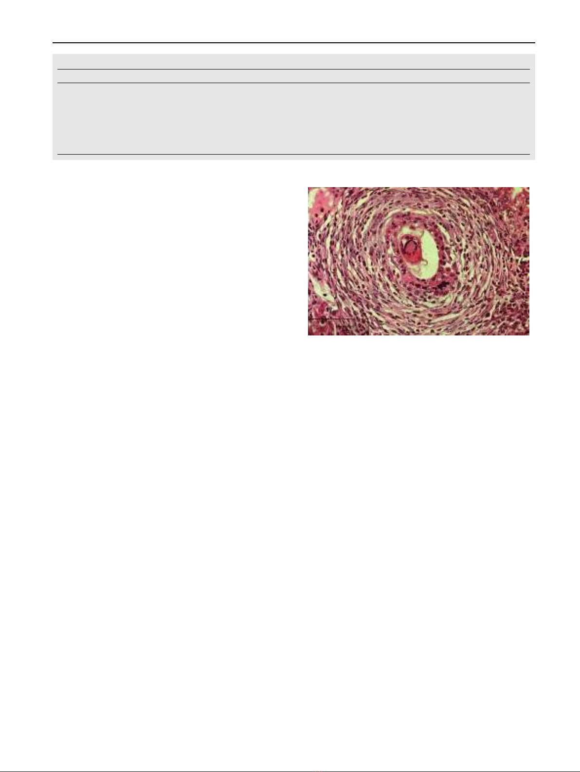
Fig. 1 Schistosomal granuloma in liver. Granuloma surround-
ing schistosomal egg in the liver. (http://www.path.cam.ac.uk/
~schisto/schistosoma/schisto_pathology_granuloma).
446 T. Elbaz and G. Esmat
gions with a high prevalence of schistosomiasis usually have a
high endemicity of chronic viral hepatitis as well. An impor-
tant cause of the high exposure to HCV was the establishment
of a large reservoir of infection as a result of extensive schisto-
somiasis control programs that used intravenously adminis-
tered tartar emetic 20–50 years ago [22]. The association
between both schistosomiasis and HCV is known to cause ear-
lier liver deterioration and more severe illness. The liver is the
principal site for both HCV replication and egg deposition,
which down-regulates the local immune responses in the liver
[23] and results in suppression of the intrahepatic bystander
immune response to HCV. This may also occur during inactive
schistosomal infection since the ova remain in the hepatic por-
tal tracts and their soluble antigens could influence the host’s
cell-mediated immunity for a considerable time [24]. In addi-
tion, this co-infection can also produce a unique clinical, viro-
logic and histologic pattern manifested by viral persistence
with high HCV RNA titers, higher necro-inflammatory and
fibrosis scores in liver biopsy specimens in addition to poor re-
sponse to interferon therapy, and accelerated progression of
hepatic fibrosis [25].
Clinical manifestations
Clinical presentation of hepatic schistosomiasis markedly dif-
fers from that of cirrhosis. Although the symptoms and signs
of portal hypertension and hypersplenism are dominant in
schistosomiasis, the counter part of hepatocellular failure is
absent. However, some patients with schistosomiasis progress
to an end stage of the disease by exhibiting muscle wasting,
hypoalbuminemia, ascites and coma. These observations led
to the concept of compensated and decompensated schistoso-
miasis to differentiate patients with the sole manifestations
of portal hypertension from those who, in addition, presented
signs of hepatocellular failure [26].
Intestinal schistosomiasis
Intestinal schistosomiasis represents another form of schisto-
somal affection. Among spectrum of intestinal lesions, polyps
are the commonest [27].
Pathogenesis
Intestinal schistosomiasis is essentially due to S. mansoni infec-
tion [28] and it has been reported as well in some S. haemato-
bium cases [29]. Egg-laying worms are present in the intestinal
micro-vasculature especially in the distribution of the inferior
mesenteric venous plexus. In the large intestine, ova are mainly
distributed in the loose submucosa, and to a lesser extent in the
subserosa where infrequently multiple granulomas are formed.
Subsequently, the muscularis mucosa becomes involved and
the overlying mucosa is either denuded forming small superfi-
cial ulcers or undergoes hyperplastic changes. Sandy patches
develop when the submucosa becomes densely thickened by fi-
brous tissue containing immense numbers of calcified eggs; the
overlying mucosa becomes atrophic and acquires a granular
dirty yellowish appearance [30].
The pathogenesis of polyp formation starts by deposition of
schistosomal eggs in the superficial layers of submucosa where
the connective tissue is loose and not bounded superficially by
firmer tissue. This allows the accumulation of large amounts of
reactive cellular debris and vascular granulation tissue. In the
submucosa, the eggs produce a cell mediated inflammatory re-
sponse with granuloma formation and necrosis. As necrotic
foci heal, fibrous connective tissue is formed and the adjacent
muscularis mucosa becomes hypertrophied. The fibrous tissue
in the submucosa and the hypertrophied muscularis mucosa
form a barrier to the usual route of ova transit from the mes-
enteric veins to the gut lumen. This entrapment of ova leads to
a foreign body reaction with progressive inflammation and
fibrosis. As this process continues, a nodule is formed that ele-
vates the hypertrophied muscularis mucosa and mucosa to
form the earliest detectable polyp [31]. This mechanism can ex-
plain the main concentration of the S. mansoni ova in the pol-
yps than in the adjacent mucosa and submucosa [32] Fig. 2.
Colonic mucosa of affected patients is usually edematous
and congested with petechial hemorrhage in acute schisto-
somal colitis cases, while shows confused vascular net with flat
or elevated yellow nodules, polyps and intestinal stricture in
chronic colitis patients. Acute and chronic inflammation could
be observed in colon segments of chronic active schistosomal
colitis patients. The most characteristic finding is the grayish
yellow or yellowish white schistosomal nodules similar to those
of pseudomembranous enterocolitis [32]. Polyps range in size
from 2 to 20 mm and may be sessile, pedunculated or showing
a cauliflower appearance. They are mainly concentrated in the
distal colon, and they count from few to very numerous pol-
yps. The covering mucosa of the polyps is usually redder than
the surrounding mucosa due to severe congestion and due to
focal hemorrhages. Ulceration is common in rectal polyps,
the ulcerated areas appear dusky to blackish gray in color
caused by superficial hemorrhage, and are frequently second-
arily infected [28,33].
Histologically, the typical polyp is composed of a stalk of
fibrous connective tissue projecting from the sub mucosa into
the lumen and partially covered with mucosa. The overlying
mucosa consists of distorted glands with varied degrees of mu-
coid activity, mucinous degeneration, and adenomatous hyper-
plasia. Focal areas of ulceration frequently interrupt the
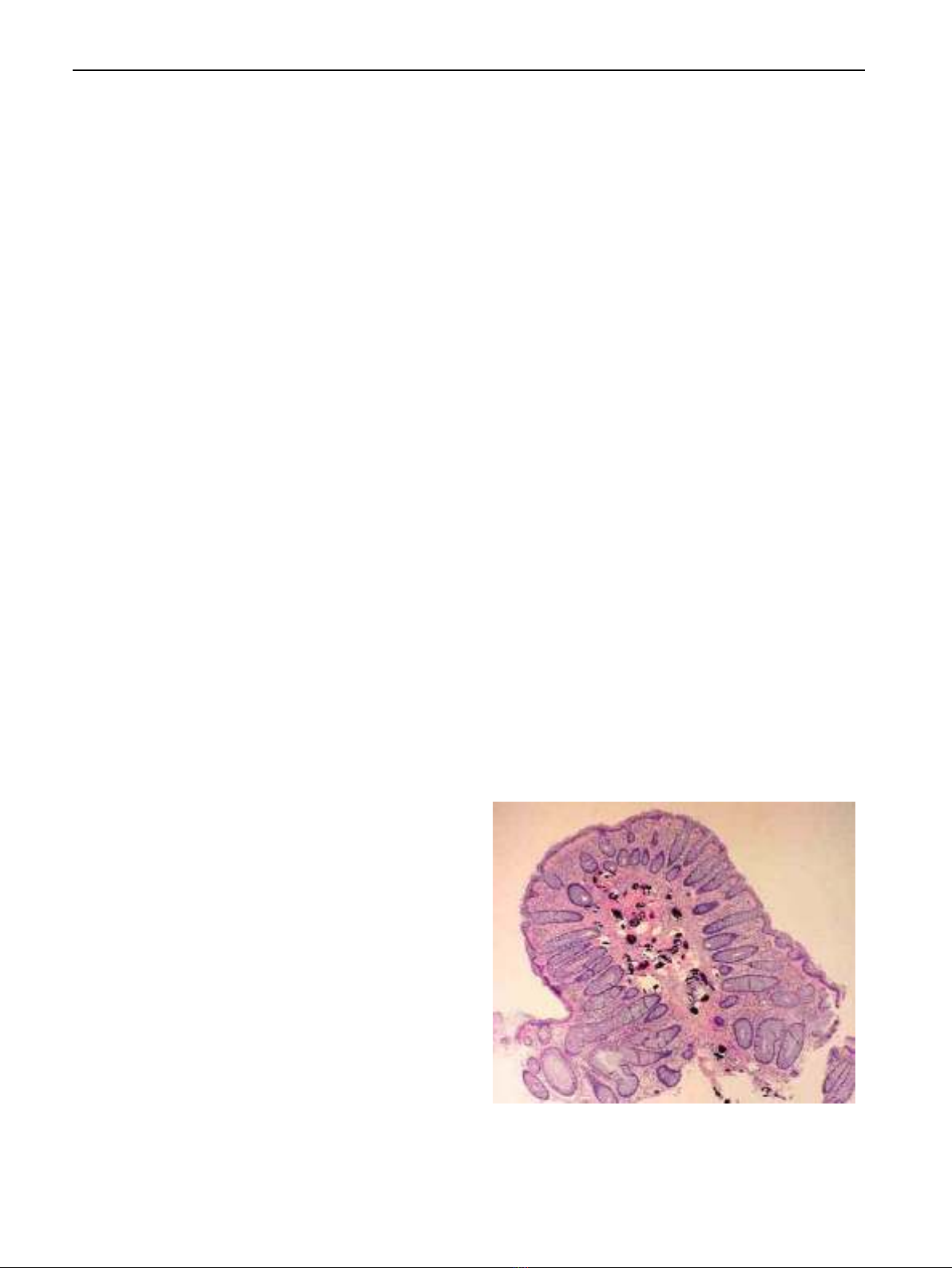
Fig. 2 Schistosomal colonic polyp. Colonic polyp with numer-
ous calcified Schisosome eggs beneath the lamina propria. In
(http://www.gastrohep.com/images/image.asp?id=1152).
Hepatic and Intestinal Schistosomiasis 447

surrounding mucosa. Larger areas of ulceration may be re-
placed by granulation tissue. Mononuclear cells, eosinophils,
and few polymorphonuclear leukocytes infiltrate the mucosa.
The supporting tissue is composed of fibrous connective tissue
and muscle derived from the muscularis mucosa. Blood vessels
may be present in large numbers but diminish as fibrosis pro-
gresses. Viable and nonviable eggs are present in all polyps
[34].
Clinical manifestations
Schistosomal colonic polyposis affects mainly adult males.
This male predominance is related to greater employment in
agricultural work and higher rates of contact with water [35].
The primary presenting symptoms are usually tenesmus and
the rectal passage of blood and mucous. Diarrhea, abdominal
pain, dyspepsia, and irreducible schistosomal papilloma pro-
truding from the anus occur in some patients [34,36]. Malnu-
trition, weight loss, nail clubbing, pitting peripheral edema,
and pericolic masses may also be present [29,36,37]. Other
manifestations include iron deficiency anemia, hypoalbumine-
mia, protein-losing enteropathy, and rectal prolapse [37,38].
The presence of polyposis does not appear to predispose pa-
tients to the development of large bowel cancer [39,40] and
many investigators even have rejected any relationship be-
tween schistosomiasis and colorectal carcinoma, although this
view is debatable if we consider S. japonicum [41–43]. How-
ever, there is a report on a patient with sigmoid cancer coexis-
ting with schistosomiasis and the authors entailed a possible
but inconclusive role for chronic schistosomiasis mansoni in
promoting carcinogenesis of colorectal neoplasms [44].
Schistosomal appendicitis is a rare complication that can
occur in 0.02–6.3% in endemic areas (representing 28.6% of
chronic appendicitis in such region) and 0.32% in developed
countries. Its main mechanism depends on mechanical appen-
diceal lumen obstruction by adult worms rather than being a
complication from egg deposition [45].
Diagnosis
Definite diagnosis of the disease depends on certain tools as
microscopy and egg identification, serology and radiologic
findings. Other non-specific findings include eosinophilia (in
relation to stage, intensity and duration of infection), throm-
bocytopenia (from splenic sequestration) and anemia (from
chronic blood loss). Liver biochemical profile is usually normal
[46]. Demonstration of parasite eggs in stool is the most com-
mon method used for making the diagnosis of schistosomiasis
and species identification. To assess intensity of infection,
quantitative sampling of defined amounts of stools (Kato Katz
technique) is applied. Concentration techniques improve the
sensitivity of egg detection. Moreover, further slide readings
from the same stool sample using the Kato Katz technique
associated with a serological test (three slides reading and
the IgG anti-Schistosoma mansoni-ELISA technique) proved
to be a useful procedure for increasing the diagnostic sensitiv-
ity [47]. Schistosomiasis can be diagnosed also by finding eggs
in tissue biopsy specimens from rectal, intestinal and liver
biopsies [48]. However, the sensitivity of these procedures is
variable due to fluctuation of egg shedding [49].
Serologic tests can detect antischistosomal antibodies in
serum samples. The main drawback is their inability to distin-
guish between past and current active infection. However, a
negative test can rule out infection in endemic population. An-
other drawback is that they remain positive for prolonged peri-
ods following therapy making them unreliable for post
treatment follow up [46]. To solve these defects, techniques
to detect parasite antigens, in sera and stools, have recently
been developed and can identify current infection and its inten-
sity [50]. Urine dipstick diagnostic tests can detect schistosome
circulating cathodic antigen (CCA). They were tested in field-
based surveys, certainly for preschool children due to the dif-
ficulty to obtain consecutive stool samples, and provided a
more sensitive and rapid testing for intestinal schistosomiasis.
This may help in future epidemiological screening studies [51].
Sensitive and specific diagnostic methods of schistosomiasis
at an early stage of infection are important to avoid egg-in-
duced irreversible pathological reactions. Detection of free cir-
culating DNA by PCR can be used as a valuable test for early
diagnosis of prepatent schistosomiasis infection [52]. Several
studies have developed polymerase chain reaction (PCR)
methods to improve the direct detection of Schistosoma anti-
gens. These tests are done on urine, stool, or organ biopsy
samples, and involve the preparation of DNA from eggs prior
to PCR amplification [53]. Only a small volume of sample can
be used for DNA extraction, and it is dependent on chance
whether the processed sample contains ova or not. Similarly,
PCR has the same limitations as microscopy and does not pro-
vide a significant clinical benefit [49]. Another study detected
S. haematobium-specific DNA in urine with similar specificity
to detection of parasite eggs but with improved sensitivity
[54]. Hopefully, an updated PCR assay has been available
for the detection of Schistosoma mansoni DNA in human stool
samples using QIAampÒDNA Stool Mini Kit. It allows the
heating of the sample (until 95 °C) to facilitate the rupture of
the egg and cellular lysis. It also includes Inhibitex, which ad-
sorbs DNA damaging substances and PCR inhibitors present
in the fecal material. For amplification, the DNA samples
are diluted only 5-fold with good reproducibility and the study
can provide high sensitivity and specificity results [55]. Another
novel diagnostic strategy is developed, following the rationale
that Schistosoma DNA may be liberated as a result of parasite
turnover and reaches the blood. Cell-free parasite DNA
(CFPD) can be detected in plasma by PCR for any stage of
schistosomiasis [49].
Radiologically, abdominal ultrasonography plays an inte-
gral role in the diagnosis of hepatosplenic schistosomiasis.
Imaging can show periportal fibrosis, splenomegaly, portal
vein dimensions and the presence of collateral vessels. In addi-
tion, ultrasonography helps to assess degree of periportal
fibrosis by measuring portal tract thickness: Grade I if thick-
ness is 3–5 mm, Grade II if it is 5–7 mm and Grade III if it
is more than 7 mm. This method reflects the hemodynamic
changes and provides a good estimate of the clinical status
of patients who have periportal fibrosis [56]. Portal hyperten-
sion is suspected when dilatation of one or more of the portal,
mesenteric and splenic veins is detected. For the collateral ves-
sels, the most commonly described are the left and right gas-
tric, the short gastric, the par umbilical and the splenorenal
veins [57,58] Fig. 3.
Lastly, the hepatic veins in schistosomiasis can be assessed
ultrasonographically. They remain patent with normal phasic
448 T. Elbaz and G. Esmat
flow as the disease evolves, which is different from liver cirrho-
sis. In advanced cirrhosis, hepatic venous outflow becomes
monophasic [57]. Colonic affection can be diagnosed by endos-
copy and biopsy from the abnormally apparent mucosa. Also,
barium enema and double contrast enema may provide a diag-
nostic tool for colonic polyps [28].
Treatment
Interventions for the control of schistosome infections involve
Mass Drug Administration (MDA) and/or chemotherapy of
individuals, as well as improved sanitation, environmental
modifications to reduce exposure to the snail intermediate
hosts and to cercariae that have been shed by snails, and edu-
cation to reduce unsafe water contact [59].
Praziquantel (PZQ) is the mainstay of chronic schistosomi-
asis treatment. It is quite safe and effective with a single oral
dose of 40–60 mg/kg bodyweight producing cure rates ranging
between 60% and 90%. Even those individuals who are not
completely cured have drastic reductions in the number of ex-
creted schistosome eggs which in turn greatly reduces the like-
lihood of long-term sequelae. Moreover, PZQ is active against
all schistosome species infecting humans, an important feature,
especially in those areas where more than a single species is
present, typically in Africa where S. mansoni and S. haemato-
bium are often co-endemic [60]. Looking for better coverage
and aiming to improve compliance of preschool children
(aged 65 years) during mass treatment campaigns targeting
schistosomiasis, the WHO provided certain areas with a syrup
formulation of PZQ. This syrup form gave very similar effica-
cies to crushed PZQ tablets in the treatment of this special age
group [61].
However, with just one drug used for individual patient
management and community-based morbidity control, resis-
tance to praziquantel may emerge and spread [62]. The second
shortcoming of PZQ is the fact that it is not active against
juvenile schistosomes [63]. When administered in the first few
days after infection, PZQ is apparently effective, but activity
rapidly drops until it reaches insignificant levels around the
fourth week and then starts rising again to attain maximal effi-
cacy around the seventh week. The practical consequence of
this phenomenon is that, in areas of very active transmission
of infection, many people are likely to harbor immature worms
at the time of treatment, with the result of correspondingly low
cure rates [64].
Some antimalarial drugs were found to have some antis-
chistosomal properties, such as the artemisinin, synthetic triox-
olanes, and mefloquine [62]. They are effective against the
juvenile stages of schistosome species but are less effective
against adult worms. From systematic review and meta-analy-
sis, the combination of an artemisinin derivatives plus prazi-
quantel showed a higher cure rate than praziquantel
monotherapy. This confirms that artemisinin derivatives used
in combination with praziquantel have the potential to in-
crease the cure rates in schistosomiasis treatment, but not
artesunate alone. However, the incorporation of artemisinin
derivatives in mass praziquantel administration has some lim-
itations such as the higher cost-effectiveness implications, the
required repeated treatments and most importantly the possi-
bility of emergence of artemisinin-resistant malaria [64].
As treatment depends on the stage of infection and the clin-
ical presentation, Deng et al. recommended a new clinical clas-
sification after reviewing the medical records of 11 092 cases of
advanced schistosomiasis. Based on the new classification
method, there were eight types: huge splenomegaly, ascites, co-
lon proliferative, dwarf, universal, bleeding, hepatic coma, and
miscellaneous. The aim of this new classification method was
to present a more comprehensive picture for clinical features,
severe complications and prognosis of advanced schistosomia-
sis [65]. The effect of antischistosomal treatment on disease
manifestations varies by stage. Early hepatomegaly is known
to resolve after specific anti schistosomal chemotherapy. Late
manifestations, such as fibrosis, do not change. Additional
management modalities are necessary for patients with com-
bined infection (e.g. schistosomiasis and HCV), such as hepa-
tocellular failure with ascites and encephalopathy which need
diuretics and anticoma measures [66].
For primary prophylaxis of variceal bleeding; beta-blockers
or endoscopic therapy could be used. For the control of acute
variceal bleeding, endoscopic therapy is effective. Ligation is
the recommended form; although sclerotherapy may be used
in the acute setting if ligation is technically difficult. Endo-
scopic therapy with tissue adhesive (e.g., N-butyl-cyanoacry-
late) is recommended for acute gastric variceal bleeding.
Interventional therapy such as TIPS, shunt surgery and
decompressive surgery should only be considered for patients
with failure of endoscopic therapy [67].
Colonoscopic polypectomy is safe and effective and may be
required along with medical therapy to achieve complete
symptom relief and prevent complications. All symptomatic
or large polyps should be removed after pharmacologic treat-
ment even before waiting for complete parasitological cure be-
cause they will not resolve with medical treatment alone
[33,34,68,69].
Vaccination
Clinical trials to develop an antischistosomal vaccine are still
in progress. A recent study identified certain issues to facilitate
its development and licensure as follows: Identification of the
human immunoprotective antigens and mechanisms, induction
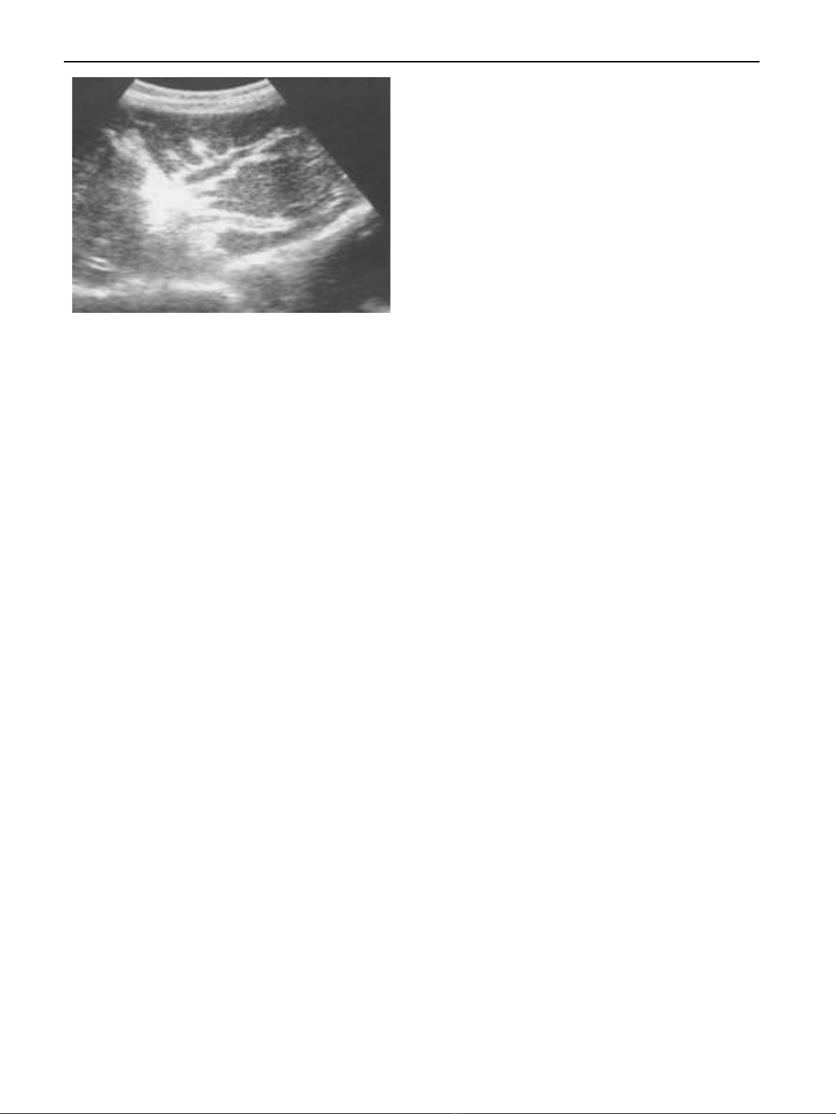
Fig. 3 Periportal fibrosis detected by ultrasonography. Thick-
ened portal tracts seen in the liver by abdominal ultrasonography
[58].
Hepatic and Intestinal Schistosomiasis 449
![Đồng thuận chẩn đoán và điều trị nhiễm Helicobacter pylori: Tài liệu [chuẩn nhất/mới nhất]](https://cdn.tailieu.vn/images/document/thumbnail/2026/20260507/baobinh_011/135x160/5151778150799.jpg)

![Chẩn đoán và điều trị nhiễm khuẩn da và mô mềm [chuẩn nhất]](https://cdn.tailieu.vn/images/document/thumbnail/2025/20250328/quyvanphi/135x160/9881743096527.jpg)


%20--%3e%3cdefs%3e%3cstyle%3e%20.st0%20{%20fill:%20%23fff;%20}%20.st1%20{%20fill:%20%237800fa;%20}%20%3c/style%3e%3c/defs%3e%3cpath%20class='st1'%20d='M117.78,12.18H43.11c2.9,3.47,4.65,7.94,4.65,12.82,0,5.6-2.3,10.66-6.01,14.29h76.02l7.22-13.56-7.22-13.56Z'/%3e%3cg%3e%3cpath%20class='st0'%20d='M53.58,26.17h-.59v-1.46h.59v-4.96h2.83c1.78,0,2.67.94,2.67,2.82v5.76c0,1.87-.89,2.81-2.67,2.81h-2.83v-4.96ZM55.36,21.37v3.34h1.1v1.46h-1.1v3.34h1.01c.61,0,.91-.37.91-1.1v-5.93c0-.74-.3-1.1-.91-1.1h-1.01Z'/%3e%3cpath%20class='st0'%20d='M65.99,31.14h-1.8l-.31-2.07h-2.19l-.31,2.07h-1.64l1.82-11.39h2.62l1.82,11.39ZM65.28,18.04c-.25.46-.51.77-.75.94-.21.15-.47.22-.79.22-.26,0-.57-.07-.92-.22l-.38-.15c-.14-.05-.26-.07-.37-.07-.3,0-.53.18-.71.54l-.91-.68c.25-.46.51-.77.75-.94.21-.14.48-.21.79-.21.26,0,.57.07.92.21l.38.15c.14.05.26.07.37.07.3,0,.53-.18.71-.54l.91.68ZM61.91,27.52h1.73l-.87-5.76-.87,5.76Z'/%3e%3cpath%20class='st0'%20d='M74.53,26.89v1.52c0,1.91-.89,2.86-2.67,2.86s-2.67-.95-2.67-2.86v-5.93c0-1.91.89-2.86,2.67-2.86s2.67.95,2.67,2.86v1.11h-1.69v-1.22c0-.75-.31-1.12-.93-1.12s-.93.37-.93,1.12v6.15c0,.74.31,1.11.93,1.11s.93-.37.93-1.11v-1.63h1.69Z'/%3e%3cpath%20class='st0'%20d='M81.4,31.14h-1.8l-.31-2.07h-2.19l-.31,2.07h-1.64l1.82-11.39h2.62l1.82,11.39ZM75.9,19.2l1.52-1.91h1.71l1.51,1.91h-1.61l-.76-.95-.75.95h-1.61ZM77.32,27.52h1.73l-.87-5.76-.87,5.76ZM83.1,15.99l-1.76,1.91h-1.26l1.17-1.91h1.86Z'/%3e%3cpath%20class='st0'%20d='M84.86,19.75c1.78,0,2.67.94,2.67,2.82v1.48c0,1.87-.89,2.81-2.67,2.81h-.85v4.28h-1.79v-11.39h2.64ZM84.01,21.37v3.86h.85c.58,0,.87-.36.87-1.08v-1.71c0-.71-.29-1.07-.87-1.07h-.85Z'/%3e%3cpath%20class='st0'%20d='M93.51,19.75c1.78,0,2.67.94,2.67,2.82v1.48c0,1.87-.89,2.81-2.67,2.81h-.85v4.28h-1.79v-11.39h2.64ZM92.66,21.37v3.86h.85c.58,0,.87-.36.87-1.08v-1.71c0-.71-.29-1.07-.87-1.07h-.85Z'/%3e%3cpath%20class='st0'%20d='M98.8,31.14h-1.79v-11.39h1.79v4.88h2.03v-4.88h1.83v11.39h-1.83v-4.88h-2.03v4.88Z'/%3e%3cpath%20class='st0'%20d='M105.36,24.55h2.46v1.62h-2.46v3.34h3.09v1.63h-4.88v-11.39h4.88v1.63h-3.09v3.18ZM108.17,17.29l-1.76,1.91h-1.26l1.17-1.91h1.86Z'/%3e%3cpath%20class='st0'%20d='M112.2,19.75c1.78,0,2.67.94,2.67,2.82v1.48c0,1.87-.89,2.81-2.67,2.81h-.85v4.28h-1.79v-11.39h2.64ZM111.35,21.37v3.86h.85c.58,0,.87-.36.87-1.08v-1.71c0-.71-.29-1.07-.87-1.07h-.85Z'/%3e%3c/g%3e%3ccircle%20class='st1'%20cx='25'%20cy='25'%20r='20'/%3e%3cpath%20class='st0'%20d='M32.78,19.27c2.92,0,4.43,2.55,5.28,5.33l.71,2.17c.14.38-.33.75-.71.75h-5.61c.19-.33.24-.71.09-1.08l-.75-2.45c-.43-1.32-.99-2.64-1.79-3.77.75-.57,1.65-.94,2.78-.94h0ZM25,18.38c3.25,0,4.9,2.78,5.89,5.89l.76,2.45c.14.42-.33.8-.8.8h-11.69c-.42,0-.94-.38-.8-.8l.75-2.45c.99-3.11,2.64-5.89,5.89-5.89h0ZM25,11.35c1.74,0,3.11,1.37,3.11,3.11s-1.37,3.11-3.11,3.11-3.11-1.41-3.11-3.11,1.41-3.11,3.11-3.11h0ZM17.27,19.27c1.08,0,1.98.38,2.73.94-.8,1.13-1.37,2.45-1.74,3.77l-.8,2.45c-.14.38-.05.75.09,1.08h-5.56c-.42,0-.9-.38-.75-.75l.71-2.17c.9-2.78,2.41-5.33,5.33-5.33h0ZM17.27,12.91c1.51,0,2.78,1.27,2.78,2.83s-1.27,2.83-2.78,2.83-2.83-1.27-2.83-2.83,1.27-2.83,2.83-2.83h0ZM32.78,12.91c1.56,0,2.78,1.27,2.78,2.83s-1.23,2.83-2.78,2.83-2.83-1.27-2.83-2.83,1.27-2.83,2.83-2.83h0ZM27.07,28.56v.09c0,.57-.24,1.08-.61,1.46h0v.05c-.38.33-.9.57-1.46.57s-1.08-.24-1.46-.61h0c-.38-.38-.61-.9-.61-1.46v-.09h1.41v.09c0,.19.05.38.19.47v.05c.09.09.28.19.47.19s.38-.09.47-.19v-.05c.14-.09.24-.28.24-.47t-.05-.09h1.41ZM30.99,28.56v.09c0,1.65-.66,3.16-1.74,4.24-1.08,1.08-2.59,1.79-4.24,1.79s-3.16-.71-4.24-1.79l-.05-.05c-1.04-1.08-1.7-2.55-1.7-4.2v-.09h1.41v.09c0,1.27.47,2.4,1.27,3.25h.05c.85.85,1.98,1.37,3.25,1.37s2.4-.52,3.25-1.37c.85-.8,1.37-1.98,1.37-3.25v-.09h1.37ZM34.99,28.56v.09c0,2.78-1.13,5.28-2.92,7.07-1.79,1.79-4.29,2.92-7.07,2.92s-5.23-1.13-7.07-2.92c-1.79-1.79-2.92-4.29-2.92-7.07v-.09h1.41v.09c0,2.4.94,4.53,2.5,6.08,1.56,1.56,3.72,2.5,6.08,2.5s4.52-.94,6.08-2.5c1.56-1.56,2.5-3.68,2.5-6.08v-.09h1.41Z'/%3e%3c/svg%3e)