doi:10.1046/j.1432-1033.2002.02942.x
Eur. J. Biochem. 269, 2727–2734 (2002) (cid:1) FEBS 2002
Importin a binds to an unusual bipartite nuclear localization signal in the heterogeneous ribonucleoprotein type I
Maria G. Romanelli and Carlo Morandi
Department of Mother and Child, Biology and Genetics, University of Verona, Italy
we demonstrate that the NLD-I is transported into the nuc- leus by cytoplasmic factor(s) with active transport modality. Binding assays using recombinant importin a show an inter- action with NLD-I similar to that of SV40 large T antigen NLS. Deletion analysis indicates that both stretches of basic residues are necessary for binding to importin a. The above experimental results lead to the conclusion that importin a acts as cytoplasmic receptor for proteins characterized by a bipartite NLS signal that extends up to 37 residues.
Keywords: heterogeneous ribonucleoprotein-I; polypyrimi-dine tract-binding protein; PTB; nuclear localization signal;
importin a.
The heterogeneous nuclear ribonucleoprotein (hnRNP) type I, a modulator of alternative splicing, localizes in the nucleoplasm of mammalian cells and in a discrete peri- nucleolar structure. HnRNP I contains a novel type of bipartite nuclear localization signal (NLS) at the N-terminus of the protein that we have previously named nuclear determinant localization type I (NLD-I). Recently, a neural counterpart of hnRNP I has been identified that contains a putative NLS with two strings of basic amino acids separated by a spacer of 30 residues. In the present study we show that the neural hnRNP I NLS is necessary and sufficient for nuclear localization and represents a variant of the novel bipartite NLS present in the NLD-I domain. Furthermore,
Transport of proteins and RNA into and out of the nucleus occurs through nuclear pore complexes (NPCs), which are plugged through the double membrane of the nuclear envelope [1,2]. Small molecules and ions may pass the NPC passively, while macromolecules larger than 40–45 kDa are actively transported through the NPC. The active nuclear import and export of proteins is mediated by specific amino- acid sequences that are referred as nuclear localization signals (NLSs) [3,4] and nuclear export signal (NESs) [5]. At least two different types of (cid:1)classical(cid:2) NLSs have been defined: a short stretch of basic amino acids, exemplified by the SV40 large T antigen NLS (T-ag P
KRPAATKKAGQAKKKK). The two sets of basic residues of bipartite-type NLS are required for sufficient nuclear localization, while the spacer is mutant-tolerant in sequence [7]. NLSs are usually recog- nized by the heterodimeric import receptor complex com- prising importins a and b, also named karyopherins [8,9]. Importins a contain the NLS-binding site and importins b
are responsible for the docking of the importin–substrate complex to the cytoplasmic side of the NPC and its translocation through the pore. Transfer subsequent through the pore of importin–NLS protein complex requires two additional soluble proteins, RanGTPase and nuclear transport factor-2 (NTF2) [1]. Once inside the nucleus, Ran-GTP binding to importin b causes the disso- ciation of the import complex and release of the cargo [10,11]. The directionality of the nuclear import is conferred by an asymmetric distribution of the GTP and GDP-bound forms of Ran between the cytoplasm and the nucleus, with the GTP-form predominant in the nucleus [12,13]. Based on the similarities of their primary structures, the importins a have been separated into three subfamilies, each of which shows distinct substrate specificity and differential expres- sion [14–16]. Importins a consist of two structural and functional domains, a short basic N-terminal importin b binding (IBB) domain, and a large NLS-binding domain comprising armadillo (Arm) repeats [17–19]. Crystal struc- tures of karyopherins a complexes with NLS peptide have revealed the determinants of specificity for the binding of NLS sequences [20–22].
A number of NLS sequences that do not conform to the classical NLS consensus motif have also been identified, such as the M9 sequence, present in the hnRNP A1, which is recognized by transportin (karyopherin-b2), rich in glycine rather than basic residues [23–25]. A unique signal, called KNS, which allows nuclear transport via a mechanism independent of soluble factors has also been described in the hnRNP type K [26]. These findings indicate that the most important level of control for nuclear protein transport is the targeting sequence.
Correspondence to M. G. Romanelli, Department of Mother and Child, Biology and Genetics, University of Verona, Strada le Grazie 8, 37134 Verona, Italy. Fax: + 39 045 8027180, Tel.: + 39 045 8027182, E-mail: mromanelli@univr.it Abbreviations: NLS, nuclear localization signal; NLD-I, nuclear localization determinant type I; T-ag, SV40 large tumor antigen; hnRNP, heterogeneous nuclear ribonucleoprotein; FITC, fluorescein isothiocyanate; NPC, nuclear pore complex; GST, glutathione S-transferase; PTB, polypyrimidine tract-binding protein; GFP, green fluorescent protein. (Received 10 January 2002, revised 10 April 2002, accepted 18 April 2002)
We have previously identified a novel bipartite NLS in the hnRNP type I protein [27], also known as polypyri- midine tract binding protein (PTB) [28,29], a member of the large set of RNA binding proteins, known as hnRNPs, that
KKKRKV) [6] and a bipartite NLS composed of two stretches of basic amino
acids separated by a spacer of 10–12 amino acids, exempli-
fied by nucleoplasmin (
2728 M. G. Romanelli and C. Morandi (Eur. J. Biochem. 269)
(cid:1) FEBS 2002
TTTTATCTTCTCCTTTACTATCATTACCATTGGCT GT-3¢) to generate the pnPTB-NLD-I D45–47; forward primer D11–13 and reverse primer D45–47 to generate the pnPTB-NLD-I D11–13; 45–47 mutant (the numbers indicate the residues deleted within nPTB amino-acid sequence). In the second PCR step, all the fragments were amplified with primers F1 and R2 that introduced the SalI and BamHI sites, respectively, and cloned in the pEGFP-C1 vector.
have been implicated in mRNA maturation and transport [30,31]. In mammalian cells, hnRNPI/PTB functions as a splicing repressor [32,33], and mediates exon skipping of several genes, such as a- and b-tropomyosin premRNAs [34,35], neuron-specific exon in the c-src, c-aminobutyric acid A c2 receptor, clathrin light chain B, N-methyl-D- aspartate premRNA [36,37]. Interestingly, a PTB homo- logue, abundantly present in the brain and in some neural cell lines, known as neural PTB (nPTB), has been recently identified [38]. Such a protein interacts with neuron-specific RNA binding proteins that participate in the control of alternative splicing in neurons [39]. nPTB is 74% identical in amino-acid sequence to PTB and contains four unusual RNA recognition motif (RRM) domains, and a putative bipartite NLS near the N-terminus.
The glutathione S-transferase (GST) fusion system was used to generate chimeric proteins [42]. HnRNP I-NLD I, and mutants D11–13, D45–47, and D11–13; 45–47 frag- ments, previously cloned in a pA1-CAT vector [27,43], were amplified by PCR with oligonucleotides that introduced EcoRI and HindIII restriction sites, and ligated into a modified pGEX-5X-1 vector (Amersham Pharmacia Bio- tech) where the XhoI had been mutated in a HindIII site.
Cell culture and transfection
Adherent HeLa cells were maintained in exponential growth in Dulbecco’s modified Eagle’s medium supplemented with 10% fetal bovine serum. Cells were grown on glass coverslips and transfected using Lipofectamine (Life Tech- nologies) according to manufacturer’s instructions.
HnRNP I localizes in HeLa cells both in the nucleoplasm and in a discrete perinucleolar structure [29,41]. We previously reported that the N-terminal sequence of hnRNP I/PTB contains a 60-amino-acid sequence that is both necessary and sufficient to target the protein to the nucleus [27]. The sequence, named nuclear localization determinant type I (NLD-I), is characterized by a NLS containing two clusters of basic amino acids (KR and KKFK) that resemble the nucleoplasmin bipartite signal, but are separ- ated by an unusually long stretch of 30 amino acids. Compared to that of hnRNP I, the nPTB bipartite NLS conserves the basic stretches whereas 14 residues differ in the 30-amino-acid spacer sequence.
Forty-eight hours after transfection, cells were washed with NaCl/Pi and fixed in 2% paraformaldehyde in NaCl/Pi at room temperature for 20 min. Cellular localization of the nPTB–GFP fusion proteins was examined under a fluores- cent microscope.
Expression of fusion proteins in Escherichiacoli
In the present work, we have characterized the NLD domain present in the neural counterpart of hnRNPI/PTB and investigated whether the hnRNPI/PTB import involves the karyopherin a/b pathway. We show that the hnRNP I NLD-I domain displays significant binding to importin a, which is diminished or eliminated by mutations in both basic stretches. The present data support the model for the recognition of bipartite NLS derived by crystallographic analysis.
E X P E R I M E N T A L P R O C E D U R E S
Plasmid construction
The pGEX-NLD-I, pGEX-NLD-I D11–13, pGEX-NLD-I D45–47 and pGEX-NLD-I D11–13; 45–47 constructs were transformed into the Escherichia coli strain BL 21(DE3). Cell culture and batch purification of the GST fusion proteins were performed essentially according to the manufacturer’s instructions (Amersham Pharmacia Biotech). All the proce- dures were carried out at 4 (cid:3)C; 1 mM EGTA and 2 mM dithiothreitol were included in the buffers throughout the purification procedures. Recombinant GST proteins, after elution, were dialyzed at 4 (cid:3)C against binding buffer (20 mM Hepes, 150 mM KOAc, 2 mM Mg (OAc)2, 2 mM dithiothre- itol). The concentration of proteins was determined by the method of Bradford [44] using the Bio-Rad dye reagent (Bio- Rad) and BSA as standard. Protein samples were aliquoted, quick frozen in liquid N2 and stored at )80 (cid:3)C.
The E. coli strain BLR containing GST fusions of a functional SV40 large T antigen nuclear localization signal (Tag NLS) or an inverse version of Tag NLS (Tag NLSinv) were cultured, and the proteins were purified as described previously [45].
Nuclear import assay
A plasmid containing the full length cDNA of human nPTB [39] was kindly provided by D. L. Black (Howard Hugnes Medical Institute, Los Angeles, CA, USA). To generate fusion constructs with the green fluorescent protein (GFP), the nPTB entire coding region, or its fragments (Fig. 2), were obtained by PCR from the original cDNA. The forward and reverse primers used for PCR included SalI and BamHI restriction enzyme site at the 5¢ and 3¢ ends, respectively. The PCR products were digested with SalI and BamHI and cloned in frame with a 5¢-GFP coding sequence into mammalian expression vector pEGFP-C1 (Clontech). To introduce deletions into the NLD-I sequence of nPTB we used a two-step PCR method. In the first step we used the following oligonucleotides: forward primer D11–13 (5¢-ATGGACGGAATCGTCACTGAAGTTGCAGTTA and GAGGATCTGACGAACTACTCTCAGGC-3¢) reverse primer R1 (5¢-ATTGGATCCTTATACACGAGA AGGAGCACC-3¢) to generate the pnPTB-NLD-I D11-13 mutant; forward primer F1 (5¢-GGCAGGCATTCAGTC GACATGGACGGAATCGTCACT-3¢) and reverse pri- mer D45-47 (5¢-TACACGAGAAGGAGCACCATCCA
Digitonin permeabilized HeLa cells were prepared essen- tially as described by Adam et al. [46]. Cells grown on coverslips were permeabilized with 55 lgÆmL)1 digitonin (Sigma) in transport buffer (TB: 20 mM Hepes pH 7.3, 110 mM potassium acetate, 5 mM sodium acetate, 2 mM magnesium acetate, 1 mM EGTA, 2 mM dithiothreitol, 1 lgÆmL)1 each of aprotinin, leupeptin, and pepstatin A).
Binding of importin a to hnRNP I bipartite NLS (Eur. J. Biochem. 269) 2729
(cid:1) FEBS 2002
bound to the GST fusion proteins was detected by fluoro- graphy using Amplify Reagent (Amersham Pharmacia Biotech).
R E S U L T S
The NLD-I of neuronal PTB is capable of targeting a heterologous protein to the nucleus
A standard 50-lg nuclear import assay was performed in transport buffer containing an energy-regenerating system (1 mM ATP, 0,5 mM GTP, 10 mM creatine phos- phate, and 0.4 UÆmL)1 creatine phosphokinase), 5 lg of GST fused proteins, and 30 lg of rabbit reticulocyte lysate (Promega) as a cytosol source. The reaction was allowed to proceed for 45 min at 30 (cid:3)C. Where indicated, wheat germ agglutinin (WGA); Sigma) at 50 lgÆmL)1, or hexokinase at 100 UÆmL)1 and glucose at 10 mM, were included. Import assays were terminated by washing the in cold NaCl/Pi cells followed by fixation in 2% paraformaldehyde for 30 min.
Immunofluorescence staining Fixed cells were permeabilized for 3 min in )20 (cid:3)C acetone, washed with NaCl/Pi and incubated for 40 min with primary antibody diluted in 3% BSA/NaCl/Pi (1 : 100 monoclonal anti-GST Ig, Santa Cruz Biotechnology), washed in NaCl/Pi and incubated for 40 min in 1 : 50 FITC-conjugated goat anti-(mouse IgG) Ig. Cells were finally washed with NaCl/Pi, coated with 90% glycerol in NaCl/Pi, and observed under a Leitz Orthoplan microscope with an epifluorescence attachment.
In vitro binding assay
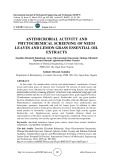
In a previous study, we identified the sequence implicated in the nuclear transport of the human hnRNPI/PTB at the N-terminal of the protein [27]. This region, which we called NLD-I, extends in the first 60 amino acids, upstream to the RRM1 (Figs 1A). The sequence contains two short basic sequences (KR and KKFK) that resemble the SV40 T-ag NLS, separated by an unusual long spacer sequence of 30 amino acids. Both short basic sequence were necessary for transport and, taken alone, were not able to target the protein to the nucleus. Sequence comparisons among NLD-I and the N-terminal region of neural PTB and PTB homologues isolated from pig, rat, mouse and Xenopus show that the basic stretches are highly conserved in all the hnRNPI/PTB homologues, whereas the sequence of the spacer, that may vary from 29 to 33 amino acids, is less conserved (Fig. 1B). The nPTB NLD-I sequence is identical in human and mouse, whereas the variations in NLD-I sequence between human nPTB and hnRNPI/PTB far exceed that among PTBs from different vertebrates.
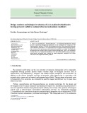
In order to characterize the novel type of bipartite NLS we first examined the ability of the NLD-I motif of the neuronal PTB to target a heterologous protein to the nucleus. GFP–nPTB fusion proteins were constructed in which NLD-I motif was deleted from nPTB or was the only sequence fused to GFP. Following transfection with GFP constructs, HeLa cells were fixed and visualized by direct fluorescence (Fig. 2). GFP is a small protein (30 kDa) that can passively diffuse through the nuclear pores and does not produce a subcellular localization bias (Fig. 2A), whereas fusion of the entire nPTB ORF to GFP led to a peptide that accumulates exclusively in the nucleus (Fig. 2B). The first 60 amino acids corresponding to the NLD-I domain are necessary and sufficient to localize the fusion protein completely in the nucleus, as demonstrated by the pGFP- nPTBD60 construct that was confined to the cytoplasm,
The plasmid pRSET-hSRP1 containing a cDNA of human importin a [47] was used to produce a [35S]methionine- labeled protein using the Promega TNT T7 Quick Coupled Translation System. Fifteen micrograms of GST–NLD-I or NLD-I mutants and 7 lg of GST–Tag NLS or GST– TagNLSinv were incubated with 40 lL of glutathione- agarose beads (Amersham Pharmacia Biotech) in 0.5 mL of binding buffer (20 mM Hepes, pH 6.8, 150 mM KOAc, 2 mM Mg (OAc)2, 2 mM dithiothreitol, 0.1% Tween 20 for 2 h at 4 (cid:3)C. The beads were collected and washed three times with binding buffer. After washing, one-twentieth of the beads were removed and the amount of immobilized GST fusion proteins were analyzed by SDS/PAGE. The rest of the beads were incubated with 90 lL of in vitro translated importin reaction mixture for 4 h at 4 (cid:3)C. The beads were then washed six times in binding buffer, boiled in 30 lL of sample buffer, and the immobilized proteins were resolved on a SDS/10% polyacrylamide gel. The 35S-labeled importin
4
(A) Diagrammatic representation of the human hnRNP I/PTB protein, with the Fig. 1. Functional domains of the human hnRNPI/PTB protein. four RNA recognition motifs (RRM1–4) and the nuclear localization determinant type I (NLD-I). (B) Amino acid alignment of the hnRNP-I NLD-I domain with homologous domains from human and mouse nPTB, and from mouse, rat, pig and Xenopus PTB. The basic clusters are in bold letters. Alignment was performed by CLUSTAL W program. Asterisks and dots show identical and similar amino acids, respectively.
2730 M. G. Romanelli and C. Morandi (Eur. J. Biochem. 269)
(cid:1) FEBS 2002
proteins. The NLS contained in the NLD-I is a new variant of bipartite NLS sequences.
whereas when the first 60 amino acids are fused to GFP (pGFP–nPTB60), the protein localized completely into the nucleus (Fig. 2D,C, respectively).
Nuclear import of GST–NLD-I is an energy-dependent process that requires soluble cytoplasmic factors and is inhibited by WGA
3
Deletions of three amino acids, including a lysine in the first basic in pGFP– stretch (deletion Gly-Val-Lys nPTBD11–13) abolished the nuclear localization and left the fusion protein to diffuse passively through the nuclear pore (Fig. 2E). A similar cellular distribution was observed when a serine and two lysines were deleted in the second basic stretch (pGFP–nPTBD45–47) or when both type of deletions were present in the basic stretches (construct pGFP–nPTBD11–13; 45–47) (Fig. 2F,G). The above obser- vations indicated that the two basic motifs in NLD-I of nPTB are interdependently required for full nuclear local- ization of the chimeric protein. Similar results were previ- ously obtained with the NLD-I present in hnRNP I/PTB that differ from that of nPTB essentially in the spacer sequence (Fig. 1). These data show that the intervening sequence does not contribute to the nuclear localization and may be modified without effect on localization, as it has been shown for the bipartite NLS of nucleoplasmin [7]. Taken together, these results indicate that the identified NLS motif is a bona fide nuclear import signal for PTB like
Due to the fact that NLD-containing NLSs motifs can be imported by different mechanisms [48], we have undertaken a study on the identification of the receptor pathway that mediates nuclear import of the PTB NLS. An in vitro nuclear transport assay was used in which the plasma membrane of HeLa cells was permeabilized with the weak nonionic detergent digitonin that leaves the nuclear envel- ope intact [46]. NLD-I motif was fused to a GST protein, that itself does not accumulate into the nucleus [49] (Fig. 3A). Nuclear transport of GST–NLD-I was examined in the presence of a transport buffer containing rabbit reticulocyte lysate as a source of cytosolic proteins and an ATP-regenerating system, to provide energy for transloca- tion. The subcellular distribution of the GST–NLD-I was determined by indirect immunofluorescence microscopy using an antibody against GST. In such experiments, we
Fig. 2. The NLD-I domain of nPTB directs the nuclear import of a heterologous protein. (A) Schematic representation of the nPTB regions used to produce GFP fusion proteins. Structural domains are diagrammed with shaded boxes representing the RRMs. Black boxes or black boxes interrupted by a white strip represent the basic stretches or the dele- ted sequences in the basic stretches, respect- ively, at the N-terminus of the protein. (B) Plasmids expressing the native GFP (a), GFP fused to the entire nPTB (b), the 60- amino-acid region at N-terminus of nPTB (c), or nPTB deleted of the first 60 amino acids (d), were transiently transfected, expressed in HeLa cells and visualized by fluorescent microscopy. Likewise, GFP fused to the 60-amino acid region containing deletions at amino acids 11–13 (e), or 44–47 (f), or both type deletions (g) were also expressed in HeLa cells.
Binding of importin a to hnRNP I bipartite NLS (Eur. J. Biochem. 269) 2731
(cid:1) FEBS 2002
peptide segments [20,50]. Importin a binding to a bipartite signal with a spacer sequence longer than 30 amino acids have not been tested thus far. The unusual sequence of the PTB NLS raised the possibility that it might not be recognized directly by importin a.
To test if NLD-I would bind to importin a, we first performed a binding assay using in vitro translated importin a and recombinant GST–NLD-I fusion protein. The GST fusions with SV40 tag NLS (GST–NLS) and an inverse version of Tag NLS (GST–NLSinv) served as positive and negative controls, respectively, for the importin a binding. As shown in Fig. 4A, importin a is able to bind NLD-I (lane GST–NLD-I). To ascertain whether the amino acid that impaired nuclear transport in NLD-I deletion mutants, mediate the importin a binding, we tested importin a binding to NLD-I D11–13, NLD-ID45–47, or NLD-ID11– 13;45–47, expressed as GFP-fusions. All mutants showed no detectable or very weak binding to importin a (Fig. 4B). These results indicate that importin a binds to NLD–I by interaction to the residues of the two short basic stretches, when both basic stretches are present in the NLS sequence.
Fig. 3. Nuclear import of NLD-I is an energy- dependent process inhibited by WGA. Digito- nin-permeabilized HeLa cells were incubated with GST, GST–TagNLS (a control for the conventional importin a/b mediated nuclear import pathway directed by SV40 NLS), GST–NLD-I, or mutated NLD-I and visual- ized by indirect immunofluorescence, using a GST monoclonal antibody. In vitro nuclear transport (see (cid:1)Material and methods(cid:2)) was carried out in the presence of cytosol and an ATP-regenerating system (A–J). The effects on nuclear transport of the ATP-regenerating system omission (D), or 4 (cid:3)C incubation (E), were examined. Transport studies were also carried out after preincubation with WGA (F). Images are representative of at least three independent experiments.
D I S C U S S I O N
found that GST–NLD-I was clearly visible within the nuclei in cells were the plasma membrane was permeabilized by digitonin, followed by incubation at 30 (cid:3)C for 45 min with rabbit reticulocyte lysate and an ATP energy-regenerating system (Fig. 3C). A similar distribution was observed for GST–TagNLS, which is transported to the nucleus by the conventional NLS-mediated nuclear protein import path- way utilizing Ran and the importin ab/heterodimer [45] (Fig. 3B). This accumulation is ATP dependent as no nuclear accumulation was observed when the permeabilized cells were incubated with hexokinase and glucose to deplete residual ATP, and when import assay was performed in the absence of an ATP-regenerating system (Fig. 3D). As expected, the transport reaction was also temperature- dependent; if the assay was carried out at 4 (cid:3)C, no transport was observed (Fig. 3E), confirming the necessity of ATP hydrolysis for nuclear protein import. When permeabilized cells were preincubated in transport buffer containing 50 lgÆmL)1 WGA, a lectin that associates with glycosylated nucleoproteins and inhibits nuclear pore complex function, the nuclear accumulation of NLD-I was inhibited (Fig. 3F). To characterize the amino-acid sequence requirement for nuclear import, we examined whether amino-acid deletions into NLD-I would perturb import. When we used GST– NLD-ID11–13, GST–NLD-ID45–47, or GST–NLD-ID11– 13;45–47 as the cargo in different import assays, no transport in the nucleus was seen (Fig. 3G,H,J).
NLD-I domain binds to importin a
In this report, we have demonstrated that nuclear translo- cation of nPTB and hnRNPI/PTB occurs via a polybasic NLS sequence present in the N-terminus NLD-I motif. This sequence functions in the nuclear import of PTB-like proteins via an active energy-dependent process and binds to the importin a. PTB NLS shares common features with the known bipartite type NLSs; in fact, it contains two clusters of basic amino acids, a smaller one of two basic amino acids (KR) and a larger one with three basic amino acids in a group of five (KKFKG); both are essential for full nuclear localization and importin a binding. However, it
The previous data suggest that the import of the PTB NLS might be mediated by cytosol receptor like the importin a/b complex. The crystal structure of importin a reveals two NLS peptide binding pockets and the distance between the two binding sites allows a 10-residue spacer to link the two
2732 M. G. Romanelli and C. Morandi (Eur. J. Biochem. 269)
(cid:1) FEBS 2002
cells, it has been shown that the segment spacer of nucleoplasmin bipartite NLS can be replaced by a sequence up to 20 alanine residues, without disrupting efficient nuclear import [8]. Recently, a bipartite NLS, containing a spacer between the basic motifs of 32 amino acids, has been functionally characterized in hypoxia inducible factors 1a [58]. We indicate a consensus sequence for bipartite long type NLS as KRx(30–32)K[K/R]xK, according to Cokol et al. [56]. Searching the PredictNLS database using this motif, we found 61 proteins, with a true nuclear protein percent of 83.6.
Nuclear import of most proteins requires both impor- tins a and b, with importin a as the adapter between importin b and the cargo protein. Some proteins undergo nuclear import via direct binding to importin b without involvement of importin a. Our results suggest that nuclear import of PTB-like proteins, and probably of all the proteins that contain the NLD-I type NLS, is an energy- dependent process mediated by importin a. Deletions in the basic regions within the N-terminal domain of hnRNP I and nPTB inhibit nuclear import and/or accu- mulation and reduces importin a binding. This conclusion fits with the in vitro binding studies, which showed that the NLD-I domain binds strongly to importin a and the bind- ing is diminished or eliminated even if only one of the two basic stretches are mutated. The affinity of the importin- targeting sequence interaction is a critical parameter in determining transport efficiency [5]. This is the first study to demonstrate that importin a recognizes and binds an NLS sequence that extends up to 37 residues. The crystal structures of importin a [20–22,50] clearly reveal two distinct binding sites that can accommodate both essential elements of the bipartite NLS. The larger binding site is structured optimally for the recognition of five lysine or arginine residues, while the smaller binding site allows specific recognition of two basic residues, and the interac- tion is simultaneous at both sites. The distance between the two binding sites allow a 10-residue spacer to link the two peptide segments, while a shorter linker would impair the simultaneous binding of the two clusters. The smaller basic cluster is required to be upstream of the larger cluster. The
represents a novel member of bipartite signal because there are differences between the critical basic residues of NLS and the consensus sequences of other bipartite signals, and because the spacer between the basic motifs is unusually longer (30 amino acids) than that of other well characterized bipartite NLSs (Table 1). Searching SWISSPROT and standard databases by PROSITE, and analyzing data deposited at the PredictNLS server (http://maple.bioc. columbia.edu/predictNLS/) [56], we find that functional bipartite NLSs are characterized by an intervening region of different lengths, but not longer than 18 amino acids, like the human androgen receptor NLS motif [57]. In transfected
Fig. 4. Binding of importin a to wild-type or mutated NLD-I. (A) Wild- type GST–NLD-I, or GST–NLS and GST–NLSinv, immobilized on glutathione sepharose 4B, were incubated with 90 lL of in vitro translated protein a labeled with [35S]methionine for 4 h at 4 (cid:3)C. The proteins were separated by SDS/10% PAGE gel, and bound importin a was analyzed by fluorography (upper panel). An amount repre- senting 1/20th of the beads incubated with GST fusion proteins, extensively washed, was resolved by SDS/PAGE and stained with Comassie-blue (lower panel, CB staining). (B) Binding of importin a was also analyzed using immobilized GST–NLD-I peptides mutated by deletions. The results are representative of two independent experiments.
Table 1. Bipartite type nuclear localization signal. The single-letter amino acid code is used. The bold letters indicate the two arms of basic residues of the bipartite NLS. aa, amino acids.
Protein Bipartite NLS Reference
Bipartite short type NLSs
KRPAATKKAGQAKKKKLDK RRRHSDENDGGQPHKRRK RKKRKTEEESPLKDKAKKSK KKYENVVIKRSPRKRGRPRK KKYIDGQKKKCGEERRRVNQ KRSAEGSNPPKPLKKLR KRALPNNTSSSPQPKKKP KK- 10–12 aa -KKK RR- -RRR
[7] [14] [54] [55] [51] [52] [53] Nucleoplasmin CBP80a N1N2b SW15c Human IL 5 Human RB Human p53 Consensus
Bipartite long type NLSs
30–32 aa
KRGSDELFSTCVTNGPFIMSSNSASAANGNDSKKFKGDS KRGSDELLSGSVLSSPNSNMSSMVVTANGNDSKKFKGED KRKMEHDGSLFQAUGIGTLLQQPDDHAATTSLSWKRVKG -K[K/R]XK KR-
a CAP-binding protein 80. b Xenopus laevis phosphoprotein. c S. cerevisiae transcription factor. d Hypoxia inducible factor 1a.
[29] [39] [58] HnRNPI/PTB_HUMAN PTB_HUMAN HIF1a d Consensus
Binding of importin a to hnRNP I bipartite NLS (Eur. J. Biochem. 269) 2733
(cid:1) FEBS 2002
spacing between a defined number of binding sites acts as molecular ruler that sets further constraints on target specificity. Binding properties of NLD-I type NLS to importin a are consistent with crystallographic structure of interaction with bipartite signal and further characterize the bipartite signals, confirming that the linker sequence does not contain a consensus, and may be as long as 30 amino acids.
13. Mattaj, I. & Englmeier, L. (1998) Nucleocytoplasmic transport: the soluble phase. Annu. Rev. Biochem. 67, 265–306.
14. Miyamoto, Y., Imamoto, N., Sekimoto, T., Tachibana, T., Seki, T., Tada, S., Enomoto, T. & Yoneda, Y. (1997) Differential modes of nuclear localization signal (NLS) recognition by three distinct classes of NLS receptors. J. Biol. Chem. 272, 26375–26381. 15. Kohler, M., Speck, C., Christiansen, M., Bischoff, F.R., Prehn, S., Haller, H., Gorlich, D. & Hartmann, E. (1999) Evidence for dis- tinct substrate specificities of importin alpha family members in nuclear protein import. Mol. Cell. Biol. 19, 7782–7791.
16. Nadler, S.G., Tritschler, D., Haffar, O.K., Blake, J., Bruce, A.G. & Cleaveland, J.S. (1997) Differential expression and sequence– specific interaction of karyopherin alpha with nuclear localization sequences. J. Biol. Chem. 272, 4310–4315.
Mammalian paralogs of importin a have recently been discovered and six genes for importin a have been found in human. The importin a that we have used in our binding experiments (hSRP1a) represents one of the three sub- families of importins a [15]. Several experiments clearly support the hypothesis that importins a might be specialized in their efficiency to transport different nuclear proteins [59]. It will be interesting to analyze the binding specificity of the different types of importin a to NLD-I NLS and to the putative NLD-I type NLSs present in other nuclear proteins.
17. Gorlich, D., Henklein, P., Laskey, R.A. & Hartmann, E. (1996) A 41 amino acid motif in importin-alpha confers binding to importin-beta and hence transit into the nucleus. EMBO J. 15, 1810–1817.
18. Moroianu, J., Blobel, G. & Radu, A. (1996) The binding site of karyopherin alpha for karyopherin beta overlaps with a nuclear localization sequence. Proc. Natl Acad. Sci. USA 93, 6572–6576.
A C K N O W L E D G E M E N T S
19. Peifer, M., Berg, S. & Reynolds, A.B. (1994) A repeating amino acid motif shared by proteins with diverse cellular roles. Cell 76, 789–791.
We wish to thank Pamela Lorenzi for excellent technical help. We thank Michael F. Rexach for his generous gift of GST–NLS and GST– NLSinv constructs and Karsten Weis for his generous gift of pRSET- hSRP a plasmid. This work was supported by grants from Ministery of Scientific Research and Technology (ex 60%). 20. Conti, E., Uy, M., Leighton, L., Blobel, G. & Kuriyan, J. (1998) Crystallographic analysis of the recognition of a nuclear locali- zation signal by the nuclear import factor karyopherin alpha. Cell 94, 193–204.
R E F E R E N C E S
21. Conti, E. & Kuriyan, J. (2000) Crystallographic analysis of the specific yet versatile recognition of distinct nuclear localization signals by karyopherin alpha. Struct. Fold Des. 8, 329–338. 22. Fontes, M.R.M., Teh, T. & Kobe, B. (2000) Structural basis of recognition of monopartite and bipartite nuclear localization sequences by mammalian importin-alpha. J. Mol. Biol. 297, 1183– 1194.
1. Gorlich, D. & Kutay, U. (1999) Transport between the cell nucleus and the cytoplasm. Annu. Rev. Cell. Dev. Biol. 15, 607–660. 2. Gant, T.M., Goldberg, M.W. & Allen, T.D. (1998) Nuclear envelope and nuclear pore assembly: analysis of assembly inter- mediates by electron microscopy. Curr. Opin. Cell. Biol. 10, 409– 415. 3. Doye, V. & Hurt, E. (1997) From nucleoporins to nuclear pore complexes. Curr. Opin. Cell Biol. 9, 401–411. 23. Bogerd, H.P., Benson, R.E., Truant, R., Herold, A., Phingbodhi- pakkiya, M. & Cullen, B.R. (1999) Definition of a consensus transportin-specific nucleocytoplasmic transport signal. J. Biol. Chem. 274, 9771–9777. 4. Dingwall, C. & Laskey, R.A. (1991) Nuclear targeting sequences – a consensus? Trends Biochem. Sci. 16, 478–481. 24. Bonifaci, N., Moroianu, J., Radu, A. & Blobel, G. (1997) Kar- yopherin beta2 mediates nuclear import of a mRNA binding protein. Proc. Natl Acad. Sci. USA 94, 5055–5060. 25. Siomi, H. & Dreyfuss, G. (1995) A nuclear localization domain in 5. Jans, D.A., Xiao, C.Y. & Lam, M.H. (2000) Nuclear targeting signal recognition: a key control point in nuclear transport? Bioessays 22, 532–544. the hnRNP A1 protein. J. Cell Biol. 129, 551–560. 6. Nigg, E.A. (1997) Nucleocytoplasmic transport: signals, mecha- nisms and regulation. Nature 386, 779–786.
7. Robbins, J., Dilworth, S.M., Laskey, R.A. & Dingwall, C. (1991) Two interdependent basic domains in nucleoplasmin nuclear tar- geting sequence: identification of a class of bipartite nuclear tar- geting sequence. Cell 64, 615–623. 26. Michael, W.M., Eder, P.S. & Dreyfuss, G. (1997) The K nuclear shuttling domain: a novel signal for nuclear import and nuclear export in the hnRNP K protein. EMBO J. 16, 3587–3598. 27. Romanelli, M.G., Weighardt, F., Biamonti, G., Riva, S. & Morandi, C. (1997) Sequence determinants for hnRNP I protein nuclear localization. Esp. Cell. Res. 235, 300–304.
28. Gil, A., Sharp, P.A., Jamison, S.F. & Garcia-Blanco, M.A. (1991) Characterization of cDNAs encoding the polypyrimidine tract- binding protein. Genes Dev. 5, 1224–1236. 8. Makkerh, J.P.S., Dingwall, C. & Laskey, R.A. (1996) Compara- tive mutagenesis of nuclear localization signals reveals the importance of neutral and acidic amino acids. Curr. Biol. 6, 1025– 1027. 9. Weis, K. (1998) Importins and exportins: how to get in and out of the nucleus. Trends Biochem. Sci. 23, 185–189. 10. Yoneda, Y. (2000) Nucleocytoplasmic protein traffic and its sig- 29. Ghetti, A., Pinol-Roma, S., Michael, W.M., Morandi, C. & Dreyfuss, G. (1992) hnRNP I, the polypyrimidine tract-binding protein: distinct nuclear localization and association with hnRNAs. Nucleic Acids Res. 20, 3671–3678. nificance to cell function. Genes Cells 5, 777–787.
30. Krecic, A.M. & Swanson, M.S. (1999) hnRNP complexes: com- position, structure, and function. Curr. Opin. Cell. Biol. 11, 363– 371.
11. Bischoff, F.R. & Ponstingl, H. (1995) Catalysis of guanine nucleotide exchange of Ran by RCC1 and stimulation of hydro- lysis of Ran-bound GTP by Ran-GAP1. Methods Enzymol. 257, 135–144. 31. Weighardt, F., Biamonti, G. & Riva, S. (1996) The roles of het- erogeneous nuclear ribonucleoproteins (hnRNP) in RNA meta- bolism. Bioessays 18, 747–756. 32. Valcarcel, J. & Gebauer, F. (1997) Post-transcriptional regulation: the dawn of PTB. Curr. Biol. 7, 705–708. 12. Izaurralde, E., Kutay, U., von Kobbe, C., Mattaj, I.W. & Gorlich, D. (1997) The asymmetric distribution of the constituents of the Ran system is essential for transport into and out of the nucleus. EMBO J. 16, 6535–6547.
2734 M. G. Romanelli and C. Morandi (Eur. J. Biochem. 269)
(cid:1) FEBS 2002
33. Wagner, E.J. & Garcia-Blanco, M.A. (2001) Polypyrimidine tract binding protein antagonizes exon definition. Mol. Cell Biol. 21, 3281–3288. 46. Adam, S.A., Marr, R.S. & Gerace, L. (1990) Nuclear protein import in permeabilized mammalian cells requires soluble cyto- plasmic factors. J. Cell Biol. 111, 807–816.
47. Weis, K., Mattaj, I.W. & Lamond, A.I. (1995) Identification of hSRP1 alpha as a functional receptor for nuclear localization sequences. Science 268, 1049–1053. 34. Mulligan, G.J., Guo, W., Wormsley, S. & Helfman, D.M. (1992) Polypyrimidine tract binding protein interacts with sequences involved in alternative splicing of beta-tropomyosin pre-mRNA. J. Biol. Chem. 267, 25480–25487.
48. Deane, R., Shafer, W., Zimmermann, H.P., Mueller, L., Gorlich, D., Prehn, S., Ponstingl, H. & Bishoff, F.R. (1997) Ran-binding protein 5 (RanBP5) is related to the nuclear transport factor importin-beta but interacts differently with RanBP1. Mol. Cell. Biol. 17, 5087–5096. 35. Gooding, C., Roberts, G.C. & Smith, C.W. (1998) Role of an inhibitory pyrimidine element and polypyrimidine tract binding protein in repression of a regulated alpha-tropomyosin exon. RNA 4, 85–100.
49. Pollard, V.W., Michael. W.M., Nakienly, S., Siomi, M.C., Wang, F. & Dreyfuss, G. (1996) A novel receptor-mediated nuclear protein import pathway. Cell 86, 985–994. 50. Dingwall, C. & Laskey, R.A. (1998) Nuclear import: a tale of two 36. Chan, R.C. & Black, D.L. (1997) The polypyrimidine tract bind- ing protein binds upstream of neural cell-specific c-src exon N1 to repress the splicing of the intron downstream. Mol. Cell. Biol. 17, 4667–4676. sites. Curr. Biol. 8, R922–R924.
37. Zhang, L., Liu, W. & Grabowski, P.J. (1999) Coordinate repres- sion of a trio of neuron-specific splicing events by the splicing regulator PTB. RNA 5, 117–130. 51. Jans, D.A., Briggs, L.J., Gustin, S.E., Jans, P., Ford, S. & Young, I.G. (1997) A functional bipartite nuclear localisation signal in the cytokine interleukin-5. FEBS Lett. 406, 315–320.
38. Ashiya, M. & Grabowski, P.J. (1997) A neuron-specific splicing switch mediated by an array of pre-mRNA repressor sites: evidence of a regulatory role for the polypyrimidine tract binding protein and a brain-specific PTB counterpart. RNA 3, 996–1015. 52. Zacksenhaus, E., Bremner, R., Phillips, R.A. & Gallie, B.L. (1993) A bipartite nuclear localization signal in the retinoblastoma gene product and its importance for biological activity. Mol. Cell. Biol. 13, 4588–4599.
53. Liang, S. & Clarke, M.F. (1999) A bipartite nuclear localization signal is required for p53 nuclear import regulated by a carboxyl- terminal domain. J. Biol. Chem. 274, 32699–32703.
54. Hu, W. & Jans, D.A. (1999) Efficiency of importin alpha/beta- mediated nuclear localization sequence recognition and nuclear import. Differential role of NTF2. J. Biol. Chem. 274, 15820– 15827.
39. Markovtsov, V., Nikolic, J.M., Goldman, J.A., Turck, C.W., Chou, M.Y. & Black, D.L. (2000) Cooperative assembly of an hnRNP complex induced by a tissue-specific homolog of poly- pyrimidine tract binding protein. Mol. Cell. Biol. 20, 7463–7479. 40. Polydorides, A.D., Okano, H.J., Yang, Y.Y.L., Stefani, G. & Darnell, R.B. (2000) A brain-enriched polypyrimidine tract- binding protein antagonizes the ability of Nova to regulate neu- ron-specific alternative splicing. Proc. Natl Acad. Sci. USA 97, 6350–6355.
55. Jans, D.A., Moll, T., Nasmyth, K. & Jans, P. (1995) Cyclin- dependent kinase site-regulated signal-dependent nuclear locali- zation of the SW15 yeast transcription factor in mammalian cells. J. Biol. Chem. 270, 17064–17067. 56. Cokol, M., Nair, R. & Rost, B. (2000) Finding nuclear localization signals. EMBO Reports 1, 411–415. 41. Matera, A.G., Frey, M.R., Margelot, K. & Wolin, S.L. (1995) A perinucleolar compartment contains several RNA polymerase III transcripts as well as the polypyrimidine tract-binding protein, hnRNP I. J. Cell Biol. 129, 1181–1193.
57. Jenster, G., Trapman, J. & Brinkmann, A.O. (1993) Nuclear import of the human androgen receptor. Biochem. J. 293, 761–768. 42. Smith, D.B. & Johnson, K.S. (1988) Single-step purification of polypeptides expressed in Escherichia coli as fusions with glu- tathione S-transferase. Gene 67, 31–40.
58. Luo, J.C. & Shibuya, M. (2001) A variant of nuclear localization signal of bipartite-type is required for the nuclear translocation of hypoxia inducible factors (1 alpha, 2 alpha and 3 alpha). Oncogene 20, 1435–1444.
59. Welch, K., Franke, J., Kohler, M. & Macara, I.A. (1999) RanBP3 contains an unusual nuclear localization signal that is imported preferentially by importin-alpha3. Mol. Cell Biol. 19, 8400–8411. 43. Weighardt, F., Biamonti, G. & Riva, S. (1995) Nucleo-cyto- plasmic distribution of human hnRNP proteins: a search for the targeting domains in hnRNP A1. J. Cell Sci. 108, 545–555. 44. Bradford, M.M. (1976) A rapid and sensitive method for the quantitation of microgram quantities of protein utilizing the principle of protein-dye binding. Anal. Biochem. 72, 248–254. 45. Rexach, M. & Blobel, G. (1995) Protein import into nuclei: association and dissociation reactions involving transport sub- strate, transport factors, and nucleoporins. Cell 83, 683–692.