Eur. J. Biochem. 269, 1006–1011 (2002) (cid:211) FEBS 2002
Invivo activation of plasma membrane H+-ATPase hydrolytic activity by complex lipid-bound unsaturated fatty acids in Ustilagomaydis
Agustı´n Herna´ ndez1, David T. Cooke2 and David T. Clarkson1 1IACR-Long Ashton Research Station, Department of Agricultural Sciences, University of Bristol, Long Ashton, UK; 2Department of Biological Sciences, University of Bristol, Bristol, UK
inhibition by vanadate and erythrosin B remain unchanged. This all indicates that activation of plasma membrane H+-ATPase by unsaturated fatty acids differs clearly from glucose-induced activation observed in yeast. Also, it is a physiologically relevant event similar to other, as yet uncharacterized, changes in plasma membrane H+-ATPase hydrolytic activity observed in plants and fungi, as part of an adaptation process to different stress conditions.
Keywords: enzyme activation; H+-ATPase; unsaturated fatty acid; Ustilago maydis; xenobiotic stress.
As an adaptation process to the growth retardation provoked by the presence of nonlethal concentrations of ergosterol biosynthesis inhibitors, Ustilago maydis alters the ratio of linoleic to oleic acid bound to plasma membrane complex lipids [Herna´ ndez, A., Cooke, D.T., Lewis, M. & Clarkson, D.T. (1997) Microbiology 143, 3165–3174]. This alteration increases plasma membrane H+-ATPase hydro- lytic activity. Activation of H+-ATPase by the linoleic/oleic acid proportion is noncompetitive, nonessential and only involves changes in the maximum velocity of the pump. Optimum pH, a(cid:129)nity to MgATP and constants for the
In previous work with Ustilago maydis, using EBI fungicides and mutations in the genes encoding enzymes targeted by them, we have presented evidence that alteration of the normal sterol profile produces changes in the stoichiometry of the proton pump. This phenomenon is accompanied by the appearance of a 5-kDa lighter ATPase-like polypeptide in Western blots probed with an antibody raised against the yeast PMA1 gene product [18]. On the other hand, another well-known effect of EBI fungicides is to provoke an increase in the unsaturation of the phospholipid-bound fatty acids [19,20]. Indeed, growth retardation in abnormal sterol-accumulating U. maydis is accompanied by changes in the linoleic/oleic acid ratio of complex lipid-bound (CLB)-fatty acids and increases in plasma membrane H+- ATPase activity [21]. However, no changes in membrane fluidity, permeability to protons or amounts of H+-ATPase polypeptide were observed [18,21]. In the present report, we show that a change in the 18 : 2/18 : 1 ratio is responsible for a promotion of ATP hydrolytic activity in U. maydis plasma membrane H+-ATPase upon disturbance of the normal membrane sterol profile. The similarity of this process with other H+-ATPase activations observed under stress conditions and its differences with glucose-induced activation will be discussed.
Several physiological factors have been reported to influence plasma membrane H+-ATPase enzyme activity. In fungi, these include salt stress [1], glucose [2], acid pH during growth [3], nitrogen starvation [4], carbon starvation [5], ethanol [6] and copper [7]. In plants, other factors have been shown to alter enzyme activity, for example, auxin [8], turgor [9], hormones [10], growth temperature [11] and toxic com- pounds, such as heavy metals or xenobiotics [12]. Mech- anisms for the modulation of H+-ATPase activity have been elucidated for some of these effectors. Thus, the characteristic changes in Km, Vmax, and Ki for vanadate and the pH optimum, associated with glucose activation of the yeast enzyme, have been shown to be the result of displacement of the autoinhibitory C-terminal domain of the protein [13], probably through phosphorylation by Ptk2p [14]. Similarly, enhancement of ATPase activity by salt in Zygosaccharomy- ces rouxii seems to be caused by an increase in the amount of polypeptide in the plasma membrane [15]; the same mech- anism has been proposed for auxin [16]. However, the basis of many others, e.g. the effects of turgor and growth temperature in plants or of ethanol, octanoic acid or copper in yeast, remains unknown. Changes in lipid composition have been studied in some of these cases [11,17] but, to date, no clear relationship can be drawn.
M A T E R I A L S A N D M E T H O D S
The fungicidal action of ergosterol biosynthesis inhibitors (EBIs) is thought to be based on changing membrane properties by depriving the plasma membrane of ergosterol and provoking the accumulation of abnormal sterols.
Strains and culture conditions
U. maydis (IMI 103761) was cultured for 48 h in minimal medium [21] on a rotatory shaker at 25 (cid:176)C. Strains and treatments used in the present study are shown in Table 1. When appropriate, 2.5 lM triadimenol (a triazole) or 0.1 lM fenpropimorph (a morpholine) as ethanolic solu- tions were added to cultures of wild-type strain at the time of inoculation (named Tri-T and Fen-T, respectively). Vehicle (ethanol 0.025%, v/v), in the absence of fungicide,
Correspondance to A. Herna´ ndez, Instituto de Recursos Naturales y Agrobiologı´ a, CSIC, Departamento de Biologı´ a Vegetal, Avda, Reina Mercedes 10, PO Box 1052, Seville 41012, Spain. Fax: + 34 95 4624002, E-mail: ahernan@cica.es Abbreviations: EBI, ergosterol biosynthesis inhibitor; CLB, complex lipid-bound; Et-C, ethanol control. (Received 18 October 2001, accepted 13 December 2001)
Stress activation of H+-ATPase by fatty acids (Eur. J. Biochem. 269) 1007
(cid:211) FEBS 2002
Table 1. Relevant biological characteristics of U. maydis strains and treatments. Genetic lesions and sterol biosynthetic steps inhibited by the fungicides used in this work. Wild-type is IMI 103761 in all cases, mutants are derivatives of it.
Strain/treatment Relevant genotype Additions to culture medium Sterol biosynthetic step affected
aEthanol final concentration: 0.025% (v/v).
was also added to wild-type sporidia as a proper control (treatment ethanol control, Et-C). Mutant strains A14 and P51 were kind gifts of J. A. Hargreaves (University of Bristol, UK) [22,23] and were cultured without additions, as was the above mentioned parental strain as a wild-type control.
briefly to aid lipid intermixing. The reaction was terminated by adding the stopping reagent used for phosphate deter- mination. Consumption of substrate by the H+-ATPase was less than 15% under any conditions. Kinetic model fitting and parameter estimation was done by nonlinear regression using an EXCEL program (Microsoft) and the accesory file ANEMONA [25].
Plasma membrane purification
Miscellaneous
U. maydis plasma membranes were isolated and purified using the aqueous two-phase polymer technique as des- cribed previously [24].
Lipid analysis
Released phosphate was determined by the method of Onishi [26]. Protein concentration was determined by the method of Bradford [27] using thyroglobulin as the standard. Except where indicated, all experiments were performed at least in triplicate.
Et-C Tri-T Fen-T Wild-type A14 P51 Wild-type Wild-type Wild-type Wild-type erg11 erg2 Ethanol (0.025%, v/v) Triadimenol (2.5 lM, in ethanola) Fenpropimorph (0.1 lM, in ethanola) None None None None Sterol 14a-demethylase Sterol D8–D7 isomerase None Sterol 14a-demethylase Sterol D8–D7 isomerase
R E S U L T S
Methyl heptadecanoate was added as an internal standard and the plasma membrane lipids were extracted as described [21]. CLB-fatty acids were quantified by GC analysis. An aliquot of the chloroform extract was evaporated to dryness under nitrogen and transmethylated with 0.5% (w/v) freshly prepared sodium methoxide dissolved in dry methanol and heated at 70 (cid:176)C for 10 min. The resultant fatty acid methyl esters were extracted with hexane, evaporated to dryness under nitrogen, dissolved in ethyl acetate and analysed by GC with a flame ionization detector, using an RSL 500-bonded capillary column and helium as the carrier gas (1 mLÆmin)1). The temperature program was 170 (cid:176)C to 200 (cid:176)C at 2 (cid:176)CÆmin)1. Injector and detector temperatures were 250 and 300 (cid:176)C, respectively.
ATPase assays
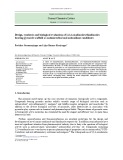
It was previously observed that changes in sterol composi- tion increased plasma membrane H+-ATPase activity and altered the fatty acid profile, but that abnormal sterols per se were probably not directly responsible for the changes observed in H+-ATPase activity. We tested the hypothesis that CLB-fatty acids could be responsible for the activation of the plasma membrane proton pump. The specific activity observed in the different strains, and when different treatments were applied to the wild-type, were plotted vs. the ratio of linoleic acid to oleic acid (18 : 2/18 : 1 ratio) found in their plasma membrane complex lipids (Fig. 1). A close correlation (r (cid:136) 0.98) was observed, and this was independent of the kind of genetic lesion or inhibitor used, suggesting that this activating effect was indeed caused by the fatty acid/lipid environ- ment of the ATPase. It must be noted that triadimenol and fempropimorph have no effect on ATPase activity in these conditions [21]. The 18 : 2/18 : 1 ratio in untreated wild-type was close to unity. When 1-palmytoyl-2-oleyl- phosphatidylcoline was added exogenously to untreated wild-type plasma membranes, a reduction in ATPase activity was found compared to a control to which a 1 : 1 mixture of oleic and linoleic acid-containing phosphat- idylcholine was added (82.9 (cid:139) 8.9% of the control). Conversely, when 1-palmytoyl-2-linoleyl-phosphatidylcho- line was added to these vesicles, an increase in ATPase activity occurred (115.4 (cid:139) 2.4% with respect to the 1 : 1 control). These results proved that the increase in plasma membrane H+-ATPase hydrolytic activity was mediated through changes in the fatty acid unsaturation of complex lipids.
The medium consisted of 100 mM Mes adjusted to pH 6.5 with Tris, 0.0125% (w/v) Triton X-100, 1 mM sodium azide, 0.1 mM sodium molybdate, 50 mM potassium nitrate, 3 mM magnesium sulphate, 3.5 mM ATP (sodium salt) and 2–5 lg of membrane protein in a total volume of 240 lL. Assays were run for 10 min at 37 (cid:176)C. Under these conditions, the concentrations of MgATP and free Mg2+ were 2.5 mM and 0.5 mM, respectively. When varying concentrations of MgATP or changes in pH were required, the appropriate amounts of MgSO4 and Na2ATP were calculated to maintain [Mg2+]free constant at 0.5 mM using the program CHELATOR (available from T. J. M. Shoenmakers, K. U. Nijmegen, the Netherlands). When appropriate, liposomes from exogenous lipids were formed by resuspending dry phospholipids in 100 mM Mes/Tris buffer, pH 6.5, and sonication until clarity was achieved. Phospholipids were added to render 50 lg in 240 lL and tubes were vortexed
1008 A. Herna´ ndez et al. (Eur. J. Biochem. 269)
(cid:211) FEBS 2002
max and V (cid:255) 1
max; m, V (cid:255) 1 max.
Fig. 2. H+-ATPase activation by the 18 : 2/18 : 1 ratio is nonessential. Plots of KmV (cid:255) 1 max vs. the ratio of bound linoleic to oleic acid in the plasma membrane of U. maydis. d, KmÆV (cid:255) 1
curve for a nonessential activation dependent on the linoleic/oleic ratio present in plasma membrane complex lipids (Fig. 2).
Glucose-induced activation of yeast plasma membrane H+-ATPase shows characteristic changes in kinetic param- eters such as pH optimum, Km for MgATP and Ki for vanadate. Although we found no glucose-induced activa- tion of ATPase activity [18] we tested whether the activation observed in these mutants and EBI-treated strains showed any similarities in its changes in kinetic parameters. Optimum pH was determined over a range of 2.5 pH units from 5.5 to 8.0. Maximum activity was found at pH 6.5 for all mutants and treatments (data not shown), thus differing from the glucose-induced activation of yeast ATPase where a shift from pH 5.8–6.5 is found upon addition of glucose to cells [2].
The effect of inhibitors on the H+-ATPase activity was determined for vanadate and erythrosin B. Surprisingly, when data from untreated wild-type membranes were plotted as a Hanes–Wolf representation, vanadate fitted an uncompetitive, instead of a noncompetitive, model. Lineweaver–Burk, Dixon [28] and Cornish–Bowden plots [29] along with nonlinear regression of raw data agreed with an uncompetitive mechanism of inhibition for vanadate (data not shown). Erythrosin B, which is believed to behave as an ATP analogue, showed a mixed-inhibition pattern (Fig. 3). These results were confirmed by nonlinear regres- sion. The same kinetic models were true for EBI-treated sporidia or the mutants (data not shown). Furthermore, the actual values for aKi for vanadate did not change appre- ciably or, in the case of Ki and aKi for erythrosin B, the changes were modest (Table 2).
Fig. 1. Correlation between the ratio of CLB-linoleic to oleic acid and H+-ATPase hydrolytic activity in plasma membrane vesicles of U. maydis. Specific activity in lmol PiÆmin)1Æmg)1 protein. Line gen- erated by linear regression (r (cid:136) 0.980).
D I S C U S S I O N
The use of sterol biosynthesis inhibitors is a usual way of evaluating the physiological effects that lipids, in particular
The affinity of the enzyme for MgATP was then tested. Substrate concentration dependence showed no sigmoidic- ity and was found to fit a Michaelis–Menten model (data not shown). Changes in activity were observed to be the result of an increase in Vmax with little changes in affinity for MgATP. Changes in Vmax correlated with increases in 18 : 2/18 : 1 ratio (r (cid:136) 0.94) (Table 2). Plots of KmV (cid:255) 1 max or V (cid:255) 1 max vs. the 18 : 2/18 : 1 ratio displayed the characteristic
Table 2. Kinetic parameters of Ustilago maydis plasma membrane H+-ATPase. Units: Km (mM); Vmax (lmol PiÆmin)1Æmg)1 protein); Ki and aKi (lM); (cid:139) SE of estimation.
MgATP Vanadate Erythrosin B
Km Vmax aKi Ki aKi 18 : 2/18 : 1 Ratio
Et-C Tri-T Fen-T Wild-type A14 P51 1.3 6.6 3.9 0.8 1.4 2.0 2.66 (cid:139) 0.11 2.00 (cid:139) 0.14 2.01 (cid:139) 0.14 1.68 (cid:139) 0.19 2.53 (cid:139) 0.19 2.31 (cid:139) 0.20 4.55 (cid:139) 0.16 7.92 (cid:139) 0.44 6.57 (cid:139) 0.37 3.60 (cid:139) 0.29 3.02 (cid:139) 0.19 6.43 (cid:139) 0.70 5.58 (cid:139) 0.93 4.74 (cid:139) 1.02 3.94 (cid:139) 0.31 6.28 (cid:139) 2.57 6.50 (cid:139) 0.41 2.89 (cid:139) 0.07 1.74 (cid:139) 030 7.37 (cid:139) 0.19 4.98 (cid:139) 0.65 3.97 (cid:139) 0.48 1.54 (cid:139) 0.07 2.58 (cid:139) 0.13 1.48 (cid:139) 0.39 3.82 (cid:139) 0.26 1.10 (cid:139) 0.21 2.17 (cid:139) 0.25 3.21 (cid:139) 0.50 5.27 (cid:139) 1.75
Stress activation of H+-ATPase by fatty acids (Eur. J. Biochem. 269) 1009
(cid:211) FEBS 2002
s (cid:136) KS
s (cid:136) KA
Fig. 4. General scheme for enzyme activation. E, enzyme; S, substrate; A, activator; P, product.
)1 and Vmax
sterols, have on membrane properties. As in past reports, the joint use of mutants and inhibitors acting on the same biosynthetic points has proved to be a useful way of i.e. sterol distinguishing the influence of direct effects, alteration, and indirect effects of EBI compounds on the plasma membrane H+-ATPase of U. maydis. Furthermore, the utilization of two different targets in the same biosyn- thetic route permitted us not only the confirmation of the direct effects but also, in this particular case, allowed us to identify a novel aspect in the indirect effects of sterol modification, namely, the activation of plasma membrane H+-ATPase through changes in the fatty acid profile.
In our case, the 18 : 2/18 : 1 ratio is an expression of the concentration of CLB-unsaturated fatty acids in contact with the membrane embedded portion of the plasma membrane H+-ATPase. A general kinetic mechanism for enzyme activation is shown in Fig. 4. This mechanism is identical to a general mechanism for inhibition except that, in this case, the enzyme is inhibited by the absence and not by the presence of the activator (A). If KS m and KA m and 0 (cid:136) k < k¢, we have noncompetitive activation, in which the observed Km is not affected but Vmax increases with increasing [A]. On the other hand, nonzero values of k give nonessential activation, in which the enzyme can catalyse the formation of product in the absence of activator. Both mechanisms would render equations which, at a fixed concentration of activator, can be fitted by simple Michaelis–Menten kinetics. In these conditions, to determine whether a noncompetitive activa- )1 can tion is essential or nonessential, KmÆVmax be plotted against [A]. Nonessential activations will give rise to lines that will curve downwards, to reach asymptotically the value of k (Fig. 2), while essential activations would produce straight lines that tend to zero [28]. Therefore, CLB-linoleic acid acts as an activator of U. maydis plasma membrane H+-ATPase which causes a noncompetitive, nonessential activation of its ATP hydrolytic activity (Table 2, Fig. 2). It could be argued that this activation may be due to other causes such as increased polypeptide amounts in membrane or changes in fluidity. We have shown previously that these two factors remain largely unchanged in the same conditions used in this study [18,21]. This regulation by CLB-unsaturated fatty acids in U. maydis differs from others described. For example, glucose-induced activation of plasma membrane H+- ATPase in yeast is one of the best characterized modifica- tions in the activity of these enzymes. Typically, on glucose addition, the pH optimum of Pma1p increases from 5.8 to 6.5, the affinity for substrate decreases from 2.1 mM to 0.8 mM and the inhibitory effect of vanadate is augmented by up to fivefold; similar changes are observed for Pma2p [34]. On the other hand, salt stress in Z. rouxii also produces activation of plasma membrane H+-ATPase activity, but in this case, it is correlated with a greater amount of enzyme in the plasma membranes [15]. In our case, present activation of H+-ATPase activity did not involve chan- ges in affinity for substrate, pH optimum or sensitivity to vanadate, but exhibits changes in the Vmax of the protein, thus differing from glucose-induced activation. As stated before, we showed that polypeptide amounts of
There have been numerous reports on the influence of lipids on membrane bound enzyme activity. However, it seems clear that each particular enzyme, and sometimes part of the function of it, responds to a different set of changes in the lipid environment (e.g [30–32]). The in vivo effect of CLB-fatty acids on H+-ATPase activity, particularly under stress, had been suggested previously [17], but lack of an appropriate experimental system probably prevented its demonstration. In our system, the modification of the fatty acid moieties was provoked by the presence of abnormal in the case of EBI-treated sporidia, other sterols plus, collateral effects of these compounds [33]. In U. maydis, changes in the plasma membrane 18 : 2/18 : 1 ratio fol- lowed the inhibition of growth rate, which was influenced not only by the biosynthetic point affected but also by the method used [21]. These changes correlated directly with increases in the H+-ATPase specific activity which could, in its turn, be mimicked by altering the 18 : 2/18 : 1 ratio of isolated plasma membrane vesicles in vitro. A14 mutant seems to depart somehow from this correlation. Both P51 and A14 mutants were obtained by UV irradiation and A14 was isolated as a partial revertant of a previous mutant [22,23]. Therefore, secondary mutations may be present, in paticular in the latter mutant, that could explain this departure from full correlation.
Fig. 3. Effects of vanadate and erythrosin B on the substrate dependence of H+-ATPase hydrolytic activity from U. maydis plasma membrane vesicles. Models of inhibition. Hanes Plot of data obtained from wild- type samples. Concentration of MgATP in mM; ATPase hydrolytic activity in lmol Pi min)1Æmg)1Æprotein. j, No additions; d, + 50 lM vanadate; m, + 30 lM erythrosin B.
1010 A. Herna´ ndez et al. (Eur. J. Biochem. 269)
(cid:211) FEBS 2002
H+-ATPase showed no changes upon EBI fungicide treatment or in the mutants [18]. On the other hand, this activation showed particular characteristics, namely, it is nonessential, and involves changes in the Vmax of the protein but other factors remain mostly unchanged.
In yeast,
4. Benito, B., Portillo, F. & Lagunas, R. (1992) In vivo activation of the yeast plasma membrane ATPase during nitrogen starvation. Identification of the regulatory domain that controls activation. FEBS Lett. 300, 271–274.
5. Amigo, L., Moreno, E. & Lagunas, R. (1993) In vivo inactivation of the yeast plasma membrane ATPase in the absence of exo- genous catabolism. Biochim. Biophys. Acta. 1151, 83–88.
6. Rosa, M. & Sa´ -Correia, I. (1991) In vivo activation by ethanol of plasma membrane ATPase of Saccharomyces cerevisiae. App. Env. Microbiol. 57, 830–835.
7. Fernandes, A. & Sa´ -Correia, I. (2001) The activity of plasma membrane H+-ATPase is strongly stimulated during Saccharo- myces cerevisiae adaptation to growth under high copper stress, accompanying intracellular acidification. Yeast 18, 511–521. 8. Serrano, R. (1989) Structure and function of plasma membrane ATPase. Ann. Rev. Plant Physiol. Mol. Biol. 40, 61–94.
between S. cerevisiae H+-ATPase
limiting free magnesium concentrations (below 0.1 mM) were reported to change the type of inhibition for vanadate from noncompetitive to mixed uncompetitive/noncompetitive [35]. In our experimental conditions (0.5 mM free Mg2+) it was surprising to find that vanadate fitted a purely uncompetitive model. The reason for this discrepancy is unknown but maybe due to species variation. It is noteworthy that U. maydis H+-ATPase is a slightly larger enzyme than that of Saccaromyces cerevisiae [18]. In our hands, erythrosin B fitted a mixed mechanism of it could have been inhibition. From previous works, expected to follow a competitive mechanism of inhibition, if this compound behaves purely as an analogue of ATP. This has been the case for the yeast H+-ATPase [36]. Again, and differences U. maydis H+-ATPase may explain this situation.
9. Reinhold, L., Seiden, A. & Volokita, M. (1984) Is modulation of the rate of proton pumping a key event in osmoregulation? Plant Physiol. 75, 846–849.
10. Martı´ nez-Cortina, C., Ros, R., Cooke, D.T., James, C.S. & Sanz, A. (1992) The lipid composition, fluidity, and Mg2+- ATPase activity of rice (Oriza sativa L. cv. Bahia) shoot plasma membranes: effects of ABA and GA3. J. Plant Growth Regul. 11, 195–201.
11. White, F.J., Cooke, D.T., Earnshaw, M.J., Clarkson, D.T. & Burden, R.S. (1990) Does plant growth temperature modulate the membrane composition and ATPase activities of tonoplast and plasma membrane fractions from rye roots? Phytochemistry 29, 3385–3393.
12. Ros, R., Cooke, D.T., Burden, R.S. & James, C.S. (1990) Effects of the herbicide MCPA, and the heavy metals, cadmium and nickel on the lipid composition, Mg2+-ATPase activity and flu- idity of plasma membranes from rice, Oryza sativa (cv. Bahı´ a) shoots. J. Exp. Bot. 41, 457–462.
13. Portillo, F., Ferna´ ndez de Larrinoa, I. & Serrano, R. (1989) Deletion analysis of yeast plasma membrane H+-ATPase and identification of a regulatory domain at the carboxyl-terminus. FEBS Lett. 247, 381–385.
14. Goosens, A., de la Fuente, N., Forment, J., Serrano, R. & Portillo, F. (2000) Regulation of yeast H+-ATPase by protein kinases belonging to a family dedicated to activation of plasma membrane transporters. Mol. Cell. Biol. 20, 7654–7661.
The effects of stress on plasma membrane H+-ATPase activity have been described for S. cerevisiae, grown in the presence of octanoic acid [37]. In this case, only Vmax was affected showing an increase that was accompanied by an increase in the presence of oleic acid in its plasma membrane lipids. It must be noted that oleic acid (and to a minor extent palmitoleic acid) is the only unsaturated fatty acid present in yeast. Similar results have been found for copper stress, where its main mode of action is believed to be lipid modification, but no relationship to a particular change in lipids was drawn [7,38]. Secale cereale also showed a greater H+-ATPase activity in plasma membrane vesicles upon acclimation to cold temperatures [11]. In this case too, changes in the H+-ATPase activity followed increases in the unsaturation of plasma membrane fatty acids, in particular, linolenic acid. Furthermore, revisiting these data, we find a direct correlation between the linolenic to linoleic acid ratio and H+-ATPase activity (r (cid:136) 0.98) similar to that found in this report. All these data suggest that, although the particular fatty acid may be different for each species, the plasma membrane H+-ATPase by activation of increases in the unsaturation of the CLB-fatty acids is a physiological and relevant effect in stress adaptation in plants and fungi.
15. Watanabe, Y., Sanemitsu, Y. & Tamai, Y. (1993) Expression of plasma membrane proton-ATPase gene in salt-tolerant yeast Zygosaccharomyces rouxii is induced by sodium chloride. FEMS Microbiol. Lett. 114, 105–108. 16. Serrano, R. (1993) Structure, function and regulation of plasma membrane H+-ATPase. FEBS Lett. 325, 108–111.
A C K N O W L E D G E M E N T S
17. Alexandre, H., Mathieu, B. & Charpentier, C. (1996) Alteration in membrane fluidity and lipid composition, and modulation of H+- ATPase activity in Saccharomyces cerevisiae caused by decanoic acid. Microbiology 142, 469–475.
We wish to thank M. Lewis for the preliminary lipid analysis. A.H. was the beneficiary of a grant (cid:212)Formacio´ n de Investigadores(cid:213) from Gobierno Vasco, Spain. 18. Herna´ ndez, A., Cooke, D.T. & Clarkson, D.T. (1998) Effects of abnormal-sterol accumulation on Ustilago maydis plasma membrane H+-ATPase stoichiometry and polypeptide pattern. J. Bacteriol. 180, 412–415.
R E F E R E N C E S
19. Van den Bossche, H., Willemsens, G., Cools, W., Marichal, P. & Lauwers, W. (1983) Hypothesis on the molecular basis of the antifungal activity of N-substituted imidazoles and triazoles. Biochem. Soc. Trans. 11, 665–667.
1. Nishi, T. & Yagi, T. (1992) A transient and rapid activation of plasma-membrane ATPase during the initial stages of osmoregu- lation in the salt-tolerant yeast Zygosaccharomyces rouxii. FEMS Microbiol. Lett. 99, 95–100. 2. Serrano, R. (1983) In vivo glucose activation of the yeast plasma 20. Burden, R.S., Cooke, D.T. & Hargreaves, J.A. (1990) Mech- anisms of action of herbicidal and fungicidal compounds on cell membranes. Pesticide Sci. 30, 125–140. membrane ATPase. FEBS Lett. 156, 11–14.
3. Eraso, P. & Gancedo, C. (1987) Activation of yeast plasma membrane ATPase by acid pH during growth. FEBS Lett. 224, 187–192. 21. Herna´ ndez, A., Cooke, D.T., Lewis, M. & Clarkson, D.T. (1997) Fungicides and sterol-deficient mutans of Ustilago maydis: plasma membrane physico-chemical properties do not explain growth inhibition. Microbiology 143, 3165–3174.
Stress activation of H+-ATPase by fatty acids (Eur. J. Biochem. 269) 1011
(cid:211) FEBS 2002
31. Mouritsen, O.G. & Bloom, M. (1993) Models of lipid–protein interactions in membranes. Annu. Rev. Biophys. Biomol. Struct. 22, 145–171. 22. Keon, J. & Hargreaves, J.A. (1996) An Ustilago maydis mutant partially blocked in P450 (14DM) activity is hypersensitive to azole fungicides. Exp. Mycol. 20, 84–88.
23. James, C.S., Burden, R.S., Loe(cid:130)er, T. & Hargreaves, J.A. (1992) Isolation and characterization of polyene-resistant mutants from the maize smut pathogen, Ustilago maydis, defective in ergosterol biosynthesis. J. Gen. Microbiol. 138, 1437–1443. 32. East, J.M., Jones, O.T., Simmonds, A.C. & Lee, A.G. (1984) Membrane fluidity is not an important physiological regulator of the (Ca2+-Mg2+)-dependent ATPase of sarcoplasmic reticulum. J. Biol. Chem. 259, 8070–8071.
24. Herna´ ndez, A., Cooke, D.T. & Clarkson, D.T. (1994) Lipid composition and proton transport in Penicillium cyclopium and Ustilago maydis plasma membrane vesicles isolated by two-phase partitioning. Biochim. Biophys. Acta. 1195, 103–109. 33. Weete, J.D., Sancholle, M., Patterson, K.A., Miller, K.S., Huang, M.Q., Campbell, F. & Van den Reek, M. (1991) Fatty acid metabolism in Taphrina deformans treated with sterol biosynthesis inhibitors. Lipids 26, 669–674.
25. Herna´ ndez, A. & Ruiz, M.T. (1997) An Excel template for calculation of enzyme kinetic parameters by non-linear regression. Bioinformatics 14, 227–228. 34. Supply, P., Wach, A. & Goffeau, A. (1993) Enzymatic properties of the PMA2 plasma membrane-bound H+-ATPase of Saccharo- myces cerevisiae. J. Biol. Chem. 268, 19753–19759.
35. Borst-Pauwels, G.W.F.H. & Peters, P.H.J. (1981) Factors affect- ing the inhibition of yeast plasma membrane ATPase by vanadate. Biochim. Biophys. Acta. 642, 173–181. 26. Onishi, T., Gall, R.S. & Mayer, M.L. (1975) An improved assay of inorganic phosphate in the presence of extra-labile phosphate compounds: Application to the ATPase in the presence of phos- phocreatin. Anal Biochem. 69, 261–267.
36. Wach, A. & Graber, P. (1991) The plasma membrane H+-ATPase from yeast. Effects of pH, vanadate and erythrosine B on ATP hydrolysis and ATP binding. Eur. J. Biochem. 201, 91–97. 27. Bradford, M.M. (1976) A rapid and sensitive method for the quantitation of microgram quantities of protein utilizing the principle of protein-dye binding. Anal. Biochem. 72, 248–254. 28. Dixon, M. & Webb, E.C. (1979). Enzymes. Longman Group Ltd, London.
29. Cornish-Bowden, A. (1974) A simple graphical method for determining the inhibition constants of mixed, uncompetitive and non-competitive inhibitors. Biochem. J. 137, 143–144. 37. Viegas, C., Almeida, P.F., Cavaco, M. & Sa´ -Correia, I. (1998) The H+-ATPase in the plasma membrane of Saccharomyces cerevisiae is activated during growth latency in octanoic acid-supplemented medium accompanying the decrease in intracellular pH and cell viability. Appl. Environ. Microbiol. 64, 779–783.
30. Grandmougin-Ferjani, A., Schuler-Muller, I. & Hartmann, M. (1997) Sterol modulation of the plasma membrane H+-ATPase activity from corn roots reconstituted into soybean lipids. Plant Physiol. 113, 163–174. 38. Fernandes, A., Prieto, M. & Sa´ -Correia, I. (2000) Modification of plasma membrane lipid order and H+-ATPase activity as part of the response of Saccharomyces cerevisiae to cultivation under mild and high copper stress. Arch. Microbiol. 173, 262–268.