Eur. J. Biochem. 269, 317–325 (2002) (cid:211) FEBS 2002
Induction of chicken ovalbumin upstream promoter-transcription factor I (COUP-TFI) gene expression is mediated by ETS factor binding sites
Ramiro Salas*, Fabrice G. Petit, Carlos Pipaon(cid:160), Ming-Jer Tsai and Sophia Y. Tsai
Department of Molecular and Cellular Biology, Baylor College of Medicine, Houston, TX, USA
to these response elements and that other ETS factors also transactivate the COUP-TFI promoter. In addition, COUP- TFI is coexpressed with some ETS factors in the mouse embryo. These results indicate that members of the ETS family can activate COUP-TFI gene expression.
Keywords: COUP-TFI; ETS; gene expression; transcription; orphan receptors.
Chicken ovalbumin upstream promoter-transcription factor I (COUP-TFI, or NR2F1) is an orphan nuclear receptor that plays a major role in the development of the nervous system. We show here that three ETS response elements in the COUP-TFI promoter mediate its transcription. A reporter gene containing these ETS binding sites is activated by Ets-1, while the same reporter with point mutations on all three ETS response elements is not. We also show that Ets-1 binds
bases, 5¢ and 3¢, of this core are important for high affinity and specific DNA binding [9]. Although ETS proteins contain activation domains [11], Ets-1, Ets-2 and other ETS proteins need to interact with other transcription factors to transactivate their target genes [12]. These factors include Fos [13], SRF [14], NF-EM5 [15], AP1 [12] and NFAT [16]. The necessity for the accessory factors is likely due to poor DNA-binding affinity of full-length Ets-1 rather than to poor potential of its activation domain. This is supported by the existence (at least in Ets-1 and Ets-2) of an auto- inhibitory domain which, in the absence of accessory factors, prevents Ets-1 binding to DNA [10,17]. When this domain is removed, Ets-1 binds DNA with higher affinity even in the absence of accessory factors. The auto-inhibitory domain is located within exon VII and an alternatively spliced form of Ets-1 that lacks this exon is constitutively active [18].
Chicken ovalbumin upstream promoter-transcription factors (COUP-TFs) are among the best characterized orphan members of the nuclear receptor superfamily [1]. COUP-TFs have been shown to be negative regulators of the transcription of many genes [1,2], but can also act as activators of gene transcription [1,3]. In mammals, two COUP-TF genes have been identified, COUP-TFI and COUP-TFII. Although they have different physiological functions [4,5], sequence analysis and molecular studies indicate that they share similar properties. The expression patterns of COUP-TFI and COUP-TFII have been exten- sively studied in a number of species [6]. In the mouse, COUP-TFI is first detected at the embryonic day 7.5 (e7.5), its expression reaches a peak at e12.5, declines before birth [6] and remains low during adulthood. COUP-TFI null mice die perinatally and exhibit neuronal defects in axonal guidance and arborization [4] and thalamocortical connections [7].
Little is known about the upstream signals that regulate COUP-TFI gene expression. Here we show that Ets-1 and other ETS factors are able to transactivate COUP-TFI expression through a cluster of ETS response elements in the promoter. In addition, several ETS factors colocalize with COUP-TFI in different tissues of the developing mouse embryo.
The ETS family of transcription factors is composed of a large number of proteins that share a similar DNA-binding domain (DBD), called the ETS domain [8]. These proteins bind as monomers to a core sequence GGAA/T and activate transcription of promoters having this ETS response element [9,10]. Besides the GGAA/T core sequence, at least three
M A T E R I A L S A N D M E T H O D S
Genomic screening
To isolate the mCOUP-TFI promoter, a genomic library (129SVJ Mouse Genomic Library in the Lambda FIX II vector, Stratagene) was screened using part of the 5¢ UTR of the mCOUP-TFI gene. The fragment was labeled with 32P-dCTP by random priming (Prime-a-gene kit, Promega). The genomic library was used to infect XL-1 blue bacteria and standard protocols were used to perform the screening [19]. After tertiary screening, the phage DNA was isolated using the k Wizard kit (Promega), cut with NotI and subcloned into the pBluescript KSII vector (Stratagene). The Genbank accession number for this sequence is AY055471.
Correspondence to S. Y. Tsai, Department of Molecular and Cellular Biology, Baylor College of Medicine, One Baylor Plaza, Houston, Texas 77030, USA. Fax: + 713 798 8227, Tel.: + 713 798 6251, E-mail: stsai@bcm.tmc.edu Abbreviations: COUP-TFI, chicken ovalbumin upstream promoter- transcription factor I; DBD, DNA-binding domain; EMSA, electro- mobility shift assay; DMEM, Dulbecco’s modified Eagle’s medium. *Present address: Division of Neuroscience, Baylor College of Medi- cine, One Baylor Plaza, Houston, Texas 77030, USA. (cid:160)Present address: Departamento de Biologia Molecular, Facultad de Medicina, Universidad de Cantabria, c/Cardenal Herrera Oria s/n 39011 Santander, Spain. (Received 28 June 2001, revised 21 September 2001, accepted 5 November 2001)
318 R. Salas et al. (Eur. J. Biochem. 269)
(cid:211) FEBS 2002
Plasmids
COUP-TFI sequences used as probes for screening were obtained previously in our lab [20]. The sequences of the COUP-TFI promoter originally cloned into pBluescript, were subcloned into pGL3-basic (Promega) or a modified version of pGL3-basic that contains a consensus TATA box (pGL3-TATA). Vectors containing the coding sequences for ETV1, ERM and PEA3 were a generous gift from Y. de Launoit and L. Coutte (Institut Curie, Paris, France). Spi-1 was a gift from F. Moreau-Gachelin (Institut de Biologie, Lille, France). Ets-1 and Ets-2 were cloned by RT-PCR from rUGM cells.
Cell culture and transfection
methods using Taq polymerase (Promega). Deletions were carried out with mixed strategies using both PCR fragments with artificial restriction sites at the ends and DNA fragments obtained by restriction digestion. The oligos used for point mutations in the three ETS response elements are listed below (the ETS core binding sequences are in bold). A forward: CGGGTACCCTCCGTTTCCCACTTCTCG; A for Mut: CGGGTACCCTCCGTTTCTCACTTCTCG; B forward: CGGGGTACCTCCCTCTTCCCCGTCTTCT CGTTCGTTCG; B for Mut CGGGGTACCTCCCTCT TCTCCGTCTTCTCGTTCGTTCG; B reverse GAA GATCTCGAACGAACGAGAAGACGGGGAAGAG GGA; B rev Mut GAAGATCTCGAACGAACGAG AAGACGGAGAAGAGGGA; C rev GAAGATCTC AAGTCAGTCACAGGAAAAGAGC; C rev Mut GAAGATCTCAAGTCAGTCACAAGAAAAGAGC.
Insituhybridization
HeLa cells were grown in 10% fetal bovine serum/ Dulbecco’s modified Eagle’s medium (DMEM) (Gibco). The day before transfection, cells were passed onto six-well plates at approximately 5 · 105 cells per well. For transfec- tion, both lipofectin (Gibco) and Fugene 6 (Boehringer) used at a 3 : 1 ratio to DNA gave similar results and both methods were used according to the manufacturer’s instructions. Cells were collected 48 h later. Luciferase activity was measured in a luminometer (Monolight 2010, Analytical Luminescence Laboratory) according to the manufacturer’s instructions. Protein content was measured using the Bradford reagent (Bio-Rad).
The (cid:212)B(cid:213) domains of the ets-1 and ets-2 genes were used to prepare probes as described previously [21]. The N-terminus of ERM, ETV1 and PEA3 genes were used to prepare probes as described previously [22]. The full length cDNA of the mCOUP-TFI gene was used to prepare the COUP-TFI probe. The templates for probes were subcloned into pBluescript and RNA probes were prepared from linearized plasmid using T3 RNA polymerase (Promega) and 100 lCi [a-35S]UTP (1000 CiÆmmol)1, ICN). In situ hybridization was performed on 14.5-day-old mouse embryos as described previously [6,23].
Electromobility shift assay
R E S U L T S
Ets-1 activates the mCOUP-TFIpromoter through a cluster of ETS response elements
constructs
containing different
For EMSA (electromobility shift assay) studies, proteins were prepared (with or without [35S]Met) from different DNA constructs using the TNT Coupled reticulocyte lysate system (Promega), according to the manufacturer’s instruc- tions. Probes for EMSA were end-labeled using a32P-dCTP (ICN) and Sequenase enzyme (Amersham USB). EMSAs were carried out as follows: 2 lL of reticulocyte lysate were incubated for 10 min at room temperature with 1 lL of labeled probe (2–3 · 104 c.p.m.) and 10 pmol of dIdC in buffer H (20 mM Hepes, 1 mM MgCl2, 100 mM KCl, 0.1 mM EDTA, 0.1% NP40, 1 mM spermidine, 5% glycer- ol). For competition experiments, 10- or 100-fold molar excess of unlabelled oligonucleotide were added to the incubation reaction. For supershift experiments, 125 ng of polyclonal anti-(Ets-1) Ig (Transduction Laboratories, San Diego, CA, USA) were added after the 10-min incubation and all tubes were then incubated on ice for an additional 15 min. Samples were subsequently loaded on a 5% acrylamide native gel and run at 30–50 mA. The gel was dried and exposed to Biomax film (Kodak).
The probe used (GTACCTCGAGCAGGAAGTTC GA) contained an Ets-1 consensus binding site (underlined).
Site-directed mutagenesis
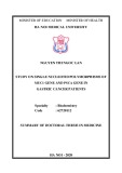
To identify possible signals that activate the COUP-TFI gene transcription, we isolated approximately 6 kb of 5¢ flanking sequences of the COUP-TFI gene (Fig. 1A). lengths of Reporter 5¢ flanking sequences linked to a Luciferase gene were used on transient transfection studies. HeLa cells were chosen for these experiments because they express high levels of COUP-TFI [24]. The activity of the promoter was the same whether constructs containing 6, 4 or 0.73 kb of 5¢ flanking sequences were used in the study. However, deletion of sequences between )734 and )387 resulted in a small but reproducible loss of promoter strength. Further deletion from )398 to )96, did not alter the activity. When the empty vector was studied, it had no significant activity (Fig. 1B). These results suggest that these two regions ()734 to )387 and )96 to +446 which includes promoter and 5¢ UTR) are important for the COUP-TFI promoter activity (Fig. 1B). Within the distal region, a putative Ets-1 response element (site C, Fig. 2A) was identified by computer search for transcription factor binding sites [25]. Surrounding this element, we identified additional sequences that resembled ETS binding sites (Fig. 2). To assess whether Ets-1 regulates COUP-TFI promoter activity on this region, we cotransfected a reporter contain- ing the )490/)259 fragment linked to a TATA-Luc reporter expression vector, in the presence or absence of an Ets-1 expression vector. Ets-1 was able to significantly
To mutate specific base pairs within the COUP-TFI promoter, primers were designed within the sequence to be mutated. Each of these primers contained a restriction site for convenient subcloning. As three Ets binding sites (site A, at )490, site B, at )460 and site C at )420) were important, the GGAA sequence of each one was mutated to AGAA. PCR reactions were performed by standard
(cid:211) FEBS 2002
ETS factors and COUP-TFI promoter (Eur. J. Biochem. 269) 319
A
-734 GTACGCGGGACCGTCCTCCTGCCTACCCCTCCTTTTGCGACCAATCACCTTCGGGAATGGGGTCTCAGTCACACACACC
CCAACACACACACACACACACACACACACACACACACACCACCACCACCACCACCACCACCACCACCACCACCACCAC
CACCACCACCACCACCACACAGCGAGTGAGAGACTCAGTCTCTTCCTCCTCCTCCTCCTCCTCCTCCTCTCCCCCTCCCC
CTCCCCTCCGTTTCCCACTTCTCGTCCCCTCCCCTCCTCCCCTCTCCCTCTTCCCCGTCTTCTCGTTCGTTCGTTTGCTCTT
ETS TTCCTGTGACTGACTTGTCCGCACTAACAGCCGCCCCACAACAATATGAGGAGTTACAAATGCTTTATTAATAATCATT
Nkx2.
Nxk2.
GAAGCATTGTTTGGAGTTTGAGCATCCTGGGAATAAAAATGATGAAAAAGGAAAAAGAGGATTGATTGGAAAGTTTAT
TTTAAGATCATCTTTGGGATGAATAGGAATCATCGATTCGGATCGAATTTGTGGCAGTAGCTGCAGTTTCATGTGTGTG
C/EBP
C/EBP
CTTTGTCGTAATTACGCCTCCGAAACTATGATATACTTCAGATTTTTAAATGAGGAGGCTTTTCATAATTATATAAAATGA
GCGGGATACAGACTAAGATTATATTGTATGAGAACTAAGATTCTAAACCAAGTAGAAAAAACAAATCATTAAAATGAT
GGAGTTTTTTTCCTGCATTAATTT
+1
B
+1
RLU 1 0
2 0
0
+ 4 4 6
ETS
-6 Kb
-4 Kb
-734
-387
-197
-96
Empty vector
activate the reporter gene containing this COUP-TFI promoter fragment. Then we subdivided that segment into two subfragments ()397/)259 and )490/)408), each carrying three putative binding sites. The subfragment )397/)259 is not responsive to Ets-1, while the )490/)408 subfragment is stimulated by Ets-1 to a similar extent as the )490/)259 fragment (Fig. 2A) in a dose-dependent manner (Fig. 2B). Interestingly, the best putative Ets-1 binding site sequence is located within this fragment.
The three putative Ets-1 response elements within )490 and )408 (ETS-RE) were then named A, B and C, in the order of 5¢ to 3¢. Point mutations (TTCC to TTCT) were introduced on each or combinations of the three putative sites. Mutations in one or two sites diminished Ets-1- dependent reporter activity (Fig. 2C), while mutations in all three sites abolished the response. These results indicate that these three Ets-1 response elements work in concert to achieve maximum Ets-1-dependent activation.
Ets-1 binds the ETS response elements in the mCOUP-TFIpromoter
elements. On electromobility
shift
We examined whether Ets-1 was able to directly bind the response assays (EMSA) Ets-1 is able to bind to its DNA response
element only if the auto-inhibitory domain is deleted [10]. Therefore, two truncations of the Ets-1 protein that lack part (Ets-1DCE, truncated from amino acids 280–331) or most (Ets-1DAE, truncated from amino acids 244–331) of the auto-inhibitory domain were made (Fig. 3A). These truncated proteins were shown to readily bind to ETS response elements [10]. To verify that our truncated proteins were active, we transfected HeLa cells with the )490/)408 luciferase reporter with increasing concentra- tions of Ets-1, Ets-1DCE or Ets-1DAE. These truncated the COUP-TFI promoter forms of Ets-1 activate (Fig. 3A). We next used in vitro translated proteins for DNA binding assays. The proteins were transcribed/ translated using [35S]Met and separated by PAGE. Figure 3B shows that Ets-1, Ets-1DCE and Ets-1DAE are all expressed to a similar level. On an EMSA using a consensus Ets-1 response element as a probe (Fig. 3C), no Ets-1 specific binding was observed when reticulocyte lysates were prepared with empty vector (lane 1) or with wild-type Ets-1 (lane 2). However, after addition of specific anti-(Ets-1) Ig to the wild-type Ets-1 lysate, a supershifted band was observed (lane 3). This was probably due to a stabilizing effect of the antibody on the Ets-1/DNA complex formation or to the possibility that the antibody may elicit a conformational change that
Fig. 1. General organization of the mCOUP- TFI promoter. (A) Sequence of the mCOUP- TFI promoter from )734 to +5. A putative Ets-1 binding site is underlined. Putative Nkx2. and c/EBP binding sites are shown underlined. The transcription initiation site is marked by an arrow. (B) Luciferase activity of 5¢ deletions of the mCOUP-TFI promoter in transfected HeLa cells (0.25 lg DNA per well). A representative experiment performed in triplicate is shown. The putative Ets-1 binding site is marked as a lane.
320 R. Salas et al. (Eur. J. Biochem. 269)
(cid:211) FEBS 2002
Ets-1 is able to bind the COUP-TFI promoter preferentially at site C. This is not surprising because site C is the most closely related to the consensus Ets-1 binding site.
ETS factors colocalize with COUP-TFI on the developing mouse embryo
at least partially relieves auto-inhibition. When lysate containing Ets-1DCE was added, a faint band corre- sponding to Ets-1DCE was detected (lane 4), and was completely supershifted by anti-(Ets-1) Ig (lane 5). When most of the auto-inhibitory domain was deleted (Ets- 1DAE), a stronger band corresponding to Ets-1DAE was detected (lane 6), and it was completely supershifted by the antibodies (lane 7).
the mesenchyme
(Fig. 4I–L),
of
We performed in situ hybridization studies on mouse embryos with COUP-TFI and different ETS factors. Mouse embryos 14.5-days-old-were chosen for these experiments because the expression levels of COUP-TFI are high at this stage of development [1]. The expression patterns of COUP- TFI, Ets-1, Ets-2, ETV1 and PEA3 were studied (Table 1). There were several areas of coexpression of COUP-TFI and Ets-1: the mesenchyme of the bladder (Fig. 4A–D), the mesenchyme of the nasal septum (Fig. 4E–H), the cerebral cortex vibrissae (Fig. 4M–P), spleen, and submandibular glands (Table 1). Ets-2 was found to colocalize with COUP-TFI on the mesenchyme of vibrissae (Figs 4M–N,Q–R) and subman- dibular glands (Table 1). PEA3 was found to colocalize with COUP-TFI in the cochlea, cerebral cortex and trigeminal ganglion (Table 1). ETV1 was found coexpressed with COUP-TFI on cells of the dorsal root ganglia and some
We next examined whether the ETS response elements in the COUP-TFI promoter were able to bind Ets-1 protein in a band-shift competition assay. The Ets-1 consensus binding site was used as a probe. Ets-1DAE was able to bind specifically (Fig. 3D, lane 3) and could be supershifted by an Ets-1 specific antibody (lane 2), but not by an unrelated antibody (lane 13). Increasing amounts (10 and 100-fold molar excess) of unlabeled site C oligos were able to compete for the binding of Ets-1DAE to the Ets-1 consensus binding site (lanes 4 and 5). In contrast, a mutation (TTCC to TACT) of site C was unable to do so (lane 6). Similar competition experiments were carried out with sites B (lanes 7, 8 and 9) and A (lanes 10, 11 and 12). Very weak competition could be detected with site A and B oligos, while oligos containing mutations of these sites did not compete at all (Fig. 3D). Taken together, these experiments suggest that
Fig. 2. Ets-1 induced the activity of the COUP- TFI promoter in HeLa cells. (A) Cells trans- fected with di(cid:128)erent portions of the promoter linked to a TATA box and a luciferase reporter gene (0.2 lg of DNA) with or with- out cotransfected Ets-1 expression vector (0.5 lg of DNA). (B) Dose–response of the Ets-1-dependent COUP-TFI promoter trans- activation. The )490 to )408 reporter con- struct was cotransfected with increasing amounts of Ets-1 expression vector (0, 0.01, 0.04, 0.06, 0.1 and 0.2 lg). (C) E(cid:128)ect of single base pair mutations on the Ets-1 response of the )490/)408 fragment of the COUP-TFI promoter. The TTCC sequence was mutated to TTCT and the activation of these mutations was assessed by cotransfection with Ets-1 expression vector (0.2 lg of reporter, 0.5 lg of expression vector). Representative experi- ments performed in triplicate are shown.
(cid:211) FEBS 2002
ETS factors and COUP-TFI promoter (Eur. J. Biochem. 269) 321
A
20
C 2 8 0
E 3 3 1
1
A 2 4 4
4 4 1
DBD
Ets-1
DBD
Ets-1D CE
10
DBD
Ets-1D AE
0
0
Ets-1 Ets-1D CE Ets-1D AE
C
B
Ets-1D C E
Ets-1
Ets-1D A E
+
+
+
V -
-
-
-
Ets-1 Ets1D CE Ets-1D AE
Antibody
kDa
Supershift
126 87
NS
64
52
Ets-1 binding
39
Ets-1D A-E
V
D
m ut
m ut
m ut
-
A A
B B
C
C
1
5
2 3
4
6 7
Competitor
10
1 0 0
1 0 0
10
Antibody
-
- -
1 0 0 1 0 0 - -
-
-
10 1 0 0 - -
1 0 0 -
- - +-
-
- U
Supershift
NS
Ets-1 binding
Fig. 3. Auto-inhibitory domain deleted Ets-1 is able to bind the COUP-TFI promoter. (A) Left panel, schematic view of the Ets-1 protein and the two deletions used in transfection and electromobility shift assay (EMSA) experi- ments. Right panel, HeLa cells were trans- fected with)490/)408 TATA reporter gene (0.2 lg) and increasing amounts of Ets-1, Ets- 1DCE and Ets-1DAE (0.1, 0.2 and 0.3 lg). (B) Analysis of in vitro translated Ets-1, Ets-1DCE and Ets-1DAE constructs. In vitro translation was performed in the presence of 35S-labeled methionine using a reticulocyte lysate system. The translated products were separated on a 10% SDS/PAGE and autoradiographed. For the deleted Ets-1, two vectors were used. (C) Electromobility shift assay (EMSA) of Ets-1, Ets-1DCE and Ets-1DAE on an Ets-1 con- sensus binding site. Lane 1 (V), vector control. Lanes 2 and 3, wild-type Ets-1 without or with anti-(Ets-1) Ig, respectively. Lanes 4 and 5, Ets-1DCE without and with antibodies, respectively. Lanes 6 and 7, Ets-1DA-E with- out and with antibodies. NS, nonspecific binding. (D) EMSA of Ets-1DAE on a con- sensus Ets-1 binding site and competition experiments (lane 1, vector control, lanes 2–13 contain Ets-1DAE). Lane 2, e(cid:128)ect of anti- (Ets-1) Ig. For competition experiments, 10x and 100x molar excess (site C, lanes 4 and 5; site B, lanes 7 and 8; site A, lanes 10 and 11) or 100x molar excess (mutated site C, lane 6; mutated site B, lane 9; mutated site A, lane 12) of unlabeled oligonucleotide were used. Lane 13, addition of unrelated antibodies have no e(cid:128)ect on Ets-1DAE binding.
2 3 4
6 7 8 9 1 0 1 1 1 2 1 3
1
5
Table 1. Expression of COUP-TFI, Ets-1, Ets-2, ETV1 and PEA3 mRNA in 14.5 day old mouse embryos.
Tissue COUP-TFI Ets-1 Ets-2 PEA3 ETV1
regions of the cerebral cortex (Table 1). In these tissues, ETS factors and COUP-TFI seemed to be localized in the same cell types. Figure 5 shows high magnification pictures of the signal for COUP-TFI and Ets-1 mRNAs in the nasal
epithelium and COUP-TFI, Ets-1, and Ets-2 in the mesenchimal cells surrounding the vibrissae. These results indicate that COUP-TFI and ETS factors are colocalized in many regions of the developing mouse embryo.
Cortex Cochlea Trigeminal ganglion Dorsal root ganglia Mesenchima of mesonephric duct Kidney Stomach muscle Bladder Spleen Mesenchima surrounding genital tubercle Mesenchima of the trachea Spinal chord, mantle layer Spinal chord, marginal layer Submandibular gland Mesenchima surrounding vibrissae +++ +++ + ++ + ++ +++ ++ ++ ++ + ++ + ++ ++ + + – +/– + + + + ++ – + – – + + +/– + – – – – +/– – +/– + +/– – + + + + +/– – +++ – – + + – + +/– – – – – + + + – – + – – – + – +/– +/– + –
322 R. Salas et al. (Eur. J. Biochem. 269)
(cid:211) FEBS 2002
DARK FIELD
BRIGHT FIELD
DARK FIELD
BRIGHT FIELD
B
A
I
J
COUP-TFI
L
C
D
K
Ets-1
E
M
F
N
COUP-TFI
G
H
O
P
Ets-1
Q
R
Ets-2
Other ETS factors are able to transactivate the COUP-TFIgene promoter
of these proteins have been studied. ETS factors are involved in processes such as development [26,27], tumor progression [8,28], specification of synaptic connectivity [29] and synapse-specific transcription [30]. Although there is a considerable body of research on ETS factors, only a few target genes have been identified.
All ETS factors bind the same consensus core sequence GGAA/T, with surrounding bases conferring additional specificity [9]. Therefore, we cotransfected HeLa cells with the )490/)408 reporter gene and increasing amounts of expression vectors for ETS factors. Ets-2, Spi-1 and ETV1 were also able to activate the promoter (Fig. 6), consistent with the fact that the response elements for these proteins are very similar. Other ETS factors, namely ERM and PEA3, were also able to activate the COUP-TFI promoter but to a lesser extent (Fig. 6).
D I S C U S S I O N
The last few years have seen a growing interest toward the ETS family and, as a result, the biological activities of some
In this paper, we have presented data indicating the coexpression of ETS proteins and COUP-TFI in the same tissues. Among these ETS factors, Ets-1, Ets-2, ETV1 and PEA3 are coexpressed with COUP-TFI in many different tissues of the developing mouse embryo suggesting that COUP-TFI may be a target gene of these factors. This would render a very complex pattern of activation of COUP-TFI as we showed that most ETS factors are able to activate the COUP-TFI promoter. Furthermore, the com- plexity of this system is also illustrated by the fact that ETS factors work together with accessory proteins [8]. Therefore, the final effect of a particular ETS factor on the promoter
Fig. 4. Coexpression of COUP-TFI and ETS mRNAs. Embryos (14.5-day-old) were hybridized with probes for COUP-TFI (panels A, E, I and M), Ets-1 (panels C, G, K and O) and Ets-2 (panel Q). Hematoxilin counterstain is shown for each hybridization (panels B, D, F, H, J, L, N, P and R). (A–D), bladder; (E–H), nasal septum; (I–L), cerebral cortex; (M–R), vibrissae.
(cid:211) FEBS 2002
ETS factors and COUP-TFI promoter (Eur. J. Biochem. 269) 323
Fig. 5. Colocalization of COUP-TFI and ETS factors on e14.5 mouse embryos. Panels A–F, COUP-TFI and Ets-1 in nasal mesenchima. (A) COUP-TFI stained section, hematoxylin stained, at 100· magnification; (B) same region, seen on dark field; (C) 20· magnification of the same section. (D) Ets-1 stained section, hematoxylin stained, at 100· magnification; (E) same region, seen on dark field; (F) 20· magnification of the same section. Panels G–O, COUP-TFI, Ets-1, and Ets-2 in mesenchima surrounding the vibrissae. (G) COUP-TFI stained section, hematoxylin stained, at 100· magnification; (H) same region, seen on dark field; (I) 20· magnification of the same section; (J) Ets-1 stained section, hematoxylin stained, at 100· magnification; (K) same region, seen on dark field; (L) 20· magnification of the same section. (M) Ets-2 stained section, hematoxylin stained, at 100· magnification; (N) same region, seen on dark field; (O) 20· magnification of the same section. Black squares on panels C, F, I, L, and O denote the regions seen at 100·. Scale bars are 20 lm for 100· pictures and 200 lm for 20· pictures.
324 R. Salas et al. (Eur. J. Biochem. 269)
(cid:211) FEBS 2002
would also depend on the availability of these accessory factors.
When transfected in HeLa cells, the activity of the COUP-TFI promoter was about the same regardless of the size of the sequence used until the )387 construct was studied. The fragment between )734 and )387 seems to be responsible for half of the activity in these cells. We believe that this is the effect of endogenous Ets-1 or other ETS factors in HeLa cells. The activity drop is small probably because the reporter amount used in transfection experi- ments is in large excess and there might not be enough endogenous protein to reach full activation. In addition, we demonstrated that all three ETS sites must be occupied and this would be even more difficult when the ETS factors are present in a limiting amount. Finally, it is also possible that sequences closer to the initiation of transcription are responsible for a high basal activity.
In transfection experiments, all the ETS factors studied activated the COUP-TFI promoter, with Ets-1 showing the strongest effect. It is interesting to note that Ets-1 is also the factor that showed more regions of coexpression with COUP-TFI. Therefore, there may be a correlation between the level of coexpression and the extent of activation in transfected cells. The fact that all the ETS factors examined activated the COUP-TFI promoter is not really surprising. As stated earlier, all ETS factors recognize the same core motif. As the neighboring sequences also affect binding, the consensus binding site is not the same for all these proteins. Therefore, it is likely that the COUP-TFI promoter might have evolved to be more highly responsive to some members of the family, in this case Ets-1, as compared to others. The putative correlation between transactivation potential and mRNA coexpression supports this hypothesis. In addition, although we studied several ETS factors, there are many more, and it is possible that some other ETS factors also colocalize with COUP-TFI. Furthermore, the temporal expression of COUP-TFI and ETS factors change during development, which can alter the effect of ETS factors on COUP-TFI transcription [6,22].
In conclusion, we have identified the COUP-TFI tran- scription factor as a new putative target of ETS proteins. To answer whether Ets-1 or other ETS factors are true physiological regulators of COUP-TFI would requiere additional studies. This would be complicated because the ETS family has so many members, and we have demon- strated that different members are able to transactivate the COUP-TFI promoter. Therefore, the usual approach of studying the levels of COUP-TFI in ETS knock-out mice might not render the expected results, because compensa- tion is very likely to occur.
A C K N O W L E D G E M E N T S
R E F E R E N C E S
We want to thank Dr Fred Pereira for critical reading of the manuscript. We would also thank Dr Francoise Moreau-Gachelin for providing the Spi-1 cDNA and Drs Yvan de Launoit and Laurent Coutte for providing the ERM, ETV1 and PEA3 cDNAs. This work was supported by grants from NIH to S. Y. T. and M.-J. T.
Fig. 6. E(cid:128)ect of other ETS family members on the COUP-TFI pro- moter. HeLa cells were transfected with the )490/)408 TATA reporter gene (0.2 lg) and increasing amounts (0, 0.1, 0.2, 0.3, 0.4, 0.5, 0.6, 0.8 and 1.0 lg) of ETS factors. (A) E(cid:128)ect of Ets-2. (B) E(cid:128)ect of ERM. (C) E(cid:128)ect of ETV1 (D) E(cid:128)ect of Spi-1 (E) E(cid:128)ect of PEA3. Representative experiments performed in triplicate are shown. 1. Tsai, S.Y. & Tsai, M.J. (1997) Chick ovalbumin upstream pro- moter-transcription factors (COUP-TFs): coming of age. Endocr Rev. 18, 229–240.
(cid:211) FEBS 2002
ETS factors and COUP-TFI promoter (Eur. J. Biochem. 269) 325
Karpinski, B. et al. (1992) cis-acting sequences required for inducible interleukin-2 enhancer function bind a novel Ets-related protein, Elf-1. Mol. Cell Biol. 12, 1043–1053.
17. Lim, F., Kraut, N., Framptom, J. & Graf, T. (1992) DNA binding is repressed by an intramolecular by c-Ets-1, but not v-Ets, mechanism. EMBO J. 11, 643–652. 2. Kimura, A., Nishiyori, A., Murakami, T., Tsukamoto, T., Hata, S., Osumi, T., Okamura, R., Mori, M. & Takiguchi, M. (1993) Chicken ovalbumin upstream promoter-transcription factor (COUP-TF) represses transcription from the promoter of the gene for ornithine transcarbamylase in a manner antagonistic to hepatocyte nuclear factor- 4 (HNF-4). J. Biol. Chem. 268, 11125–11133.
3. Pipaon, C., Tsai, S.Y. & Tsai, M.J. (1999) COUP-TF upregulates NGFI-A gene expression through an Sp1 binding site. Mol. Cell Biol. 19, 2734–2745.
18. Jorcyk, C.L., Watson, D.K., Mavrothalassitis, G.J. & Papas, T.S. (1991) The human ETS1 gene: genomic structure, promoter characterization and alternative splicing. Oncogene 6, 523–532. 19. J., Sambrook, E.F.F. & Maniatis, T. (1989) Molecular Cloning. A Laboratory Manual, 2nd edn. Cold Spring Harbor Laboratory Press, Cold Spring Harbor, New York.
4. Qiu, Y., Pereira, F.A., DeMayo, F.J., Lydon, J.P., Tsai, S.Y. & Tsai, M.J. (1997) Null mutation of mCOUP-TFI results in defects in morphogenesis of the glossopharyngeal ganglion, axonal pro- jection, and arborization. Genes Dev. 11, 1925–1937.
20. Qiu, Y., Krishnan, V., Zeng, Z., Gilbert, D.J., Copeland, N.G., Gibson, L., Yang-Feng, T., Jenkins, N.A., Tsai, M.J. & Tsai, S.Y. (1995) Isolation, characterization, and chromosomal localization of mouse and human COUP-TF I and II genes. Genomics 29, 240–246. 5. Pereira, F.A., Qiu, Y., Zhou, G., Tsai, M.J. & Tsai, S.Y. (1999) The orphan nuclear receptor COUP-TFII is required for angio- genesis and heart development. Genes Dev. 13, 1037–1049.
21. Maroulakou, I.G., Papas, T.S. & Green, J.E. (1994) Di(cid:128)erential expression of ets-1 and ets-2 proto-oncogenes during murine embryogenesis. Oncogene 9, 1551–1565.
6. Qiu, Y., Cooney, A.J., Kuratani, S., DeMayo, F.J., Tsai, S.Y. & Tsai, M.J. (1994) Spatiotemporal expression patterns of chicken ovalbumin upstream promoter-transcription factors in the devel- oping mouse central nervous system: evidence for a role in seg- mental patterning of the diencephalon. Proc. Natl Acad. Sci. USA 91, 4451–4455. 22. Chotteau-Lelievre, A., Desbiens, X., Pelczar, H., Defossez, P.A. & de Launoit, Y. (1997) Di(cid:128)erential expression patterns of the PEA3 group transcription factors through murine embryonic develop- ment. Oncogene 15, 937–952.
23. Qiu, Y., Krishnan, V., Pereira, F.A., Tsai, S.Y. & Tsai, M.J. (1996) Chicken ovalbumin upstream promoter-transcription factors and their regulation. J. Steroid Biochem. Mol Biol. 56, 81–85. 7. Zhou, C., Qiu, Y., Pereira, F.A., Crair, M.C., Tsai, S.Y. & Tsai, M.J. (1999) The nuclear orphan receptor COUP-TFI is required for di(cid:128)erentiation of subplate neurons and guidance of thalamo- cortical axons. Neuron 24, 847–859.
24. Wang, L.H., Tsai, S.Y., Cook, R.G., Beattie, W.G., Tsai, M.J. & O’Malley, B.W. (1989) COUP transcription factor is a member of the steroid receptor superfamily. Nature 340, 163–166. 8. Wasylyk, B., Hahn, S.L. & Giovane, A. (1993) The Ets family of transcription factors [published erratum appears in Eur. J. Biochem. (1993) 215, 907]. Eur. J. Biochem. 211, 7–18.
9. Nye, J.A., Petersen, J.M., Gunther, C.V., Jonsen, M.D. & Graves, B.J. (1992) Interaction of murine ets-1 with GGA-binding sites establishes the ETS domain as a new DNA-binding motif. Genes Dev. 6, 975–990. 25. Heinemeyer, T., Wingender, E., Reuter, I., Hermjakob, H., Kel, A.E., Kel, O.V., Ignatieva, E.V., Ananko, E.A., Podkolodnaya, O.A., Kolpakov, F.A., Podkolodny, N.L. & Kolchanov, N.A. (1998) Databases on transcriptional regulation: TRANSFAC, TRRD and COMPEL. Nucleic Acids Res. 26, 362–367.
10. Wasylyk, C., Kerckaert, J.P. & Wasylyk, B. (1992) A novel modulator domain of Ets transcription factors. Genes Dev. 6, 965–974.
26. Queva, C., Leprince, D., Stehelin, D. & Vandenbunder, B. (1993) p54c-ets-1 and p68c-ets-1, the two transcription factors encoded by the c-ets-1 locus, are di(cid:128)erentially expressed during the devel- opment of the chick embryo. Oncogene 8, 2511–2520.
11. Seneca, S., Punyammalee, B., Bailly, M., Ghysdael, J. & Crabeel, M. (1991) Ets1, when fused to the GAL4 DNA binding domain, e(cid:129)ciently enhances galactose promotor dependent gene expression in yeast. Oncogene 6, 357–360.
27. Kola, I., Brookes, S., Green, A.R., Garber, R., Tymms, M., Papas, T.S. & Seth, A. (1993) The Ets1 transcription factor is widely expressed during murine embryo development and is associated with mesodermal cells involved in morphogenetic processes such as organ formation. Proc. Natl Acad. Sci. USA 90, 7588–7592. 12. Wasylyk, B., Wasylyk, C., Flores, P., Begue, A., Leprince, D. & Stehelin, D. (1990) The c-ets proto-oncogenes encode transcrip- tion factors that cooperate with c-Fos and c-Jun for transcrip- tional activation. Nature 346, 191–193.
13. Dalton, S. & Treisman, R. (1992) Characterization of SAP-1, a protein recruited by serum response factor to the c-fos serum response element. Cell 68, 597–612. 28. Paul, R., Schuetze, S., Kozak, S.L., Kozak, C.A. & Kabat, D. (1991) The Sfpi-1 proviral integration site of Friend erythroleu- kemia encodes the ets-related transcription factor Pu.1. J. Virol. 65, 464–467.
14. Hipskind, R.A., Rao, V.N., Mueller, C.G., Reddy, E.S. & Nordheim, A. (1991) Ets-related protein Elk-1 is homologous to the c-fos regulatory factor p62TCF. Nature 354, 531–534.
29. Lin, J.H., Saito, T., Anderson, D.J., Lance-Jones, C., Jessell, T.M. & Arber, S. (1998) Functionally related motor neuron pool and muscle sensory a(cid:128)erent subtypes defined by coordinate ETS gene expression. Cell 95, 393–407.
15. Pongubala, J.M., Nagulapalli, S., Klemsz, M.J., McKercher, S.R., Maki, R.A. & Atchison, M.L. (1992) PU.1 recruits a second nuclear factor to a site important for immunoglobulin kappa 3¢ enhancer activity. Mol. Cell Biol. 12, 368–378. 30. Fromm, L. & Burden, S.J. (1998) Synapse-specific and neuregulin- induced transcription require an ets site that binds GABPalpha/ GABPbeta. Genes Dev. 12, 3074–3083. 16. Thompson, C.B., Wang, C.Y., Ho, I.C., Bohjanen, P.R., Petryniak, B., June, C.H., Miesfeldt, S., Zhang, L., Nabel, G.J. &