doi:10.1046/j.1432-1033.2002.03307.x
Eur. J. Biochem. 269, 5861–5870 (2002) (cid:1) FEBS 2002
Inhibition of the MEK/ERK signaling pathway by the novel antimetastatic agent NAMI-A down regulates c-mycgene expression and endothelial cell proliferation
Gianfranco Pintus1,2, Bruna Tadolini1,2, Anna Maria Posadino1,2, Bastiano Sanna1,2, Marcella Debidda1,2, Federico Bennardini2,3, Gianni Sava4 and Carlo Ventura1,2 1Department of Biomedical Sciences, Division of Biochemistry, Laboratory of Cardiovascular Research, 2Division of Cell Biology, National Institute of Biostructures and Biosystems, and 3Department of Drug Sciences, University of Sassari, Italy; 4Callerio Foundation, Institutes for Biological Research, Trieste, Italy
serum stimulated- and completely suppressed phorbol 12-myristate 13-acetate (PMA)-triggered MAPK/ERK kinase activity. NAMI-A was also able to inhibit the phos- phorylation of MEK, the upstream activator of ERK, and, similar to both the protein kinase C (PKC) inhibitor GF109203X and the MAPK/ERK (MEK) inhibitor PD98059, it completely counteracted PMA-induced ERK phosphorylation. Finally, NAMI-A and PD98059 down regulated c-myc gene expression to the same extent in serum- cultured cells and dose-dependently counteracted, and ultimately abolished, the increase in c-myc gene expression elicited by PMA in serum-free cells. These results suggest that inhibition of MEK/ERK signaling by NAMI-A may have an important role in modulating c-myc gene expression and ECV304 proliferation.
Keywords: ruthenium compound; signal transduction; gene expression; cell proliferation; cancer. Imidazolium trans-imidazoledimethyl sulfoxide-tetrachlo- roruthenate (NAMI-A) is a novel ruthenium-containing experimental antimetastatic agent. Compelling evidence ascribes a pivotal role to endothelial cells in the orchestration of tumor angiogenesis and metastatic growth, suggesting antiangiogenic therapy as an attractive approach for anticancer treatment. In this context, activation of the mitogen-activated protein kinase (MAPK)/extracellular signal-regulated kinase (ERK) signaling pathway has been found fundamental in transducing extracellular stimuli that modulate a number of cellular process including cell prolif- eration, migration and invasion. Here we show that expo- sure of the transformed endothelial cell line ECV304 to NAMI-A significantly inhibited DNA synthesis, as well as the expression of the proliferating cell nuclear antigene (PCNA). These responses were associated with a marked down-regulation of ERK phosphorylation in serum- cultured cells. In addition, NAMI-A markedly reduced
Uncontrolled cell proliferation, as well as neoplastic growth, consistently associate with the functional abrogation of different intracellular checkpoint pathways. Among these, the mitogen-activated protein kinase (MAPK)/extracellular signal-regulated kinase (ERK) pathway has been proposed to play a crucial role in the modulation of cellular process such as proliferation, differentiation and development [1]. Interestingly, a MAPK-related pathway has also been reported to be involved in neoplastic transformation [2], and an increase in MAPK expression and activity reported in
Correspondence to G. Pintus, Department of Biomedical Sciences, Division of Biochemistry, Laboratory of Cardiovascular Research, University of Sassari, Viale San Pietro 43/B, 07100 Sassari. Fax: + 39 079228120, E-mail: gpintus@uniss.it Abbreviations: NAMI-A, imidazolium trans-imidazoledimethyl sulfoxide-tetrachlororuthenate; MAPK, mitogen-activated protein kinase; ERK, extracellular signals-regulated kinase; MEK, MAPK/ ERK kinase; GPCR, G-protein coupled receptor; RTK, receptor tyrosine kinase; GAPDH, glyceraldehyde-3-phosphate dehydrogenase; PCNA, proliferating cell nuclear antigen; PMA, phorbol 12-myristate 13-acetate. (Received 25 June 2002, revised 3 September 2002, accepted 11 October 2002)
carcinoma cells suggest that its overexpression may be of critical relevance in the maintenance of tumor cell growth [3]. The finding that the ERK signaling pathway is involved in tumor cell migration and invasion [4,5], as well as in tubular formation induced by insulin in human endothelial cells [6], suggests a crucial role for the MAPK/ERK pathway in the modulation of extracellular stimuli leading to angiogenesis and metastatic growth. Within this context, the c-myc protooncogene emerged as a major conductor among the different genes involved in both primary and metastatic tumor growth. This protooncogene is intimately implicated in the control of cell proliferation and its overexpression is detected in many tumor cell types [7]. Amplified c-myc expression has also been reported to correlate with metastatic growth [8,9], and an involvement of the Ras/Raf signaling pathway in the modulation of its expression has been proposed [10]. These findings, and the discovery that transfection of constitutively active MAPK/ ERK kinase confers tumorigenic and metastatic potentials to NIH3T3 cells [11], suggest that pharmacological mani- pulation of this signal transduction pathway may represent a promising strategy for handling of human diseases including cancer and metastasis.
New blood vessel formation is a prerequisite step for metastasis dissemination, a phenomenon that mainly occurs
5862 G. Pintus et al. (Eur. J. Biochem. 269)
(cid:1) FEBS 2002
Immunoblot analysis
through tumor-induced angiogenesis [12]. Therefore, tar- geting endothelial cells with antiangiogenic drugs may represent a useful implementation in the current antineo- plastic approaches. In this regard, newly synthesized molecules are frequently proposed for being added to the antitumoral or antimetastatic arsenal. NAMI-A (imidazo- lium trans-imidazoledimethyl sulfoxide-tetrachlororuthe- nate) is a new ruthenium-based compound active against lung metastasis in vivo [13] and tumor cell invasion in vitro [14]. So far, the molecular mechanisms by which this novel ruthenium complex exerts its antimetastatic activity are largely unknown.
In this study, we attempted at elucidating the molecular target(s) and the possible mechanism(s) involved in NAMI-A action, by assessing its effects on cell proliferation, ERK1/2 activation and c-myc gene expression in ECV304, a spontaneously transformed human endothelial cell line.
M A T E R I A L S A N D M E T H O D S
Cell culture
ECV304 is a spontaneously transformed, immortal endo- thelial cell line established from the vein of an apparently normal human umbilical cord. This line displays high proliferation rates along with the capability to induce tumor in nude mice and has been proposed as a suitable model for providing novel insights into the mechanisms governing angiogenesis under both physiological and pathological conditions [6,15]. ECV304 were provided by the European Collection of Animal Cell Cultures (Salisbury, UK). Cells were grown in medium M199 supplemented with 10% fetal bovine serum (Life Technologies, Paisley, UK), 100 UÆmL)1 penicillin, and 100 lgÆmL)1 streptomycin (Sigma, St Louis, MO, USA). Cells were maintained in a standard culture incubator with humidified air containing 5% CO2 at 37 (cid:2)C. In selected experiments, cells were serum- starved by incubation in a serum-free medium M199 containing antibiotics for 24 or 48 h before use.
phospho-MEK1/2 and Determination of DNA synthesis
Serum-starved or growing ECV304 cells were treated as described in the figure legends. Immunoblotting analysis was performed as previously described [17]. At the end of each experimental point, the medium was removed and cells were detached with 0.1% trypsin plus 0.02% EDTA in NaCl/Pi, pH 7.3, and pelleted by centrifugation at 1000 g for 5 min. The pellet was washed with NaCl/Pi, centrifuged as above and then resuspended in 100 lL of a chilled lysis buffer (50 mM Hepes, pH 7.5, 150 mM NaCl, 1% Nonidet P-40, 0.5% sodium deoxycholate, 1 mM sodium vanadate, 50 mM sodium fluoride, 20 mM 2-glycerophosphate, 0.1 lM okadaic acid, 1 mM phenylmethanesulfonyl fluoride, 20 lgÆmL)1 aprotinin, 50 lgÆmL)1 leupeptin, and 10 lM pepstatin). The samples were sonicated for 10 s (Branson, sonifer B-12, setting 3) and incubated at 4 (cid:2)C for 15 min. Lysates were then centrifuged at 10 000 g for 15 min (4 (cid:2)C) and analyzed for the protein content. Each sample was added with Laemmli sample buffer and boiled for 4 min. Equal amounts of sample protein (5–10 lg per lane) and prestained molecular mass markers (Santa Cruz Biotech- nology, Inc., Santa Cruz, CA, USA) were fractionated by SDS/PAGE with a 12% acrylamide gel. Proteins were transferred to nitrocellulose in 25 mM Tris/HCl, 192 mM glycine, and 10% methanol at 4 (cid:2)C for 12–16 h at a constant current of 50 mA or for 2 h at 300 mA with similar results. Nitrocellulose membranes were incubated in 20 mM Tris/HCl, pH 7.6, 137 mM NaCl, 0.2% Tween 20 with 5% nonfat dried milk for 1 h, washed three-times in Tween 20 (3, 3, 5 min) and incubated for 1 h with primary antibody in Tween 20 containing 1% milk. Incubation was performed at room temperature for nonphospho-antibodies and overnight at 4 (cid:2)C for phospho-specific antibodies. Proteins of interest were detected using specific antibodies against PCNA, c-myc (Santa Cruz Biotechnology), MEK1/ 2, ERK-1/2, phospho-MEK1/2 and phospho-ERK1/2 (New England Biolabs, Inc. Beverly, MA, USA). The following dilutions were used for individual antibodies against different proteins: MEK1/2 and ERK1/2 (1 : 1600), phospho-ERK1/2 (1 : 1000), PCNA and c-myc (1 : 1000). After further washing in Tween 20, membranes were incubated for 1 h with horse- radish peroxidase-linked anti-IgG secondary Ig diluted 1 : 5000 (Bio-Rad, Hercules, CA, USA) and immunoreac- tive proteins were detected by ECL as described by the supplier (Amersham Pharmacia Biotech, Buckinghamshire, UK). The intensities of autoradiographic bands were measured with a laser densitometer (ImageQuant Compu- ting Densitometer 300/325, Molecular Dynamics, Sunny- valle, CA, USA) data are representative of three or more independent experiments.
Assessment of MAPK/ERK1/2 activity
ECV304 cells were stimulated to growth in 24-well plates (Falcon, Oxnard, CA, USA) for the indicated times in the presence of medium M199, containing 10% fetal bovine serum in the absence or presence of 100 lM NAMI-A. During the last 24 h, cells from each experimental group were added with 1 lCiÆmL)1 [methyl-3H]thymidine (specific activity 5 CiÆmmol)1, Amersham Pharmacia Biotech, Buck- inghamshire, UK). At the end of each experiment, the medium was removed and the cell monolayer in each well was washed twice with phosphate buffered saline (NaCl/Pi) (120 mM NaCl, 2.5 mM KCl, 8.5 mM NaH2PO4, 1.5 mM KH2PO4) pH 7.3, exposed to 5% trichloroacetic acid (500 lL) for 5 min and then fixed in methanol (500 lL). Finally, the cells were digested by the addition of 25 M formic acid (500 lL). Each formic acid digest was trans- ferred with one rinse of NaCl/Pi (1 mL) to a scintillation vial containing 3.5 mL of INSTA-GEL scintillation fluid (Pack- ard instrument Co., Meriden, CT, USA), and radioactivity was determined by liquid scintillation counting using a Wallac 1215 RackBeta liquid scintillation counter (LKB Instrument Inc., Gaithersburg, MD, USA) [16]. Serum-starved ECV304 cells were treated as described in the figure legends. At the end of each experiment, cells were washed in ice-cold NaCl/Pi and removed from the flask by gentle scraping in lysis buffer (50 mM Hepes, pH 7.5, 150 mM NaCl, 1% Nonidet P-40, 0.5% sodium deoxy- cholate, 1 mM sodium vanadate, 50 mM sodium fluoride, 20 mM 2-glycerophosphate, 0.1 lM okadaic acid, 1 mM phenylmethanesulfonyl fluoride, 20 lgÆmL)1 aprotinin,
NAMI-A affects c-myc gene expression and MEK/ERK phosphorylation (Eur. J. Biochem. 269) 5863
(cid:1) FEBS 2002
for c-myc and GAPDH and PCR conditions were as previously described [20].
Analysis of c-myc gene transcription by nuclear runoff assay
6 lM pepstatin), leupeptin,
50 lgÆmL)1 leupeptin, and 10 lM pepstatin). After 15 min on ice, insoluble material was removed by sedimentation for 20 min at 100 000 g, and ERK1/2 was recovered by immunoprecipitation with anti-(ERK1/2) Ig [17]. Briefly, each cell extract (400 lg) was mixed with 10 lL of anti- (ERK1/2) Ig for 1 h, and then 30 lL of 50% protein A– Sepharose in lysis buffer was added for an additional 1 h. MAPK/ERK activity was estimated as previously described [18], using the serine/threonine kinase SPA assay kit (Amersham Pharmacia Biotech). The immune complex was recovered by sedimentation for 5 min in a micro- centrifuge, washed three times with 0.5 mL NaCl/Pi containing 1% Nonidet P-40 and 2 mM sodium vanadate and then dissolved in the reaction mixture (25 mM Hepes, pH 7.5, 10 mM MgCl2, 20 mM 2-glycerophosphate, 0.5 mM sodium vanadate, 0.5 mM EDTA, 10 mM dithiothreitol, 10 lgÆmL)1 containing [c-33P]ATP (specific activity 2500 CiÆmmol)1, Amersham Pharmacia Biotech). Samples were then incubated for 1 h at 37 (cid:2)C, pelleted by centrifugation at 14 000 g and the resulting pellet was transferred to a scintillation vial. The radioactivity was determined by liquid scintillation counting.
Determination of c-myc gene expression by RT-PCR analysis
counts of
To prepare ECV304 nuclei, cells were washed with ice-cold NaCl/Pi and lysed with 0.5% Nonidet P-40 solution [10 mM Tris HCl, 10 mM NaCl, 3 mM MgCl2, and 0.5% NP-400 (v/v), pH 7.4]. Nuclei were isolated by centrifugation and resuspended in a 40% glycerol buffer [50 mM Tris HCl, 40% (v/v] glycerol, 5 mM MgCl2, and 0.1 mM EDTA, pH 8.3]. In vitro nascent transcription was performed as described in detail elsewhere [21], with a minor modification. Briefly, 90 lL of nuclei suspension were added with 100 lL of 2 · reaction buffer (10 mM Tris/HCl, pH 7.5, 5 mM MgCl2, 0.3M KCl, 5 mM dithiothreitol, 1 mM each of [a-32P]UTP ATP, GTP, and CTP), and 5 lL of (3000 CiÆmmol)1), followed by incubation at room tem- perature for 15 min. DNA was digested by incubating the transcription mixture for 5 min at room temperature in the presence of 1 lL of 20 000 UÆmL)1 RNase-free DNase. Nuclear RNA was isolated by using the Trizol reagent (Amersham Pharmacia Biotech, Buckinghamshire, UK), followed by purification on RNAMATRIXTM (BIO 101, inc. Vista, CA, USA). Radiolabeled nuclear RNA was then subjected to a solution hybridization RNase protection assay, as previously described [21]. Briefly, a 354-base pair fragment amplified from the human genomic c-myc gene [20] was inserted into pCRII-TOPO (Invitrogen Ltd, 32P-labeled RNA Paisley, UK). Equal ((cid:1) 5 · 106 c.p.m.) were hybridized for 12 h at 55 (cid:2)C in the presence of an unlabeled antisense c-myc RNA probe generated by transcription of the plasmid linearized with BamHI. Samples were then incubated with a combination of RNase A and T1 and exposed to proteinase K. The protected fragments were recovered after phenol chloro- form extraction and electrophoretically separated in a gel. Autoradiographic polyacrylamide nondenaturing exposure was performed for 48 h on Kodak X-Omat film with an intensifying screen. 32P-labeled nuclear RNA was also hybridized with unlabeled antisense GAPDH mRNA synthesized from a SacI-linearized pCRII-TOPO vector, containing a 788-base pair fragment amplified from the human genomic GAPDH gene [20]. GAPDH mRNA was utilized as a constant mRNA for control.
Statistical analysis
dehydrogenase The statistical analysis of the data was performed using the unpaired Student’s t-test, assuming a P < 0.05 as the limit of significance. All values are given as means ± SE of at least three independent experiments.
R E S U L T S
NAMI-A inhibits ECV304 proliferation and PCNA expression
To test whether NAMI-A may affect cell proliferation, we first examined the rate of DNA synthesis by following [3H]thymidine incorporation in ECV304 cells. Figure 1A shows that 100 lM NAMI-A significantly inhibited DNA Serum-starved or growing ECV304 cells cultured in T25 culture flasks (Falcon, Oxnard, CA, USA), were treated as described in figure legends. At the indicated time points, total RNA was extracted, reverse transcribed and amplified according to a previously described procedure [19]. Total RNA (1 lg) from ECV304 cells was reverse-transcribed for 45 min at 37 (cid:2)C. The reaction was performed in a solution of 25 lL containing, 50 mM Tris/HCl pH 8.3, 75 mM KCl, 3 mM MgCl2, 10 mM dithiothreitol, 0.2 mM of each dNTP, 0.1 lg of oligo dT, 200 units of M-MLV reverse transcrip- tase (Life Technologies). The reaction mixture was then heated at 95 (cid:2)C for 5 min to inactivate the enzyme. PCR amplification was performed in 25 lL of a reaction mixture containing, 5 lL of the reverse transcribed cDNA, 20 mM Tris/HCl pH 8.3, 50 mM KCl, 1.5 mM MgCl2, 2.5 U of Taq polymerase (Life Technologies, Paisley, UK), 0.2 mM of each dNTP, and 50 pmol of each sense and antisense primer that had previously dissolved in Tris/EDTA solutions (Tris 10 mM pH 8.0, EDTA, 1 mM pH 8.0). The number of amplification cycles was determined experimentally for each primer pairs by using 0.5 lCi of [a-32P]dCTP (specific activity 3000 CiÆmmol)1, Amersham Pharmacia Biotech) and establishing the point at which exponential accumula- tion plateaus. Using 30 PCR cycles, the products of glyceraldehyde-3-phosphate (GAPDH) and c-myc amplification were all within the linear phase of the reaction. Indeed, similar conditions have been previ- ously reported for semiquantitative analysis of gene expres- sion [19]. The position of PCR fragments was evaluated by comparison with a DNA molecular mass marker (Gibco BRL). GAPDH mRNA was used for each sample as an internal control for mRNA integrity and equal loading. The levels of radioactivity incorporated into c-myc product were normalized by comparison with the levels of radioactivity incorporated into the GAPDH product from the same sample. Specific primers directed against human sequences
5864 G. Pintus et al. (Eur. J. Biochem. 269)
(cid:1) FEBS 2002
NAMI-A inhibits both ERK1/2 activation and activity
(PMA)- 12-myristate
The MAPK/ERK cascade, including ERK1/2, normally promotes cell proliferation, as indicated by the strong correlation between ERK1/2 activation and both DNA synthesis and PCNA expression [22,23]. Here, we assessed both ERK1/2 activation and activity in either serum- or phorbol stimulated 13-acetate ECV304 cells that had been exposed to NAMI-A. Cells were treated with 100 lM NAMI-A for the times indicated and the activation of ERK1/2 was assessed by immunoblot analysis with antibodies that recognize the activated phos- phorylated form of this kinase. Drug treatment was able to down regulate serum-induced ERK1/2 phosphorylation, compared to untreated cells. Such an inhibitory effect was already detectable after 0.5 h incubation, increased up to 6 h, then decreased at 48 h (Fig. 2A). In ECV304 cells, ERK1/2 phosphorylation was also enhanced by exposure of serum-free cells to 100 nM PMA (Fig. 2B). NAMI-A or the selective MEK/ERK1/2 inhibitor PD98059 completely prevented PMA-generated ERK1/2 phosphorylation in serum-free cells, indicating the capability of both drugs to selectively inhibit the PMA-dependent signaling leading to the phosphorylation of ERK1/2 isoenzymes (Fig. 2B).
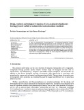
Fig. 1. NAMI-A inhibits DNA synthesis and PCNA expression in ECV304 cells. (A) Growing cells were treated for the indicated times with medium M199, containing 10% fetal bovine serum in the absence (j) or presence (s) of 100 lM NAMI-A. *, significantly different from fetal bovine serum. (B) Growing cells were treated for 72 h with medium M199, containing 10% fetal bovine serum in the absence (0 lM) or presence (s) of the indicated concentration of NAMI-A. *, Significantly different from fetal bovine serum. (C) Growing cells were treated for the indicated times with medium M199, containing 10% fetal bovine serum in the absence (j) or presence (s) of 100 lM NAMI-A. Equal amounts of protein from each sample were subjected to SDS/PAGE and analyzed by immunoblotting. The upper and lower part of panel C show, respectively, the PCNA immunoreactivity and the quantitative immunodensity expressed as percentage of control (time 0). *, significantly different from fetal bovine serum.
To verify the involvement of a PKC-dependent pathway in both serum- and PMA-generated ERK1/2 phosphoryla- tion, the selective PKC inhibitor GF109203X was used in separate experiments. As reported in the Fig. 3A and B, the exposure of ECV304 cells to GF109203X was able to partially suppress serum-induced ERK phosphorylation while completely inhibiting that elicited by the PMA. In addition, NAMI-A inhibited MEK1/2 phosphorylation with a magnitude similar to that of ERK1/2 inhibition (Fig. 3C,D), suggesting that NAMI-A-mediated inhibition of ERK1/2 phosphorylation may be lying upstream of ERK1/2.
Using a serine/threonine kinase SPA assay kit, the capability of NAMI-A to affect the ERK1/2 phosphoryla- tion activity was also assessed. Consistent with immunoblot analyses, a 15-min exposure to 100 lM NAMI-A signifi- cantly but not completely inhibited serum-stimulated kinase activity, which was still detected after 1 h of drug treatment. In contrast, the exposure of ECV304 cells to 100 lM NAMI-A completely prevented the phorbol ester-induced ERK1/2 activity at all times assessed (Fig. 4A). The effect elicited by NAMI-A on both serum- and PMA-induced ERK1/2 activity was dose-dependent and 100 lM was the most effective concentration in down regulating ERK1/2 activity (Fig. 4B,C).
synthesis in cells cultured under standard conditions. This effect was evident after 24 h of treatment and was still consistent in cells exposed to the ruthenium compound for 48 and 72 h, indicating the capability of NAMI-A to markedly affect cell proliferation. In these time course experiments we utilized 100 lM NAMI-A since it proved to be the most effective concentration in counteracting DNA synthesis over a 1–200 lM range (Fig. 1B).
NAMI-A-induced ERK1/2 inhibition down regulates c-myc gene expression
Further analysis of the inhibitory response elicited by NAMI-A on ECV304 growth was accomplished by inves- tigating the effect of NAMI-A on proliferating cell nuclear antigen (PCNA), a cell growth-related protein. Growing cells were exposed to the drug for the indicated times and PCNA expression was evaluated by Western blotting analysis using specific anti-PCNA Ig. The current experi- mental data show that cell exposure to NAMI-A time-dependently inhibited PCNA protein expression, as compared to untreated cells This effect was detectable after 24 h of cell treatment and reached the maximal amplitude at 72 h (Fig. 1C). To investigate whether NAMI-A-induced inhibition of ERK1/2 signaling could affect the expression of a cell growth-related gene, we assessed c-myc gene expression in growing cells treated either with NAMI-A or the MAPK/ ERK inhibitor PD98059. The addition of 100 lM NAMI-A to the incubation medium significantly inhibited serum- elicited c-myc gene expression as compared with untreated cells. This effect was already evident 1 h after NAMI-A exposure and reached a maximum at 3 h (Fig. 5A). A down regulation of c-myc mRNA expression was also observed in
NAMI-A affects c-myc gene expression and MEK/ERK phosphorylation (Eur. J. Biochem. 269) 5865
(cid:1) FEBS 2002
Fig. 2. NAMI-A and PD98059 down regulate serum-induced ERK1/2 phosphorylation. (A) Growing cells were stimulated for the indicated times with medium M199, containing 10% fetal bovine serum in the absence (j) or presence (s) of 100 lM NAMI-A. * Significantly different from fetal bovine serum. (B) Serum-starved ECV304 cells were stimulated for the indicated times with medium M199 containing 100 nM PMA (j), or pretreated for 1 h with either 50 lM PD98059 or 100 lM NAMI-A and then stimulated with medium M199 con- taining 100 nM PMA in the presence of either 50 lM PD98059 (h) or 100 lM NAMI-A (s). *, Significantly different from PMA; § signifi- cantly different from time 0. The upper part of each panel shows representative autoradiograms corresponding to ERK1/2 and phos- pho ERK1/2 immunoreactivity while the lower part of each panel reports the quantitative analysis of phospho ERK1/2 immunodensity expressed as percentage of control (individual densities from ERK1 and 2 bands were added up to generate one single ERK1/2 value).
Fig. 3. NAMI-A inhibits MEK1/2 activation and GF109203X down regulates ERK1/2 phosphorylation. (A,B) Serum-starved ECV304 cells were pretreated for 1 h with 5 lM GF109203X (+) and then stimu- lated for 1 h with medium M199 containing 10% fetal bovine serum (A) or 100 nM PMA (B) in the presence (+) or absence (–) of 5 lM GF109203X. (C,D) Serum-starved ECV304 cells were pretreated for 1 h with 100 lM NAMI-A (+) and then stimulated for 1 h with medium M199 containing 10% fetal bovine serum (C) or 100 nM PMA (D) in the presence (+) or absence (–) of 100 lM NAMI-A. * Significantly different from fetal bovine serum; §, significantly different from PMA. The upper part of each panel shows representative auto- radiograms corresponding to the immunoreactivity of ERK1/2 and phospho ERK1/2 (A,B), and MEK1/2 and phospho MEK1/2 (C,D). The lower part of each panel reports the quantitative analysis of phospho ERK1/2 (A,B), and phospho MEK1/2 (C,D) immunodensity expressed as percentage of control (individual densities from ERK1 and 2 bands were added up to generate one single ERK1/2 value).
PD98059 might have exerted their effect by following the same signal transduction pathway (Fig. 5C).
We next investigated whether NAMI-A may also induce c-myc down-regulation in serum-free cells exposed to PMA, an experimental condition, which has been previously shown to elicit a NAMI-A inhibitable increase in ERK1/2 activity. Figure 6A shows that in serum-free cells, 100 nM PMA increased c-myc gene expression. Such an effect was evident at 1 h, peaked after 3 h of treatment, thereafter progressively declined and returned to the control value at 12 h. PMA-induced increase in c-myc gene expression serum-cultured ECV304 cells exposed to PD98059 (Fig. 5B). Interestingly, both the inhibition of MEK/ERK pathway and the exposure of ECV304 cells to NAMI-A resulted in down regulation of c-myc gene expression with superimposable magnitudes and kinetics profiles. Figure 5C shows the dose–response effect of NAMI-A on ECV304 c-myc mRNA expression. As reported for DNA synthesis and ERK1/2 activity, 100 lM NAMI-A resulted to be the concentration most effective in inhibiting c-myc gene expression (Fig. 5C). Furthermore, NAMI-A addition to PD98059-treated cells failed to produce any additive inhibition of the residual c-myc gene expression induced by serum, suggesting the possibility that both NAMI-A and
5866 G. Pintus et al. (Eur. J. Biochem. 269)
(cid:1) FEBS 2002
Fig. 4. NAMI-A partially inhibits serum-induced ERK 1/2 activity but completely counteracts the activity induced by PMA. (A) Serum-starved ECV304 cells were left untreated (U) or pretreated for 1 h with 100 lM NAMI-A (N). Cells were then stimulated for the indicated times with medium M199 containing either 10% fetal bovine serum (open bars) or 100 nM PMA (hatched bars) in the absence (–) or presence (+) of 100 lM NAMI-A. ERK1/2 activities are expressed as c.p.m. 32P incorporated per mg protein per hour. *, Significantly different from fetal bovine serum; §, significantly different from PMA; #, significantly different from time 0. (B,C) Serum-starved ECV304 cells were left untreated (U) or pretreated for 1 h with the indicated NAMI-A con- centration (NAMI-A). Cells were then stimulated for 1 h with medium M199 containing either 100 nM PMA (B) or 10% fetal bovine serum (C), in the presence of the indicated concentrations of NAMI-A. ERK1/2 activities are expressed as percentage of control. * Signifi- cantly different from U.
Fig. 5. NAMI-A and PD98059 down regulate serum-induced c-myc gene expression with superimposable magnitudes and kinetic profiles. (A) Growing cells were stimulated with medium M199 containing 10% (v/v) fetal bovine serum in the absence (j) or presence (s) of 100 lM NAMI-A. (B) Growing cells were stimulated with medium M199 containing 10% fetal bovine serum in the absence (j) or presence (half shaded square) of 50 lM PD98059. (C) Growing cells were stimulated for 3 h with medium M199 containing 10% fetal bovine serum in the absence (0 lM) or presence (d) of the indicated concentration of NAMI-A. Cells were also treated with 100 lM NAMI-A + 50 lM PD98059 for 3 h. The upper part of Panel A shows representative ethidium bromide stained gels of the reaction products obtained using 5 lL of the RT products after 30 cycles of PCR amplification. The lower part of panel A, and panels B, C report the expression of c-myc mRNA levels detected by[32P]dCTP-PCR. Individual results were normalized to GAPDH mRNA detected in each sample and expressed as a ratio to GAPDH. *, Significantly different from fetal bovine serum.
occurred in a dose-dependent fashion, reaching the maximal amplitude at 100 nM over a concentration range of 10–200 nM PMA (Fig. 6B). Both NAMI-A and PD98059 dose-dependently counteracted and ultimately abolished the increase in c-myc mRNA expression elicited by PMA (Fig. 6C,D), suggesting a close relationship between the inhibitory effect of NAMI-A on ERK1/2 activation and NAMI-A- induced down regulation of c-myc gene expres- sion.
NAMI-A inhibits c-myc gene transcription and protein expression
To establish whether the decrease in c-myc mRNA expres- sion elicited by NAMI-A might have occurred at the transcriptional level, we assessed the rate of transcription of the c-myc gene by using an in vitro nuclear run-off transcription assay. A remarkable decrease in c-myc gene transcription was observed in nuclei isolated from serum- stimulated ECV304 cells that had been exposed for the times indicated to 100 lM NAMI-A, as compared with nuclei from untreated control cells (Fig. 7A). The transcriptional decrease induced by NAMI-A exhibited a time-course that overlaid that observed in RT-PCR experiments, being evident at 1 h and reaching a maximum after 3 h (Fig. 7A). In addition, the down-regulatory effect of NAMI-A on gene transcription was paralleled by a decrease in c-myc protein expression, as confirmed by immunoblotting experiments (Fig. 7B).
NAMI-A affects c-myc gene expression and MEK/ERK phosphorylation (Eur. J. Biochem. 269) 5867
(cid:1) FEBS 2002
Fig. 7. NAMI-A inhibits c-myc gene transcription and protein expres- sion. (A) Nuclear run-off transcription assay. Growing cells were sti- mulated for the indicated times with medium M199 containing 10% (v/v) fetal bovine serum in the absence (–) or presence (+) of 100 lM NAMI-A and then processed as described in materials and methods. Representative autoradiograms of the ribonuclease protection analysis of c-myc mRNA are shown in panel A. Autoradiographic exposure was for 2 days on Kodak X-Omat film with an intensifying screen. Row a, transcription of the c-myc gene. Row b, GAPDH mRNA. On the right are indicated the position of 350- or 800-bp radiolabeled DNA markers, showing that the single protected fragments migrated with a molecular size comparable to c-myc (bp 354) or GAPDH (bp 788) mRNA. (B) Growing cells were treated for the indicated times with medium M199, containing 10% (v/v) fetal bovine serum in the absence (–) or presence (+) of 100 lM NAMI-A. Equal amounts of protein from each sample were subjected to SDS/PAGE and analyzed by immunoblotting. Representative autoradiograms of the immuno- blotting analysis correspond to c-myc immunoreactivity are shown in the panel B.
Fig. 6. NAMI-A and PD98059 completely inhibits PMA-induced c-myc gene expression. (A) Serum-starved ECV304 cells were stimulated for the indicated times with medium M199 containing 100 nM PMA. *, Significantly different from time 0. (B) Serum-starved ECV304 cells were stimulated with the indicated concentrations of PMA for 3 h. *, Significantly different from 0 nM PMA. (C,D) Serum-starved ECV304 cells were left untreated (U) or pretreated for 1 h with the indicated concentration of PD98059 (PD) or NAMI-A (N). Cell were then stimulated for 3 h in medium M199 containing 100 nM PMA, in the abesence (U), or presence of the indicated concentration of either PD98059 (PD) or NAMI-A (N). *, significantly different from PMA. The upper part of panels A and B, shows representative ethidium bromide stained gels of the reaction products obtained using 5 lL of the RT products after 30 cycles of PCR amplification. The lower part of panels A, B and panels C, D report the expression of c-myc mRNA levels detected by [32P]dCTP-PCR (30 cycles). Individual results were normalized to GAPDH mRNA detected in each sample and expressed as a ratio to GAPDH.
D I S C U S S I O N
ECV304 cells and that such a condition plays a key role in the alteration of the growth behavior of these cells [24]. Constitutively active components of the ERK pathway have been shown to be associated with neoplastic growth [3] and an increase in ERK-related signaling has been found to represent a common outcome for a number of molecular events leading to angiogenesis [6] and metastatic growth [25,26]. These observations prompt the hypothesis that pharmacological manipulation of the ERK pathway may prove rewarding in counteracting such pathological process. A number of extracellular stimuli, including serum, have been reported to activate ERK via Ras/Raf/MEK/signaling [27,28]. This activation is mediated, at least in part, by G-protein coupled receptors (GPCRs), stimulation of membrane phospholipid hydrolysis, and activation of the diacylglycerol sensitive isoforms of protein kinase C (PKC) [28]. Thus, phorbol esters such PMA also powerfully activated the ERK cascade either by c-raf-mediated phos- phorylation [29] or in a Ras-dependent manner [30]. The The process of angiogenesis involves the formation of new blood vessels from established vasculature and is essential for progressive tumor growth and metastasis. The finding that such a pathological condition mainly depends on endothelial cell proliferation, migration and invasion [12] has sparked an interest in identifying cellular models of angiogenesis as well as molecular mechanisms amenable to endothelial cell proliferation. In this context, ECV304 a spontaneously transformed endothelial cell line, has been recently proposed as a suitable model for the investigation of the signaling mechanisms governing angiogenesis under both normal and pathological conditions [6,15]. A recent investigation reports that ERK1/2 is constitutively active in
5868 G. Pintus et al. (Eur. J. Biochem. 269)
(cid:1) FEBS 2002
serum-cultured cells time-course and by superimposable magnitudes. Third, NAMI-A failed to further decrease c-myc mRNA expres- sion in PD98059-treated, serum-cultured cells. Since both PD98059 and NAMI-A could not completely inhibit c-myc gene expression in serum-cultured cells, it is possible that protooncogene expression may also have been induced by serum through signal(s) unrelated to ERK1/2.
indicates that The finding that, in serum-free cells PD98059 completely suppressed PMA-induced c-myc mRNA expression, strongly indicates a crucial role of the Ras/Raf/MEK/ ERK pathway in modulating phorbol ester-activated mechanism(s) leading to c-myc gene expression. Consistent with the present results is the finding that the c-myc gene promoter contains conserved binding sites for the Ets family [34] and that both phorbol ester and Ras are able to induce expression from Ap-1/Ets-driven promoters via the upstream MEK/ERK effector Raf [35]. The finding that NAMI-A completely inhibited PMA-induced ERK1/2 activation and activity and totally counteracted phorbol ester-primed c-myc gene expression heavily suggests that the inhibitory effect of NAMI-A on c-myc gene expression may have occurred by suppressing ERK1/2 activation and activity elicited by PMA-generated signals.
to be an important
present results showing the ability of NAMI-A to com- pletely abolish ERK1/2 activation elicited by PMA in serum-free cells, clearly indicate the specificity of the drug in inhibiting phorbol ester-generated signals leading to ERK1/2 phosphorylation. Whereas, the finding that expo- sure of to NAMI-A markedly, although not completely, inhibited ERK1/2 phosphoryla- tion indicates that the wide-scale profiling of intracellular signals leading to ERK1/2 activation may have not been entirely encompassed by the drug-treatment. Such a hypo- thesis is further confirmed by the observation that, differ- ently from the effect of serum, the stimulatory effect of PMA on ERK1/2 activity could also be totally abolished by NAMI-A. Within this context, the present observation that the level of phospho-ERK1/2, as well as the peak increase in ERK1/2 activity, was significantly lower in the presence of PMA than in serum-stimulated cells suggests, as previously reported [31,32], that multiple signaling mechanisms occurred in ERK1/2 activation induced by serum. The finding that PKC inhibition only partially down regulated serum-generated ERK1/2 phosphorylation, but abolished PMA-primed activation, serum-induced ERK1/2 phosphorylation may both result from the activa- tion of PKC-dependent and -independent pathways, while that evoked by PMA mainly proceeded through PKC/Ras/ Raf/MEK-dependent signals. In addition, the finding that PMA-induced MEK1/2 activation was completely preven- ted by the drug treatment strongly suggests that NAMI- A-mediated inhibition of ERK1/2 phosphorylation may be lying upstream of ERK1/2.
Unlike the ras gene family, mutations within the coding feature in sequence appear not converting c-myc from a protooncogene to oncogene. Rather, abnormally high transcription of c-myc, at an inappropriate stage of the cell cycle or during differenti- ation, leads to oncogenic transformation [36]. In this regard, our results showing the ability of NAMI-A to down regulate c-myc gene transcription as well as protein expres- sion may be of particular relevance. Interestingly, human melanoma cells derived from metastatic lymph node displayed cisplatin resistance, higher tumorigenic ability and increased c-Myc expression, as compared with cell lines derived from the primary tumor. The finding that the treatment with c-myc antisense oligodeoxynucleotides abrogated cisplatin resistance and induced apoptosis in a metastatic lymph node cell line suggests that pharmacolo- gical manipulation of c-myc gene expression may have an important role in metastasis control [37]. Within this context, NAMI-A has displayed a better therapeutic effect on the growth of mouse lung metastases, when compared to the reference drugs, including cisplatin [38].
invasion of matrigel
The exact molecular sequelae of events underlying the observed effects of NAMI-A on ERK1/2 activation as well as c-myc gene expression remain to be elucidated and are the subject for future investigations. Nevertheless, the cellular targets and signaling mechanisms uncovered in the present study are associated with the modulation of critical growth regulatory decisions in different cell types. Moreover, growing evidence supports the view that unplugging the currently dissected pathways from regulatory control may associate with abnormalities in the architectural plans of cell growth and differentiation, ultimately ensuing in the appearance of a malignant phenotype [5,11,25,26]. These findings, along with the recent observation that NAMI-A [14], strongly inhibited tumor cell suggest that the down regulation of ERK1/2 currently elicited by NAMI-A may be a part of the molecular mechanism by which this drug exerts its antimetastatic activity in vivo. Such a view may be further inferred by the following observations. The NAMI-induced inhibition of Recent observations indicate that activation of Raf by PMA may trigger the same signaling pathway as oncogenic Raf, or Raf activation by Ras in combination with tyrosine phosphorylation [30]. Moreover, it is now established that PKC activation may both result from GPCR- and receptor tyrosine kinase (RTK)-mediated signaling and that the activation of ERK by GPCRs closely parallels that employed by RTK [33]. On the whole, these observations suggest that the inhibitory effect elicited by NAMI-A on ERK signaling might have occurred by affecting a serum- generated PKC-dependent Ras/Raf/MEK signaling upstream to ERK1/2. Such a hypothesis is supported by the finding that, similar to NAMI-A, exposure of PMA- stimulated cells to either PD98059 or GF109203 completely counteracted ERK1/2 activation, indicating that PMA- generated signals leading to ERK1/2 phosphorylation are under a tight control of the PKC/Ras/Raf/MEK pathway. The present results showing the capability of NAMI-A to drastically reduce c-myc mRNA levels indicate c-myc gene expression as a part of the molecular patterning affected by this new compound. The present observation that the selective MEK/ERK inhibitor PD98059 significantly reduced c-myc mRNA level in serum-cultured cells indicates that c-myc gene expression may be at least in part mediated by the activation of this signal transduction pathway. In the current study, a number of interrelated observations suggest that the inhibitory effect elicited by NAMI-A and PD98059 on c-myc mRNA expression might have occurred through a common intracellular pathway. First, both ERK1/2 activa- tion and c-myc gene expression were markedly inhibited following the exposure of serum-feeded cells to NAMI-A. Second, both the MEK/ERK inhibitor PD98059 and NAMI-A inhibited c-myc gene expression with a similar
NAMI-A affects c-myc gene expression and MEK/ERK phosphorylation (Eur. J. Biochem. 269) 5869
(cid:1) FEBS 2002
expression in normal breast tissue and in invasive and noninvasive breast carcinoma. Cancer Res. 52, 2597–2602.
10. Kerkhoff, E., Houben, R., Loffler, S., Troppmair, J., Lee, J.E. & Rapp, U.R. (1998) Regulation of c-myc expression by Ras/Raf signaling. Oncogene 16, 211–216.
11. Welch, D.R., Sakamaki, T., Pioquinto, R., Leonard, T.O., Goldberg, S.F., Hon, Q., Erikson, R.L., Rieber, M., Rieber, M.S., Hicks, D.J., Bonventre, J.V. & Alessandrini, A. (2000) Transfec- tion of constitutively active mitogen-activated protein/extracellu- lar signal-regulated kinase kinase confers tumorigenic and metastatic potentials to NIH3T3 Cells. Cancer Res. 60, 1552–1556. 12. Stetler-Stevenson, W.G. & Corcoran, M.L. (1997) Tumor angio- genesis, functional similarities with tumor invasion. Experientia Supplementa 79, 413–418.
ERK1/2 activation and activity was attained in a trans- formed endothelial cell line and it is now evident that the endothelial cell plays a crucial role in angiogenesis [39] and that it might affect the growth and differentiation of neighboring cells [40]. The endothelial cell is also the target cell for tumor neovascularization, a process that closely associates with tumor growth and metastasis [12], and in this context, NAMI-A has been shown to inhibit angiogen- esis [41] and to induce apoptosis in endothelial cells [42]. In conclusion, the inhibitory effects of NAMI-A on ERK1/2 activation and c-myc gene expression observed here may represent a prominent feature in the control of pathological processes of the blood vessel wall and in the modulation of angiogenic stimuli leading to neoplastic growth.
A C K N O W L E D G E M E N T S
13. Bergamo, A., Gagliardi, R., Scarcia, V., Furlani, A., Alessio, E., Mestroni, G. & Sava, G. (1999) In vitro cell cycle arrest, in vivo action on solid metastasizing tumors, and host toxicity of the antimetastatic drug NAMI-A and cisplatin. J. Pharmacol. Exp. Ther. 289, 559–564.
14. Zorzet, S., Bergamo, A., Cocchietto, M., Sorc, A., Gava, B., Alessio, E., Iengo, E. & Sava, G. (2000) Lack of In vitro cytotoxicity, associated to increased G(2)-M cell fraction and inhibition of matrigel invasion, may predict in vivo-selective anti- metastasis activity of ruthenium complexes. J. Pharmacol. Exp. Ther. 295, 927–933.
This study represents work done within the frame of the COST D20/ 0005/01 and supported by grants from (cid:2)Ministero della Sanita` (Attivita` di Ricerca Finalizzata – 2000)(cid:3), (cid:2)Ministero dell(cid:3)Istruzione, dell’Univer- sita` e della Ricerca (Fondo Integrativo Speciale per la Ricerca – 2001; Programmi di Ricerca Cofinanziati – 2001)’ and University of Sassari (cid:2)(cid:2)Young Researchers Grant(cid:3). We thank Ms Annalisa Cossu for her technical assistance.
R E F E R E N C E S
15. Hughes, S.E. (1996) Functional characterization of the sponta- neously transformed human umbilical vein endothelial cell line ECV304: use in an in vitro model of angiogenesis. Exp. Cell. Res. 225, 171–185.
1. Ferrell, J.E. Jr (1996) MAP kinases in mitogenesis and develop-
ment. Curr. Top. Dev. Biol. 33, 1–60.
16. Pintus, G., Tadolini, B., Maioli, M., Posadino, A.M., Bennardini, F., Bettuzzi, S. & Ventura, C. (1998) Heparin inhibits phorbol expression in ester-induced ornithine decarboxylase gene endothelial cells. FEBS Lett. 423, 98–104.
2. Plattner, R., Gupta, S., Khosravi-Far, R., Sato, K.Y., Der Perucho, M., C.J. & Stanbridge, E.J. (1999) Differential con- tribution of the ERK and JNK mitogen-activated protein kinase cascades to Ras transformation of HT1080 fibrosarcoma and DLD-1 colon carcinoma cells. Oncogene. 18, 1807–1817.
17. Kumar, A., Middleton, A., Chambers, T.C. & Mehta, K.D. (1998) Differential roles of extracellular signal-regulated kinase-1/2 and p38 (MAPK) in interleukin-1beta- and tumor necrosis factor- alpha-induced low density lipoprotein receptor expression in HepG2 cells. J. Biol. Chem. 273, 15742–15748.
3. Oka, H., Chatani, Y., Hoshino, R., Ogawa, O., Kakehi, Y., Terachi, T., Okada, Y., Kawaichi, M., Kohno, M. & Yoshida, O. (1995) Constitutive activation of mitogen-activated protein (MAP) kinases in human renal cell carcinoma. Cancer Res. 55, 4182–4187.
18. McDonald, O.B., Chen, W.J., Ellis, B., Hoffman, C., Overton, L., Rink, M., Smith, A., Marshall, C.J. & Wood, E.R. (1999) A scintillation proximity assay for the Raf/MEK/ERK kinase cas- cade: high-throughput screening and identification of selective enzyme inhibitors. Anal. Biochem. 268, 318–329.
19. Pintus, G., Tadolini, B., Maioli, M., Posadino, A.M., Gaspa, L. & Ventura, C. (1999) Heparin down-regulates the phorbol ester- induced protein kinase C gene expression in human endothelial cells: enzyme-mediated autoregulation of protein kinase C-alpha and -delta genes. FEBS Lett. 449, 135–140.
4. Nguyen, D.H., Webb, D.J., Catling, A.D., Song, Q., Dhake- phalkar, A., Weber, M.J., Ravichandran, K.S. & Gonias, S.L. (2000) Urokinase-type plasminogen activator stimulates the Ras/ Extracellular signal-regulated kinase (ERK) signaling pathway and MCF-7 cell migration by a mechanism that requires focal adhesion kinase Src, and Shc. Rapid dissociation of GRB2/ Sps-Shc complex is associated with the transient phosphorylation of ERK in urokinase-treated cells. J. Biol. Chem. 275, 19382– 19388.
20. Hsieh, S.C., Huang, M.H., Tsai, C.Y., Tsai, Y.Y., Tsai, S.T. & Sun, K.H., Yu, H.S., Han, S.H. & C.L. (1997) The expression of genes modulating programmed cell death in normal human polymorphonuclear neutrophils. Biochem. Biophys. Res. Commun. 233, 700–706.
5. Kobayashi, H., Suzuki, M., Tanaka, Y., Hirashima, Y. & Terao, T. (2001) Suppression of urokinase expression and Invasiveness by urinary trypsin inhibitor is mediated through inhibition of protein kinase C- and MEK/ERK/c-Jun – dependent signaling pathways. J. Biol. Chem. 276, 2015–2022.
21. Ventura, C., Pintus, G., Fiori, M.G., Bennardini, F., Pinna, G. & Gaspa, L. (1997) Opioid peptide gene expression in the primary hereditary cardiomyopathy of the Syrian hamster. I. Regulation of prodynorphin gene expression by nuclear protein kinase C. J. Biol. Chem. 272, 6685–6692.
6. Shigematsu, S., Yamauchi, K., Nakajima, K., Iijima, S., Aizawa, T. & Hashizume, K.D. (1999) Glucose and insulin stimulate migration and tubular formation of human endothelial cells in vitro. Am. J. Physiol. 40, E433–E438.
7. Spencer, C.A. & Groudine, M. (1991) Control of c-myc regulation
in normal and neoplastic cells. Adv. Cancer Res. 56, 1–48.
22. Ishizuka, S., Yano, T., Hagiwara, K., Sone, M., Nihei, H., Ozasa, H. & Horikawa, S. (1999) Extracellular signal-regulated kinase mediates renal regeneration in rats with myoglobinuric acute renal injury. Biochem. Biophys. Res. Commun. 254, 88–92.
8. Bover, L., Barrio, M., Bravo, A.I., Slavutsky, I., Larripa, I., Bolondi, A., Ayala, M. & Mordoh, J. (1998) The human breast cancer cell line IIB-BR-G has amplified c-myc and c-fos oncogenes in vitro and is spontaneously metastatic in vivo. Cell. Mol. Biol. 44, 493–504.
23. Sergeant, N., Lyon, M., Rudland, P.S., Fernig, D.G. & Delehedde, M. (2000) Stimulation of DNA synthesis and cell proliferation of human mammary myoepithelial-like cells by hepatocyte growth factor/scatter factor depends on heparan and sustained phosphorylation of sulfate proteoglycans
9. Pavelic, Z.P., Pavelic, L., Lower, E.E., Gapany, M., Gapany, S., Barker, E.A. & Preisler, H.D. (1992) c-myc, c-erbB-2, and Ki-67
5870 G. Pintus et al. (Eur. J. Biochem. 269)
(cid:1) FEBS 2002
mitogen-activated protein kinases p42/44. J. Biol. Chem. 275, 17094–17099.
33. Luttrell, L.M., Daaka, Y. & Lefkowitz, R.J. (1999) Regulation of tyrosine kinase cascades by G-protein-coupled receptors. Curr. Opin. Cell. Biol. 11, 177–183.
24. Shin, E.Y., Lee, J.Y., Park, M.K., Chin, Y.H., Jeong, G.B., Kim, S.Y., Kim, S.R. & Kim, E.G. (1999) Overexpressed alpha3beta1 and constitutively activated extracellular signal-regulated kinase modulate the angiogenic properties of ECV304 cells. Mol. Cells. 9, 138–145.
34. Roussel, M.F., Davis, J.N., Cleveland, J.L., Ghysdael, J. & Hiebert, S.W. (1994) Dual control of myc expression through a single DNA binding site targeted by ets family proteins and E2F-1. Oncogene. 2, 405–415.
35. Bruder, J.T., Heidecker, G. & Rapp, U.R. (1992) Serum-, TPA-, and Ras-induced expression from Ap-1/Ets-driven promoters requires Raf-1 kinase. Genes Dev. 6, 545–556.
25. Seddighzadeh, M., Zhou, J.N., Kronenwett, U., Shoshan, M.C., Auer, G., Sten-Linder, M., Wiman, B. & Linder, S. (1999) ERK signalling in metastatic human MDA-MB-231 breast carcinoma cells is adapted to obtain high urokinase expression and rapid cell proliferation. Clin. Exp. Metastasis 17, 649–654.
36. Lang, J.C., Whitelaw, B., Talbot, S. & Wilkie, N.M. (1988) Transcriptional regulation of the human c-myc gene. Br. J. Cancer. 9, 62–66.
26. Zhu, X., Price-Schiavi, S.A. & Carraway, K.L. (2000) Extra- cellular regulated kinase (ERK)-dependent regulation of sialo- mucin complex/rat Muc4 in mammary epithelial cells. Oncogene 19, 4354–4361.
37. Leonetti, C., Biroccio, A., Candiloro, A., Citro, G., Fornari, C., Mottolese, M., Del Bufalo, D. & Zupi, G. (1999) Increase of cis- platin sensitivity by c-myc antisense oligodeoxynucleotides in a human metastatic melanoma inherently resistant to cisplatin. Clin. Cancer Res. 5, 2588–2595.
27. Flamigni, F., Facchini, A., Giordano, E., Tantini, B. & Stefanelli, C. (2001) Signaling pathways leading to the induction of ornithine decarboxylase: opposite effects of p44/42 mitogen-activated protein kinase (MAPK) and p38 MAPK inhibitors. Biochem. Pharmacol. 61, 25–32.
38. Sava, G., Gagliardi, R., Bergamo, A., Alessio, E. & Mestroni, G. (1999) Treatment of metastases of solid mouse tumours by NAMI-A: comparison with cisplatin, cyclophosphamide and dacarbazine. Anticancer Res. 19, 969–972.
28. Sugden, P.H. & Clerk, A. (1997) Regulation of the ERK subgroup of MAP kinase cascades through G protein-coupled receptors. Cell Signal. 9, 337–351.
39. DeLisser, H.M., Christofidou-Solomidou, M., Strieter, R.M., Burdick, M.D., Robinson, C.S., Wexler, R.S., Kerr, J.S., Garlanda, C., Merwin, J.R., Madri, J.A. & Albelda, S.M. (1997) Involvement of endothelial PECAM-1/CD31 in angiogenesis. Am. J. Pathol. 151, 671–677.
29. Marquardt, B., Frith, D. & Stabel, S. (1994) Signalling from TPA to MAP kinase requires protein kinase C, raf and MEK: recon- stitution of the signalling pathway in vitro. Oncogene 11, 3213– 3218.
40. Bobik, A. & Campbell, J.H. (1993) Vascular derived growth factors: cell biology, pathophysiology, and pharmacology. Pharmacol. Rev. 45, 1–42.
41. Vacca, A., Bruno, M., Boccarelli, A., Coluccia, M., Ribatti, D., Bergamo, A., Garbisa, S., Sartor, L. & Sava, G. (2002) Inhibition of endothelial cell functions and of angiogenesis by the metastasis inhibitor NAMI-A. Br. J. Cancer 6, 993–998.
30. Chiloeches, A., Paterson, H.F., Marais, R., Clerk, A., Marshall, C.J. & Sugden, P.H. (1999) Regulation of Ras. GTP loading and Ras–Raf association in neonatal rat ventricular myocytes by G protein-coupled receptor agonists and phorbol ester. Activation of the extracellular signal-regulated kinase cascade by phorbol ester is mediated by Ras. J. Biol. Chem. 274, 19762–19770.
31. Han, X.B. & Conn, P.M. (1999) The role of protein kinases A and C pathways in the regulation of mitogen-activated protein kinase activation in response to gonadotropin-releasing hormone receptor activation. Endocrinology 5, 2241–2251.
42. Sanna, B., Debidda, M., Pintus, G., Tadolini, B., Posadino, A.M., Bennardini, F., Sava, G. & Ventura, C. (2002) The anti-metastatic agent imidazolium trans-imidazoledimethylsulf- oxide-tetrachlororuthenate induces endothelial cell apoptosis by inhibiting the mitogen-activated protein kinase/extracellular sig- nal-regulated kinase signaling pathway. Arch. Biochem. Biophys. 403, 209–218.
32. Lerosey, I., Pizon, V., Tavitian, A. & de Gunzburg, J. (1991) The cAMP-dependent protein kinase phosphorylates the rap1 protein in vitro as well as in intact fibroblasts, but not the closely related rap2 protein. Biochem. Biophys. Res. Commun. 175, 430–2436.