doi:10.1046/j.1432-1033.2003.03833.x
Eur. J. Biochem. 270, 4426–4438 (2003) (cid:2) FEBS 2003
Mutations in the C-terminal domain of ALSV (Avian Leukemia and Sarcoma Viruses) integrase alter the concerted DNA integration process invitro
Karen Moreau1, Claudine Faure1, Se´ bastien Violot2,3, Ge´ rard Verdier1,3 and Corinne Ronfort1,3 1Universite´ Claude Bernard, Centre National de la Recherche Scientifique, Institut National de la Recherche Agronomique, Lyon, France; 2Institut de Biologie et Chimie des Prote´ines, Centre National de la Recherche Scientifique, Laboratoire de Bio-Cristallographie, Universite´ Claude Bernard, France; 3IFR 128 ‘BioSciences Lyon Gerland’, Lyon, France
resulted in a decrease of integration efficiency that was concomitant with a decrease of IN dimerization; (b) the V239A, V249A and K266A mutants preferentially mediated non-concerted DNA integration rather than concerted DNA integration although they were found as dimers. Other mutations (V260E and Y246W/DC25) highlight the role of the C-terminal domain in the general folding of the enzyme and, hence, on its activity. This study points to the important role of residues at the IN C-terminal domain in the folding and dimerization of the enzyme as well as in the concerted DNA integration of viral DNA ends.
Keywords: concerted DNA integration; integrase; multi- merization; mutations; retroviruses.
Integrase (IN) is the retroviral enzyme responsible for the integration of the DNA copy of the retroviral genome into the host cell DNA. The C-terminal domain of IN is involved in DNA binding and enzyme multimerization. We previ- ously performed single amino acid substitutions in the C-terminal domain of the avian leukemia and sarcoma vir- uses (ALSV) IN [Moreau et al. (2002). Arch. Virol. 147, 1761–1778]. Here, we modelled these IN mutants and ana- lysed their ability to mediate concerted DNA integration (in an in vitro assay) as well as to form dimers (by size exclusion chromatography and protein–protein cross-linking). Muta- tions of residues located at the dimer interface (V239, L240, Y246, V257 and K266) have the greatest effects on the activity of the IN. Among them: (a) the L240A mutation
gaps are repaired, creating a short duplication (4–6 bp) of host sequence. The integration process is defined as concerted because it enables the concomitant integration of two viral DNA ends at the same site of the host cell DNA generating a complete provirus flanked by short host DNA repeats [1]. Steps of 3¢-processing and strand transfer are catalysed by the viral IN enzyme whereas repairing DNA gaps most probably involves cellular enzymes [2–5].
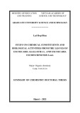
Integration of the retrotranscribed viral DNA into a host cell chromosome, an essential requirement for viral gene expression and hence retroviral replication, is mediated by the viral integrase (IN). Integration also requires short specific DNA sequences at the viral DNA ends, designated att sequences [1]. Using in vitro assays, it has been shown that the integration process occurs in three steps as illustrated in Fig. 1A. Firstly, two terminal nucleotides are removed from both 3¢ viral ends to generate the CA-3¢OH ends, with a two-base 5¢ overhang (3¢-processing step). Secondly, during the strand transfer reaction, the 3¢ viral ends are linked to the host DNA in a single cleavage– ligation reaction. The host DNA is asymmetrically cleaved and the insertion of the two viral DNA ends typically occurs 4–6 bp apart, according to the retrovirus [1]. In the third step (gap filling), the 5¢ overhanging dinucleotides of the viral DNA ends are removed and single-stranded DNA
Concerted DNA integration has been reconstituted in vitro using a short linear DNA flanked by viral att sequences at its ends as donor DNA, a suitable plasmid as acceptor DNA and the IN enzyme supplied either as preintegration complex purified from infected cells or as a recombinant protein. This system has been developed with Avian Leukaemia and Sarcoma Viruses (ALSV) [6–13], HIV [12,14–19], Simian Immunodeficiency Virus [20] and more recently Murine Leukemia Virus [21] integrases. Such an in vitro assay has allowed reproduction of the integration process as observed in vivo, with the cleavage of the two terminal nucleotides of viral DNA ends and the duplication of a short acceptor DNA sequence.
The IN enzyme, which consists of three domains, is rather well conserved among the different retroviruses [22–24]. The C-terminal domain is the least conserved and contains no recognizable active site, but is necessary for both 3¢-processing and strand transfer activities in vitro [25,26]. It is involved in binding to both viral DNA and nonspecific target DNA [27–29]. Several experiments have shown that the C-terminal domain is also involved in the oligomeriza- tion of IN. Indeed, ASLV and HIV INs are present as
Correspondence to C. Ronfort, Laboratoire (cid:1)Retrovirus et Pathologie Compare´ e(cid:2), UCBL-INRA-ENVL, Universite´ Claude Bernard. 50, avenue Tony Garnier, 69366 Lyon cedex 07, France. Fax: +33 437 287 605, Tel.: +33 437 287 629, E-mail: ronfort@univ-lyon1.fr Abbreviations: ALSV, Avian Leukemia and Sarcoma Viruses; att, attachment sequence; HMG, high mobility group; IN, integrase; RSV, Rous Sarcoma Virus; RF, recombinant form; DSS, disuccinimidyl suberate. (Received 25 July 2003, revised 9 September 2003, accepted 12 September 2003)
Mechanism of integration of retroviral IN mutants (Eur. J. Biochem. 270) 4427
(cid:2) FEBS 2003
IN proteins result in proteins deficient in multimerization [31,34] and specific mutations in the C-terminal domain inhibit the oligomerization of HIV-1 IN [39,40]. Conversely, the ALSV IN 201–286 fragment was shown to self-associate [31] and NMR analysis revealed that the C-terminal domain of HIV IN form dimers in solution [41]. The formation of multimeric molecules is essential for correct IN function, as shown by trans-complementation experiments in vitro [25,26] and in vivo [42,43]. It has been suggested that IN may function as a dimer, a tetramer or even as an octamer complex during the integration process [23,32–35,37,38,44]. We have previously introduced specific changes in selected amino acid in the C-terminal domain of an ALSV IN [24] and analysed the effects of these mutations on the catalytic activities of the resulting proteins [3¢-processing, strand transfer and disintegration (reversal of strand transfer)]. These assays of catalytic activities relied on the use of short oligonucleotides carrying a unique viral end. In the present study, our aim was to test effects of several mutations on integration of two viral ends (concerted DNA integration) in an in vitro assay, as well as on oligomeriza- tion of IN. Recently, a two-domain structure of the Rous Sarcoma Virus (RSV) IN was published [23]. We used this structure to model the structure of the mutants. Our analyses focussed on proteins mutated at conserved residues or on residues shown to be involved in the dimer interface. These analyses allow us to identify the important role of specific residues within the C-terminal domain of ALSV IN.
Experimental procedures
DNA manipulation
The DNA pBSK-Zeo acceptor plasmid was constructed as follows: Plasmid pBSK+ (Stratagene) was digested with SmaI and SacII restriction enzymes, treated with Klenow DNA polymerase and reclosed by ligation to generate plasmid pBSK+DBamHI. This was then digested with HindIII and EcoRV, filled by Klenow enzyme and reclosed by ligation to generate plasmid pBSK+D2. These mani- pulations removed the BamHI and EcoRV restriction sites, respectively. Then, plasmid pBSK+D2 was amplified by PCR using Pfu turbo polymerase (Stratagene) and primers BU (5¢-CCGATATCATACTCTTCC-3¢) and BL (5¢-CC GATATCAGACCAAGTTTAC-3¢). In the same way, the zeo gene was amplified from plasmid pHook (Invitrogen) using primers Z1 (5-CCGATATCGTGTTGACAATT AATC-3¢) and Z2 (5¢-CCGATATCCAGACATGATAA GATAC-3¢). All primers contain an EcoRV restriction site and resulting PCR products, pBSK+D2 and zeo gene, were digested by the EcoRV restriction enzyme and ligated together to produce plasmid pBSK-zeo. This plasmid, which carries the zeocin resistance gene, was amplified in E. coli DH5a (Invitrogen).
The donor DNA was obtained as follows: supF gene was amplified by PCR from piVX plasmid (ATCC) using primers H-sup1 (5¢-GAGAAGCTTAACGTTGCCCGG ATCCGGTC-3¢) and P-sup2 (5¢-GAGCTGCAGTAGTC CTGTCGGGTTTCGCC-3¢) containing HindIII and PstI restriction sites, respectively. The amplification product was digested with HindIII and PstI restriction enzymes and ligated into the pBSK+ plasmid digested by the same
monomers, dimers and tetramers in solution, as shown by exclusion chromatography and analytical ultracentrifuga- tion [30–38]. Within the C-terminal domain, deletion of residues 208–286 of ALSV IN or residues 218–288 of HIV
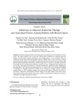
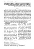
Fig. 1. Schematic representation of the retroviral integration process the in vitro concerted DNA integration assay. and principle of (A) Retroviral integration. The viral DNA made by reverse tran- scription is linear and blunt-ended. In the first step of integration (3¢-processing), two nucleotides are removed from each 3¢ end of the viral DNA. In the second step (strand transfer), the hydroxyl groups at the 3¢ ends of the processed viral DNA attack a pair of phosphodiester bonds in the target DNA. In the last step (gap filling), completion of the integration process requires removal of the two unpaired nucleo- tides at the 5¢ ends of the viral DNA and filling in the gaps between target and viral DNAs, generating a duplication of target DNA. (B) In vitro assay. Representation of the donor DNA with 15 bp of the U3 viral end and 12 bp of the U5 viral end. The highly conserved CA dinucleotides are underlined. The closed rectangle represents the supF tRNA transcription unit. (C) In vitro assay. Schematic representation of the reconstituted integration reaction with the donor DNA, acceptor plasmid, purified integrase and HMGI proteins. Concerted DNA integration products include those that result from use of both ends from a single donor (product a) and from use of different ends from two donors (product b). Note that when two donors are inserted at the same site, a linear product is synthesized. Non-concerted DNA integration products result from one-ended integration of a single donor (product c), or two-ended integration of a single donor with insertion at different sites on the acceptor DNA (product d), or one- ended integration of two or more donors at different sites on the acceptor DNA (product e). Auto-integrants result from integration of a donor DNA in a second donor DNA (product f). Adapted from [13].
4428 K. Moreau et al. (Eur. J. Biochem. 270)
(cid:2) FEBS 2003
NaCl, 50 mM NaH2PO4 pH 7.4 and the proteins were eluted with a gradient of 0.5–1.5 M NaCl. Each fraction was analysed by Bradford quantification and Western blot.
Integration reaction
restriction enzymes, giving pBSK-supF plasmid. The donor DNA was then amplified from pBSK-supF plasmid by PCR using pfu turbo polymerase, and primers U3 (5¢-GA TGTAGTCTTATACGTTGCCCGGATCCGG-3¢) and U5bi (5¢-AATGAAGCCTTCTGCTTTGAGCGTCGAT TTTTG-3¢). The PCR product was purified from agarose gel using the Qiaex II kit (Qiagen). The final donor DNA contained 15 bp of the U3 end sequence of Avian Erythro- blatosis Virus and 12 bp of the U5 end.
Modelling of the mutants
Purified IN protein (60 ng) was incubated overnight at 4 (cid:3)C with 100 ng pBSK-zeo plasmid, 10 ng donor DNA and 100 ng purified HMGI protein in a final volume of 5 lL. The volume of reaction was then increased to 20 lL with a final concentration of 20 mM Hepes, pH 7.5, 1 mM dithio- threitol, 30 mM MgCl2, 15% dimethyl sulfoxide, 8% PEG 8000 and 50 mM NaCl, and the integration mixture was incubated at 37 (cid:3)C for 90 min.
Gel analysis of the integration reaction
For gel analysis of the integration reaction, the DNA donor was radiolabelled by including 8 lCi [32P]dCTP[aP] in the PCR amplification mixture. After the integration reaction was performed, the volume was increased to 50 lL by the addition of 4.25 mM EDTA, 0.44% SDS and 20 ng prote- inase K (Roche Diagnostics) and samples were incubated for 1 h at 55 (cid:3)C. The DNAs were deproteinized by phenol/ chloroform extraction and purified by ethanol precipitation. Samples were then loaded on 1.2% agarose gel in 0.5 · Tris/ borate/EDTA electrophoresis buffer. After electrophoresis, the gels were fixed in 5% trichloroacetic acid for 30 min and dried for 3 h at 45 (cid:3)C. Lastly, the gels were exposed to autoradiographic film overnight at )80 (cid:3)C. Integration products were quantified using a phosphoimager (Biorad).
Construction of IN mutants has been reported elsewhere [24]. Two two-domain structures of RSV IN, containing the core and the C-terminal region, have been solved in space groups P21 and P1 at 3.1-A˚ and 2.5-A˚ resolution, respect- ively [23]. No structure containing also the N-terminal domain has yet been published. In consequence, the 2.5 A˚ two-domain structure of RSV IN was used to model the structure of mutants. Modelling was performed on the dimer. Each structure of a single mutant was generated using the program CALPHA [45] and minimized with the program CNS using a conjugate gradient method [46]. Resulting models were displayed and analysed on a graphic station using the program TURBO-FRODO [47]. Contact distances were computed with CNS around each mutated residue. In parallel, a BLAST search [48] was performed against the SWISS-PROT and the TrEMBL sequences data- bases [49] to detect homologous proteins. A multiple sequence alignment was performed in turn with CLUSTAL [50]: the eight studied substitutions are unique in retrovirus as well as in lentivirus integrases.
Cloning and sequencing of two-ended integration products
Purification of proteins
IN mutants [24] were expressed in BL21 bacteria (Invitro- gen) and purified as described by others [40].
To clone integration products for sequencing, products of the integration reaction were purified on a Qiaquick column (Qiagen) as described by the supplier. The whole reaction was introduced into MC1060/P3 E. coli (Invitrogen) as described by others [9]. MC1061/P3 E. coli carry ampicillin, tetracyclin and kanamycin resistance genes. Both ampicillin and tetracyclin resistance genes carry an amb mutation. These proteins are thus expressed only in the presence of the supF gene products. Integration clones carrying both zeocin-resistant and supF genes were therefore selected in the presence of 40 lgÆmL)1 ampicillin, 10 lgÆmL)1 tetra- cyclin, 15 lgÆmL)1 kanamycin and 25 lgÆmL)1 zeocin. Plasmids were isolated from quadruply resistant colonies and donor–acceptor DNA junctions were sequenced using SL primer (5¢-ACTCTAAATCTGCCGTCATCG-3¢) for the U3 junction and SU primer (5¢-ATCATATCAA ATGACGCGCCG-3¢) for the U5 junction. SL and SU primers are located on the donor DNA.
Size exclusion chromatography
All proteins were centrifuged for 10 min at 14 000 r.p.m. to remove IN aggregates. A total of 100 lL integrase solution at a concentration of 30 lM was loaded on a Superoz 12 column (Pharmacia) equilibrated previously with 1 M NaCl, 25 mM Hepes pH 7.5, 0.1 mM EDTA, 1 mM b-mercapto- ethanol. Size exclusion chromatography was performed at
The HMGI(Y) proteins (high mobility group; now referred as HMGa1) consist of two proteins (HMGI and HMGY) which are expressed from the same gene and differ by altenative mRNA splicing. The pET15b-HMGI vector (generously donated by T. H. Kim, Harvard University, Cambridge, MA, USA) expresses HMGI [51]. The HMGI protein was expressed in BL21(DE3) pLysS bacteria (Invitrogen) in the presence of 100 lgÆmL)1 ampicillin and 34 lgÆmL)1 chloramphenicol upon induction with 1 mM of isopropyl-thio-b-D-galactopyranoside for 3 h. Purification was carried out as follows. The bacterial pellet was resuspended in NaCl/Pi containing 0.1% Triton X-100 and sonicated. Then 5% of perchloric acid was added and the solution was incubated for 30 min at 4 (cid:3)C. The lysate was then centrifuged for 10 min at 12 000 g. A total of 25% of trichloroacetic acid was added to the supernatant which was incubated for 1 h on ice. After 10 min centrifugation at 12 000 g, the pellet was rinsed once with acetone and 0.2% HCl ()20 (cid:3)C), twice with acetone 70%/ethanol 20%/20 mM Tris/HCl pH 8 ()20 (cid:3)C), and once with acetone ()20 (cid:3)C). The pellet was dried at room temperature before being resuspended in 250 lL Tris/EDTA, pH 8.0. The solution was passed through a Hitrap Heparin column (Pharmacia), which had been equilibrated with 0.5 M NaCl, 50 mM NaH2PO4 pH 7.4. The column was washed with 0.5 M
Mechanism of integration of retroviral IN mutants (Eur. J. Biochem. 270) 4429
(cid:2) FEBS 2003
4 (cid:3)C. The column was calibrated with molecular mass markers. Protein elution was monitored at A280 nm at a flow rate of 0.3 mLÆmin)1.
Protein–protein cross-linking
integrases were
Wild-type or mutant treated with 40 lgÆmL)1 disuccinimidyl suberate (Pierce). Reactions included 2 lg protein in a final volume of 10 lL 20 mM Hepes pH 7.5, 60 mM NaCl, 0.7 mM EDTA, 10% glycerol, 4.5 mM Chaps. After 30 min at 22 (cid:3)C reactions were quenched by the addition of 3 mM lysine and 25 mM Tris/ HCl pH 8. After a further 10 min at 22 (cid:3)C, reactions were boiled for 10 min in sample buffer and separated by SDS/ PAGE (10% acrylamide). Products were revealed by Western blot using anti-His-tag Ig (Roche Diagnostics).
Results
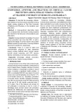
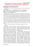
a mix of circular forms (Recombinant Form RFII products: a, c and d), the middle band correspond to the linear form b (RFIII products) and the fastest band correspond to auto- integration products (form f). Product e, which migrates more slowly because two or more donors are inserted into the target, is observed on some gels, but not all. A recombinant, identified by an asterisk in Fig. 3A, and which migrated slightly faster than the RFII recombinants has been observed by others [6,10,16,18,19]; its structure is unknown [18]. Total integration products were cleaved with either BamHI (which cleaves the donor DNA) or XhoI (which cleaves in the acceptor DNA). Structures of diges- tion products were fully consistent with the above assign- ment of the DNA forms (data not shown). As controls, reactions were performed in the absence of IN (Fig. 3A, lane 1) or with an IN mutated in its catalytic site, the D121E mutant (lane 2) [24]. No integration product was observed demonstrating that the products observed with wild-type protein resulted from IN enzymatic activity.
Reconstitution of the concerted DNA integration assay invitro
The in vitro retroviral concerted DNA integration system (Fig. 1B,C) has previously been described by others [9,12,13]. It is composed of a linear donor DNA, a plasmid acceptor DNA and recombinant IN. HMGI protein is added to the reaction because it has been found to enhance the concerted DNA integration reaction [12]. HMGI is a component of the HMGI(Y) protein (now referred as HMGa1). HMGI(Y) is a DNA binding protein that has been found in HIV preintegration complexes isolated from infected cells [52]. HMGI(Y) might stimulate concerted DNA integration by bending the donor DNA and helping to bring the two ends into close proximity; alternatively, the unwinding activity of HMG proteins could facilitate binding of IN proteins to DNA ends and their subsequent distortion [12,53].
Gel analysis permits the quantification of integration efficiency but does not distinguish one-ended from two- ended integration products, as product a is not resolved independently of other RFII forms (c and d products). However, integration products can also be cloned into MC1061/P3 E. coli, which contain drug resistance markers with amber mutations. Only DNA products carrying the amber mutation suppressor gene (supF) should be able to replicate and form colonies under drug selection. Among the different integration products, one-ended or multiple one-ended donor integration products (c and e) and linear product (b) should be lost upon cloning into E. coli. Only the circular two-ended integration products (forms a and d) should be able to replicate into bacteria [14,15]. Thus, the cloning analysis enables estimation of the efficiency of IN proteins to perform two-ended donor integration (concer- ted, form a; or not, form d). Following cloning, donor DNA–acceptor plasmid junctions of isolated integration products have to be sequenced in order to check the accuracy of the integration reaction (cleavage of viral ends and duplication of short acceptor DNA sequence).
In the present report, we used the IN protein from Rous Associated Virus type 1, and a donor DNA of 326 bp containing 15 bp of the U3 att sequence at one end and 12 bp of the U5 att sequence at the other end (Fig. 1B). Products of the integration reaction can arise from concer- ted or non-concerted DNA integration (Fig. 1C) [9,12,13]. Two-ended concerted DNA integration products include those that result from integration of both viral ends from a single donor (product a) or those that result from integra- tion of two viral ends from two donors at the same integration site (generating the linear product b). Non-con- certed DNA integration products result from one-ended integration of a single donor (product c), from two-ended integration of a single donor with insertion at different sites on the acceptor DNA (product d), or from one-ended integration of two or more donors at different sites on the acceptor DNA (product e). Auto-integration products, which are the results of the integration of donor DNA in a second donor DNA are also observed (product f).
Integration products generated with wild-type IN were cloned. Between 98 and 324 colonies were observed according to the experiments. Thirty-one clones were isolated and sequenced (Table 1). Sixteen clones exhibited a target DNA duplication of 6 bp and 11 clones a duplication of different size (from 4 or 5 bp). In vivo, the 6-bp duplication is a hallmark of ALSV viruses [54,55] although some size variations have been reported [56]. In in vitro assays, shorter duplications have often been observed [9,12,13]. Four clones exhibited a deletion of acceptor DNA. However, as these clones were correctly cleaved at both ends and integrated between the canonical TG and CA viral dinucleotides, they were interpreted as the result of an IN mediated process but with incorrect cleavage of the acceptor DNA. Integration products with acceptor DNA deletion could arise from either two independent one-ended donor integration events (form e) or from nonconcerted DNA integration of the two ends of one donor (form d). Assuming that only circular integration products (a and d, Fig. 1C) could be amplified in bacteria [14,15], we speculate that these clones were most probably the result of a non-concerted DNA
By using labelled donor DNA, the integration of the small donor DNA into larger acceptor DNA can be visualized by autoradiography after separation on agarose gel. Under these conditions, three characteristic bands were revealed in presence of IN (Fig. 3A, lane 3). As described previously by others [7,8,16], the slowest band correspond to
4430 K. Moreau et al. (Eur. J. Biochem. 270)
(cid:2) FEBS 2003
Table 1. Sequencing of donor–target junctions from clones produced by wild-type and V239A INs. Square brackets, number of clones har- bouring incorrect cleavage of att sequences (deletion of more than the 2 nucleotides expected).
Products obtained with [n (%)]
WT V239A
Duplication size 7 bp 6 bp 5 bp 4 bp 0 16 (51.5) [2] 8 (26) [1] 3 (9.5) [1] 1 (3.5) 18 (60) [1] 3 (10) [1] 1 (3.5) [1]
Deletion 4 (13)a 7 (23)b
a Deletions range from 150 to 948 bp, b deletions range from 33 to 503 bp.
Total 31 30
[57,58]
integration of the two viral ends of one donor DNA at two different sites on acceptor plasmid DNA (form d, Fig. 1C). Deletion in the acceptor DNA by two-ended nonconcerted DNA integration events has already been described [12–15]. Regarding the viral DNA ends, we observed deletion of more than the two expected nucleo- tides at one or the other att sequence in four clones. These four clones exhibited a duplication of acceptor DNA at the integration site, which led us to conclude that they have indeed arisen from a mechanism of integration mediated by IN. In other works describing the ALSV concerted DNA integration, neither deletions of acceptor DNA nor the use of internal cleavage sites on the donor DNA were observed using the wild-type enzyme unless the viral sequences were mutated [13]. These assays with wild-type IN have typically used 15 bp of viral sequence at each end, while we used 12 bp of U5 instead of 15. Therefore, it is possible that the structures we observed were generated due to the small U5 att site. IN may have used a larger U5 att site, recognizing the nonviral sequence covalently linked to the att site, which would represent a mutant att site. However, the number of such clones is rather low and does not impair the following analyses since all mutants were systematically compared with wild-type IN.
Description and modelling of IN mutants
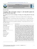
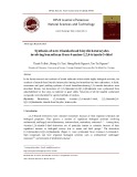
[23]. Strands b1¢, b2¢ and b5¢ of the proximal monomer and strands b2¢, b3¢ and b4¢ of the distal monomer are involved in the dimer interface (Fig. 2B). It is noteworthy that the two-domain structures of HIV-1 and Simian show different INs Immunodeficiency Virus arrangements of the C-terminal domains. The biological relevance of this is unclear: it may indicate considerable flexibility in the linkage between the core and C-terminal domains [59].
Modelling of IN mutants. We used the two-domain structure of RSV [23] to model the mutants studied here.
First, five of the mutants studied herein carried mutations on residues involved in the dimer interface (Fig. 2, Table 2). This includes the V239 and K266 residues of the proximal monomer and the L240, Y246, V257 residues of the distal monomer. Mutations of these residues were supposed to affect the dimeric interface.
the C-terminal domain. We have Arrangement of previously constructed mutants, each containing single amino acid substitutions in the C-terminal domain of the Rous Associated Virus type 1 IN [24] (Fig. 2A). In the meantime, a 2.5-A˚ structure of the closely related RSV IN was published [23] containing both the core and C-terminal domains. In this structure, the two core domains are related by a twofold symmetry axes, whereas the two C-terminal domains have a similar fold but associate asymmetrically, giving rise to a (cid:1)proximal(cid:2) and a (cid:1)distal(cid:2) domain (close to the core domain or away from it, respectively; Fig. 2B). Therefore, equivalent residues of the (cid:1)proximal(cid:2) and (cid:1)distal(cid:2) domains have a different environment at interface regions [23]. The C-terminal domain is composed of six strands forming a b-barrel fold resembling an SH3 domain (Fig. 2)
V239 is located in strand b2¢ at the dimeric interface. Based on multiple sequence alignments, this residue is well conserved among INs [24]. The proximal V239 residue is involved in the interface with the second C-terminal domain and has an intermolecular long contact distance (4.1 A˚ ) with residues V241 and W259 of the distal domain. The
Fig. 2. Description of the IN mutants analysed. (A) Sequence of the ALSV C-terminal domain (residues 219–286) is shown. Above are indicated b-strands (large arrows) [23]. Arrows indicate residues mutated in the present study. Arrows with asterisks indicate residues at the C dimer interface. Longer arrow at position 261 indicates end of Y246W/DC25 IN mutant. (B) Ribbon representation of the dimeric two-domain structure of RSV integrase (residues 54–268). Green and red molecules represent (cid:1)proximal(cid:2) and (cid:1)distal(cid:2) subunits, respectively. Labels on the green subunit correspond to the eight mutated residues discussed in this paper. Labels on the red subunit indicate b-strands, strand b2¢ being designated as two shorter strands b2¢* and b2¢ (adapted from [23]).
Mechanism of integration of retroviral IN mutants (Eur. J. Biochem. 270) 4431
(cid:2) FEBS 2003
Table 2. Contacts between residues in the monomers and dimers of the wild-type and mutants INs. The two-domain structure [23] was used to model the mutants. #, Residues of the distal subunit. In bold, residues at the interface of the dimer. Maximum contact distance is 5 A˚ , residues in italic type have contact distances < 3.2 A˚ .
ALSV Location Contacts between side chains in wild-type Contacts between side chains in mutant
P222, V224, #V241, W242, A247, V249, #W259, V265 L55, L218, V241, A248, K250, V257
V224, I226, W237, V239, A247, I258, V260, V265 L55, L240, A248, K250, W259 I226, A247, V249, I258, P261, K264, V265 Proximal K225H V239A L240A Y246W V249A V257A V260E K266A W233, K235, D268 P222, V224, W242, V265 L55, V241, A248 R53, W259, P261 I226, W237, I258 L55, K250 I226, W246, I258, P261, K264 W233, P267, #R244 Strand b1¢ W233, K235, K266, D268 Strand b2¢ Strand b2¢ Strand b3¢ R53, W259, P261 Strand b3¢ Strand b4¢ Strand b4¢ Strand b5¢ K225, W233, P267, #R244 Distal
V239A mutation removes these two intermolecular contacts as well as several other intramolecular contacts within each monomer (with A247 and V249).
Finally, three other mutants were studied too: K225 is a nonconserved residue of the b1¢ strand. The conservative K225H mutation makes closer con- tact with the D268 residue within the monomer and removes an intramolecular contact with the K266 residue (Table 2).
V249 is a moderately well conserved residue of strand b3¢ which is not involved in intermolecular contacts. Mutation V249A removes several contacts in the monomers, especi- ally with the V260 residue.
L240 is also located in strand b2¢. This residue is well conserved in retroviruses [24]. The distal L240 is at the dimeric interface between C-terminal domains, and its side chain makes van der Waals’ contacts with residues L218 and P222 in strand b1¢ of the proximal monomer. The L240A mutation does not remove these contacts at the interface of the dimer. However, the mutation decreases the number of intramolecular contacts within both monomers. Y246 is located at the beginning of strand b3¢. The distal Y246 is involved in intermolecular contacts in the dimer through an interaction with P267 in strand b5¢ of the proximal monomer. Nevertheless, the mutation Y246W does not remove this contact. It only reinforces the contact with P261 in each monomer.
V260 is a highly conserved residue of strand b4¢. V260 in HIV-1 IN is potentially involved in the formation of multimeric complexes [39]. The V260E mutation was the same as that performed on HIV IN [39]. The V260E mutation replaces several contacts inside both monomers (W246 instead of A247, V249 and V265 in the proximal monomer, W237 and I258 instead of K264 in the distal monomer). It also makes closer contact with the I226 residue in each monomer (Table 2).
V257 is located at the beginning of strand b4¢. The distal V257 is involved in an intermolecular contact in the dimer through an interaction with P223 in strand b1¢ of the proximal monomer. Residue V257 is also involved in a contact with the above-mentioned L240 residue within each monomer. The V257A mutation removes the intermole- cular contact between monomers as well as several intra- molecular contacts.
K266 is a well conserved residue located in strand b5¢. At the dimeric interface, the proximal K266 is in contact with R244 of the distal monomer, a residue located in a turn between strands b2¢ and b3¢. Nevertheless, the K266A mutation does not remove this contact in the dimer. The mutation only removes a contact with K225 within each monomer.
Catalytic activities of IN mutants. In the preliminary study [24], 3¢-processing and strand transfer catalytic activities of wild-type protein and of each mutant were examined in vitro, using a 15-bp long oligonucleotide corresponding to the U5 att terminal sequence (Table 3). Briefly, mutant K266A was as efficient as wild-type protein for both activities. K225H, V239A, L240A and V249A mutants displayed a slightly reduced efficiency for 3¢ processing while strand transfer activity was close to that of the wild-type protein. Y246W, Y246W/DC25, V257A and V260E mutants had 3¢-processing activity that was drastically reduced compared to that of wild-type IN, while strand transfer activity was either correct or reduced (V260E). With the exception of V260E, all other mutants displayed a correct disintegration activity. Furthermore, mutants bound DNA with an efficiency similar to that of the wild-type protein [24] (Table 3).
Secondly, mutant Y246W/DC25, missing the 25 C-ter- minal residues was studied as well to evaluate the effect of deleting the terminal end of the C-terminal domain. The protein ends at P261, just after strand b4¢ (Fig. 2A) and lacks the b5¢ strand.
#W233, #K235, #K266, #D268 #P222, #V224, #W242, #A247, #V249, #V265 L218, #E220, P222, #V241, #A248, #K250, #V257 #R244, #W259, #P261,#S262, P267 #V224, #I226, #W237, #V239, #A247, #I258, #V260, #V265 #L240, #K250, P223 #I226, #V249, #P261, #K264, #V265 #K225, #W233, #P267 #W233, #K235, #D268 #P222, #V224, #W242 L218, P222, #V241, #A248, #R244, #W259, #P261, #S262, P267 #V224, #I226, #W237, #I258 #K250 #I226, #W237, #V249, #I258, #P261, #V265 #W233, #P267 #K225H Strand b1¢ Strand b2¢ #V239A Strand b2¢ #L240A Strand b3¢ #Y246W Strand b3¢ #V249A Strand b4¢ #V257A Strand b4¢ #V260E Strand b5¢ #K266A
4432 K. Moreau et al. (Eur. J. Biochem. 270)
(cid:2) FEBS 2003
Table 3. Compilation of data obtained for each mutant. Catalytic activity data from unpublished observations and from [24]. DNA binding data from [24]. Integration efficiency results from Fig. 3B (integration efficiencies as revealed on gel, and in comparison with wild-type IN efficiency). 1- and 2-ended results from Fig. 3C. Oligomeric status results from Figs 4 and 5. C, residues conserved among INs (as shown by sequence alignments [24] and checked by comparing crystallographic structures of the INs); 3¢-P, 3¢-processing; S.t., strand transfer; dis, disintegration; +, 0–30% activity of the wild-type IN; ++, 30–60% activity of the wild-type IN; +++, 60–90% activity of the wild-type IN; ++++ > 90% activity of the wild-type IN; 1 = 2, level of 1- and 2-ended DNA integration events comparable to those of wild-type IN; 1 > 2, 1-ended DNA integration events are favoured over 2-ended DNA integration events, as revealed in E. coli; D, dimers; M, monomers; mis, misfolded; ND, not determined.
Catalytic activities Concerted integration
Mutation Conservation S.t. dis Integration efficiency 1- and 2- ended Oligomeric status DNA binding 3¢-P
1 ¼ 2 1 (cid:2) 2
1>2
a Residues at the dimer interface.
Analysis of integration efficiency of IN mutants
mutants, which led us to conclude that there were no relevant differences between these mutants and wild-type IN regarding the ratio of the product b.
Second, integration products were cloned into E. coli. Integration efficiency was determined by comparing the number of clones obtained for each tested mutant to the one obtained with wild-type IN (Fig. 3C). For each mutant, the experiment was repeated at least twice and the independent experiments gave similar results (integration efficiencies relative to that of wild-type IN). The K225H mutant had an activity close to that of the wild-type protein and the V249A mutant presented a slightly reduced activity (118 and 62%, respectively). V260E and Y246W/DC25 mutants were totally defective (< 2% of the wild-type IN activity). All other mutants (V239A, L240A, Y246W, V257A and K266A) exhibited reduced activity, from 10 to 40% of wild-type IN activity.
The IN mutants were analysed in the context of the concerted DNA integration assay in vitro. Integration reactions were performed in the presence of labelled donor DNA, and integration products were separated by electrophoresis (Fig. 3B). For each mutant, integration efficiency (Fig. 3C, black bars) was determined by calculating the intensity of bands corresponding to RFII and RFIII integration pro- ducts (forms a + b + c + d) and in comparison with the wild-type protein. The experiment was repeated at least twice (according to the mutants) and results (integration efficien- cies relative to wild-type IN) were similar in these experi- ments. The integration activity of V249A (lane 6) and K266A (lane 9) mutants was roughly similar to that of the wild-type IN (lane 1). The K225H (lane 2) and V239A (lane 3) mutants were slightly more efficient than wild-type IN. L240A (lane 4), Y246W (lane 5), V257A (lane 7), V260E (lane 8) and Y246W/DC25 (lane 10) mutants exhibited low activities as deduced from gel analyses (Fig. 3B,C). It is noteworthy that mutants which displayed 3¢-processing reduced to a level 30% of that of wild-type IN (e.g. K225H, V239A, V249A) were nevertheless able to perform concerted DNA integra- tion with high efficiency (Table 3). Only mutants displaying a strong reduction in 3¢-processing activity (< 20% that of wild-type IN) such as Y246W, V257A and V260E did not perform concerted DNA integration with high efficiency. Afterwards, we focussed on the ability of IN mutants to perform two-ended integration.
For some mutants, the gel analysis (black bars) was in agreement with cloning analysis (white bars). Thus, the K255H mutation did not modify the integration efficiency as observed by electrophoresis and after cloning into E. coli. L240A, Y246W, V257A, V260E and Y246W/DC25 muta- tions modified the integration efficiency both on gels and after cloning into E. coli. On the contrary, V239A, K266A mutants, and to a lesser extent V249A, were found to be at least as efficient as the wild-type protein for integration by electrophoresis but they were less efficient for two-ended donor integration, as revealed by cloning. For these mutants, this result suggests that among the integration products observed on the gels, there was a lower proportion of two-ended integration products as compared to the wild- type protein (Table 3). Thus, these three mutations (V239A, K266A and V249A) appear to alter specifically the two- ended integration process.
Molecular characterization of integration products
First, the RFIII products containing the linear b form were quantified as this form was supposed to result from one event of two-ended concerted DNA integration. For each mutant, results are given as percentage of b products relative to total integration products (RFII/RFII + RFIII) (Fig. 3B, bottom). Product b represents 28% of total integration products generated by wild-type IN. For four mutants (L240A, Y246W, V260E and Y246W/DC25), product b was too low and was not quantified. For all others, product b represents 21–35% according to the
After cloning, we sequenced integration products of mutants which displayed a reduced efficiency for two-ended
+++ +++ +++ +++ +++ +++ ++ ++++ ++++ Same ++++ ++++ Increased +++ ++++ Reduced ++++ ++++ Reduced ++++ ++++ Same ++++ ++++ Reduced ++++ Reduced ++ 1 (cid:2) 2 K225H V239Aa L240Aa Y246Wa V249A V257Aa V260E K266Aa Y246W/D25 – C C – C – C C – ++ ++ ++ + ++ + + +++ ++++ ++++ ++++ Same +++ + Reduced +++ ND D D D+M D D D mis D mis
Mechanism of integration of retroviral IN mutants (Eur. J. Biochem. 270) 4433
(cid:2) FEBS 2003
For the two other mutants specifically defective in two- ended integration (Fig. 3C) (K266A and V249A), about 10 clones were sequenced (data not shown). These clones did not display any differences with products obtained from wild-type protein. For them, the sequencing analysis was not extended.
In conclusion, these analyses show that V239A, V249A and K266A mutants performed a correct integration process, roughly comparable to that of the wild-type protein, with correct cleavage of viral ends and small size duplication of acceptor DNA.
Multimeric forms of IN proteins
It has been reported that IN acts as a multimeric com- plex during integration, and this complex is at least a
integration (Fig. 3C). For V239A, 30 clones were sequenced (Table 1). Eighteen clones exhibited a 6-bp duplication of acceptor DNA, and five a duplication of another size (4–7 bp). Among these clones, three exhibited incorrect cleavage of the U3 att sequence with more than two nucleotides deleted, although they exhibited short duplica- tion of acceptor DNA. These structures were also observed with the wild-type IN in similar proportion and therefore were not characteristics of this mutant. Seven clones exhibited acceptor DNA deletion. As previously suggested for wild-type IN, these structures might be the result of a nonconcerted DNA integration of both viral ends at two different sites of acceptor DNA (form d, Fig. 1). Neverthe- less, these structures seemed to be generated more by the V239A mutant (23%) than by the wild-type IN (13%), but the difference was not statistically significant (P < 0.05).
Fig. 3. Analysis of the integration products. (A) Integration reactions performed in absence of IN, with D121E IN mutant and the wild-type IN. –IN, reaction without IN. DNA products were analysed by gel electrophoresis. *Structure of this recombinant is unknown. (B) Integration reactions performed with wild-type IN and the C-terminal domain mutants. Letters above indicate the mutation: the first letter is the original residue, the number its position in the protein, and the second letter the residue that it was substituted into. Bottom: percentage of b product [RFIII forms relative to total integration products (RFII + RFIII)]. Nd, not determined. (C) Quantification of integration products shown in (B), corresponding to RFII plus RFIII products (in black) and total number of colonies recovered after the reaction products were introduced into bacteria (in white). Integration efficiency of wild-type protein was set as 100%. For cloning analyses, 100% correspond to 98–320 colonies per plate (according to the experiments) derived from reaction products with wild-type IN. Results for mutants are the mean of at least two experiments.
4434 K. Moreau et al. (Eur. J. Biochem. 270)
(cid:2) FEBS 2003
dimer [25,26]. Some mutations studied herein involved residues at the dimer interface (Table 2). To test whether these substitutions altered the ability of IN to form dimers, the wild-type and IN mutants were analysed by size exclusion chromatography and protein–protein cross- linking.
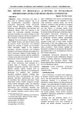
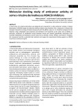
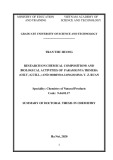
In protein–protein cross-linking experiments (Fig. 5), INs were incubated with the disuccinimidyl suberate (DSS) cross-linker. Reaction products were separated by SDS/ PAGE and revealed by Western blot. As expected, in the absence of IN, we did not observed any product (Fig. 5, lane 1); in the absence of cross-linker, we observed only the monomeric form of IN (lane 2). With wild-type IN and in presence of DSS, we detected products at the expected molecular mass of integrase monomers and dimers (lane 3). K225H (lane 4), V239A (lane 5), Y246W (lane 7), V249A (lane 8) and V257A (lane 9) mutants were observed as monomeric and dimeric forms in similar proportions to that of the wild-type protein (lane 3). On the contrary, L240A (lane 6), V260E (lane 10), K266A (lane 11) and Y246W/ DC25 (lane 12) mutants were not cross-linked as efficiently as wild-type protein by DSS and the dimeric form was less represented for mutants than for the wild-type protein. These results confirm those from size exclusion chromato- graphy analysis for L240A mutants. For V260E and Y246W/DC25, these analyses are in accordance with our these two mutants have a misfolded hypothesis that structure rather than being formed of stable dimers and tetramers. By contrast, the K266A mutant was able to form dimers as shown by size exclusion chromatography. How- ever, DSS is reactive towards amino groups. Therefore, the most likely explanation is that the lysine to alanine mutation
In size exclusion chromatography (Fig. 4), wild-type protein eluted at a position consistent with the molecular size of a dimer. In similar conditions, others [31] also observed dimers of ALSV IN. Mutants V239A, Y246W and K266A (Fig. 4) as well as mutants K225H, V249A and V257A (data not shown) had the same elution profiles as wild-type protein and were complexed in a dimeric form. Conversely, L240A, V260E and Y246W/DC25 exhibited different profiles. The elution peaks were smaller. The L240A profile exhibited a large and a small peak, which could correspond to a mix of dimers and monomers. The V260E profile exhibited two peaks consistent with dimer and higher-molecular forms, while the Y246W/DC25 elution profile exhibited three peaks which correspond to monomers, dimers and higher molecular size products (Fig. 4). However, regarding size of the peaks, we inter- preted these two last mutants as being misfolded rather than structured as stable dimers and tetramers. The same interpretation has been made previously for the counterpart V260E mutation of HIV IN [39,40].
Fig. 5. Protein–protein cross-linking of wild-type integrase and mutants. Proteins were incubated in the presence of disuccinimidyl suberate (DSS). Reaction products were analysed on 10% polyacrylamide gels and revealed by Western blotting using anti-His-tag Ig. The migration of cross-linked species, monomers and dimers, are marked. (A) Con- trols, mutants K225H and V239A. –IN, Without integrase; –DSS, without DSS. (B) Other mutants. Fig. 4. Size exclusion chromatography of wild-type integrase and mutants. Elution profiles of wild-type IN as well as V239A, L240A, Y246W, V260E, K266A and Y246W/DC25 mutants are shown. The molecular size of monomeric form of all INs is 36.7 kDa except for the Y246W/DC25 mutant which is 33.9 kDa. For reference, the elution positions of three globular standard proteins are indicated by dotted vertical lines. Retention times in minutes are indicated on x-axis. Other mutants (K225H, V249A, V257A, which had the same profiles than the wild-type protein) are not shown.
Mechanism of integration of retroviral IN mutants (Eur. J. Biochem. 270) 4435
(cid:2) FEBS 2003
renders the mutant unable to be cross-linked by DSS in this position, although it was associated as a dimer.
Discussion
The C-terminal domain of IN is able to bind DNA [27–29], is required for the 3¢-processing and strand transfer activities of IN [25,26], and is essential for the formation of IN oligomers [30–38]. In this study, we analysed several points mutants in the C-terminal domain of ALSV IN and examined their ability to mediate the concerted DNA integration in an in vitro assay as well as to form dimers. Our analysis focused on mutations at the C-terminal dimer interface. Similar analyses have been performed on residues of the core domain [60].
similar proportion; (b) whereas, in the same reaction, the quantity of product a (and product d) may decrease in an independent manner. Therefore, discrepancies between gels and bacteria may be due to an increase in one-ended integration events (which are not amplified in bacteria) or to a specific decrease in two-ended integration events, or to both. Further, these observations strongly suggest that product b and product a are generated by different mechanisms. We propose that product b should be consid- ered as the result of two non-independent events of one- ended DNA integration with two donors rather than the result of two-ended integration with two donors. Alternat- ively, product b could be a mix of several products: the expected product b and other products generated by non-concerted events of integration whose structures are unknown. Thus, to estimate the two-ended concerted DNA integration efficiency, quantification of product b on a gel would not be as stringent as quantification of product a by cloning and sequencing.
Data obtained for each C-terminal domain mutant studied here and in the previous study [24] are shown in Table 3.
We observed that V260E and Y246W/DC25 mutants were drastically misfolded and completely defective in the concerted DNA integration assay. In the case of the Y246W/DC25 mutant, this misfolding was most probably due to deletion of the last 25 residues of the C domain, as the single Y246W mutant was not so significantly impaired. The loss of strand b5¢ could locally destabilize the C domain by disrupting intramolecular interactions with strand b1¢ (Fig. 2B). Alternatively, this defect might be due to the combination of both the Y246W mutation and the deletion of the 25 terminal residues. Regarding the V260E mutation, it has been shown previously that mutant V260E in HIV-1 IN was mainly misfolded as well [40]. V260 is a highly conserved residue of strand b4¢. The V260E mutation could prevent the formation of this strand as glutamate acts as a strand breaker [61]. Altogether, these data suggest a strong structural role for the terminal part of the C-terminal domain of ALSV integrase in the general folding of the enzyme and, hence, in its activity in the concerted DNA integration assay.
According to the structure proposed by Yang et al. [23], residues V239 and K266 of the proximal monomer and residues L240, Y246 and V257 of the distal monomer are directly involved in the C domain dimer interface (Table 2). Three mutations at this dimer interface (L240A, Y246W and K266A) do not remove contacts between monomers (Table 2). Accordingly, mutants Y246W and K266A were present exclusively in dimeric forms (Figs 4 and 5). How- ever, and to our surprise, the L240A mutant had a reduced ability to form dimers. As mutating this residue reduces intramolecular interactions within the monomers (Table 2), it is possible that the conformation of the whole monomeric molecule is destabilized rendering the monomer unable to associate as dimers. Alternatively, it is noteworthy that this residue is well conserved among INs and that the homo- logue HIV IN residue (L242) has been involved in the formation of tetramers [40]. Therefore, it is possible that this residue is involved in other intermolecular interactions not seen in the dimeric structure proposed for ALSV IN. The two other mutations of residues at the dimer interface
In the concerted DNA integration assay, we could evaluate the ability of IN to catalyse the two-ended concerted DNA integration in two ways: (a) by quantifying the linear product b, since this product is supposed to be generated by a two-ended concerted DNA integration of two DNA donors [9,12–15]; and (b) by quantifying the number of colonies recovered after cloning of integration products into bacteria which allow selective amplification of two-ended circular integration products [a (concerted) and d (nonconcerted)]. The products a and d are subsequently distinguished by sequencing the integration products, and gross deletions of target DNA are assigned to the two-ended nonconcerted DNA integration (class d) [13–15]. In our experiments with wild-type IN, most products (87%) were of type a (without deletion of target DNA) (Table 1). Therefore, cloning of integration reactions into bacteria give a relevant estimation of the product a and, subsequently, of the two-ended concerted DNA integration events. Accord- ing to these assays, if an IN mutant performed two-ended integration less efficiently than wild-type IN, we would expect a concomitant decrease both in the proportion of product b among the total integration products and in the number of recovered colonies from bacteria. Unexpectedly, we found that the quantity of product b did not systemati- cally match the recovered number of colonies (Fig. 3B,C). This is particularly striking for mutant V239A which produced total integration products (RFII plus RFIII) in ratios as high as 170% that of wild-type, and the ratio of product b was found close to that of wild-type proteins (25 and 28% of product b, respectively). By contrast, the proportion of two-ended integration products amplified in bacteria was reduced to less than 30% that of wild-type IN. Such a discrepancy is also evident for the mutant K266A and, to a lesser extent, for mutant V249A. Similar obser- vations have been made previously by others [6,13–15]. For example, the ability of a U5 mutated-donor DNA to undergo concerted DNA integration in vitro was 1.5–2-fold greater than observed with a wild-type donor substrate. This stimulation of integration concerned both the RFII (a + c + d) and RFIII products (b). However, when integrants were introduced into bacteria, the number of colonies recovered was reduced to 25% relative to the wild-type donor. Even more, a reduction to 4% was observed in the presence of HMGI despite an increase in the RF products on gels [13]. Altogether, these independent observations show that: (a) when the quantity of the total integration products increases, the quantity of product b increases in a
4436 K. Moreau et al. (Eur. J. Biochem. 270)
(cid:2) FEBS 2003
(V239A and V257A) abrogate a contact between the two monomers (Table 2) but mutants were not impaired in dimer formation (Figs 4 and 5). For these last two mutants, it is possible that mutating these two residues was not sufficient by itself to impair the formation of the dimer.
C-terminal part of this C-terminal domain in the general folding of the enzyme. They reinforce the role of the IN dimers, as a mutant deficient in dimerization is similarly deficient in concerted DNA integration. Even more, they predict that high-order IN complexes are required to perform two-ended concerted DNA integration. Finally, they con- firm the importance of residues within the C-terminal domain dimer interface in concerted DNA integration. This part of the protein may constitute a new target for the development of antiviral drugs against integrases.
Acknowledgements
All the mutations of residues at the dimer interface caused a decrease in the concerted DNA integration process (Fig. 3; Table 3). For the Y246W and V257A mutants, this decrease in concerted DNA integration is most probably due to a strong defect in 3¢-processing activity (Table 3). It is possible that these mutations induce local conformational changes in the region of the b3¢ strand rendering the molecule less efficient in 3¢-processing.
The L240A mutant is less efficient than wild-type IN in performing all types of integration events (one- and two- ended, concerted and not) as revealed on gels and in bacteria. We speculate that the decrease in integration efficiency is directly related to the decrease in the proportion of dimers that this mutant is able to form.
This work was supported by research grants from the Centre National de la Recherche Scientifique and the Institut National de la Recherche Agronomique. We acknowledge the French Ministry of Research and the Agence Nationale de Recherche contre le SIDA (ANRS) for fellowships (K.M. and S.V.). We thank Dr T.H. Kim (Cambridge) for providing the pET15b-HMGI plasmid. Special thanks to Dr S. Carteau for helpful discussions and to Dr E. Derrigton for helpful discussions and for correcting the English. We also thank Dr P. Gouet for valuable scientific support and Pr J. L. Darlix for critical comment on the manuscript. Thanks are also due to Dr S. Arnaud and M.-F. Grasset (Dr G. Mouchiroud’s laboratory) for their help with size exclusion chromatography. We gratefully acknowledge Pr P. Boulanger for putting his laboratory at our disposal for some parts of this work.
References
1. Brown, P.O.
(1997) Integration. Retroviruses (Coffin, J.M., Hugues, S.H. & Varmus, H.E., eds), pp. 161–204. Cold Spring. Harbor Laboratory Press, Cold spring Harbor, New York. 2. Daniel, R., Katz, R.A. & Skalka, A.M. (1999) A role for DNA- PK in retroviral DNA integration. Science 284, 644–647.
3. Daniel, R., Kao, G., Taganov, K., Greger, J.G., Favorova, O., Merkel, G., Yen, T.J., Katz, R.A. & Skalka, A.M. (2003) Evi- dence that the retroviral DNA integration process triggers an ATR-dependent DNA damage response. Proc. Natl Acad. Sci. USA 100, 4778–4783.
4. Gaken, J.A., Tavassoli, M., Gan, S.U., Vallian, S., Giddings, I., Darling, D.C., Galea-Lauri, J., Thomas, M.G., Abedi, H., Schreiber, V., Menissier-de Murcia, J., Collins, M.K., Shall, S. & Farzaneh, F. (1996) Efficient retroviral infection of mammalian cells is blocked by inhibition of poly (ADP-ribose) polymerase activity. J. Virol. 70, 3992–4000.
5. Siva, A.C. & Bushman, F. (2002) Poly (ADP-ribose) polymerase 1 is not strictly required for infection of murine cells by retroviruses. J. Virol. 76, 11904–11910.
6. Vora, A.C., Chiu, R., McCord, M., Goodarzi, G., Stahl, S.J., Mueser, T.C., Hyde, C.C. & Grandgenett, D.P. (1997) Avian retrovirus U3 and U5 DNA inverted repeats. Role of non- symmetrical nucleotides in promoting full-site integration by purified virion and bacterial recombinant integrases. J. Biol. Chem. 272, 23938–23945.
7. Vora, A.C., McCord, M., Fitzgerald, M.L., Inman, R.B. & Grandgenett, D.P. (1994) Efficient concerted integration of retro- virus-like DNA in vitro by avian myeloblastosis virus integrase. Nucleic Acids Res. 22, 4454–4461.
We observed that mutations V239A, K266A and, to a lesser extent, V249A affected the two-ended integration process specifically, but did not reduce (K266 and V249A), and even enhanced (V239A) the one-ended integration process (Fig. 3, Table 3). However, when mutants catalysed concerted DNA integration, it was performed correctly as revealed by sequencing of integration products. These observations suggest that a few molecules are able to assemble in a complex competent in performing concerted DNA integration and that most molecules performed one- ended non-concerted DNA integrations rather than two- ended concerted DNA integration. Many data have suggested that at least a tetramer is necessary to catalyse concerted DNA integration [23,33,35–38,62]. DNAse pro- tection studies [8] have even suggested that a dimer of ALSV IN is required for the one-ended donor insertion reaction and that for two-ended donor concerted DNA integration a tetramer is assembled on the ALSV U5 end and a higher- order multimer forms on the ALSV U3 end. Therefore, as we observed that K266A, V239A and V249A were less efficient at performing two-ended concerted DNA integra- tion in the absence of alterations in dimer formation of IN (Figs 4 and 5), it is tempting to speculate that mutations of the K266, V239 and V249 residues might prevent the formation of a higher molecular size complex such as a tetramer. It is possible that these mutations either induce local conformational changes that prevent the formation of a tetramer or have distal effects in the protein affecting its global structure. Alternatively, these residues could be directly involved in the formation of the tetramer. Accord- ingly, the HIV L241A IN mutant (L241 of HIV IN is homologous to V239 of ALSV IN), has been shown to be unable to form tetramers [40]. Furthermore, in the tetra- meric model of HIV-1, residue L241 is located at the interface between dimers [59]. Unfortunately, this mutant has not been yet tested in the concerted DNA integration assay. However, it is tempting to speculate that the distal V239, which is accessible and is located away from the dimeric interface (Fig. 2), could be part of the putative tetrameric interface in the ALSV IN.
8. Vora, A. & Grandgenett, D.P. (2001) DNase protection analysis of retrovirus integrase at the viral DNA ends for full-site integration in vitro. J. Virol. 75, 3556–3567.
Our results provide news insights into the multiple structure–function relationships of IN for concerted DNA integration. They show a strong structural role of the most
9. Aiyar, A., Hindmarsh, P., Skalka, A.M. & Leis, J. (1996) Con- certed integration of linear retroviral DNA by the avian sarcoma virus integrase in vitro: dependence on both long terminal repeat termini. J. Virol. 70, 3571–3580.
Mechanism of integration of retroviral IN mutants (Eur. J. Biochem. 270) 4437
(cid:2) FEBS 2003
29. Mumm, S.R. & Grandgenett, D.P. (1991) Defining nucleic acid- binding properties of avian retrovirus integrase by deletion ana- lysis. J. Virol. 65, 1160–1167. 10. Chiu, R. & Grandgenett, D.P. (2000) Avian retrovirus DNA internal attachment site requirements for full- site integration in vitro. J. Virol. 74, 8292–8298.
30. Jones, K.S., Coleman, J., Merkel, G.W., Laue, T.M. & Skalka, A.M. (1992) Retroviral integrase functions as a multimer and can turn over catalytically. J. Biol. Chem. 267, 16037–16040. 11. Chiu, R. & Grandgenett, D.P. (2003) Molecular and genetic determinants of rous sarcoma virus integrase for concerted DNA integration. J. Virol. 77, 6482–6492. 31. Andrake, M.D. & Skalka, A.M.
(1995) Multimerization determinants reside in both the catalytic core and C terminus of avian sarcoma virus integrase. J. Biol. Chem. 270, 29299– 29306. 12. Hindmarsh, P., Ridky, T., Reeves, R., Andrake, M., Skalka, A.M. & Leis, J. (1999) HMG protein family members stimulate human immunodeficiency virus type 1 and avian sarcoma virus concerted DNA integration in vitro. J. Virol. 73, 2994–3003.
32. Coleman, J., Eaton, S., Merkel, G., Skalka, A.M. & Laue, T. (1999) Characterization of the self association of Avian sarcoma virus integrase by analytical ultracentrifugation. J. Biol. Chem. 274, 32842–32846. 13. Hindmarsh, P., Johnson, M., Reeves, R. & Leis, J. (2001) Base- pair substitutions in avian sarcoma virus U5 and U3 long terminal repeat sequences alter the process of DNA integration in vitro. J. Virol. 75, 1132–1141.
33. Bao, K.K., Wang, H., Miller, J.K., Erie, D.A., Skalka, A.M. & Wong, I. (2003) Functional oligomeric state of avian sarcoma virus integrase. J. Biol. Chem. 278, 1323–1327. 14. Brin, E. & Leis, J. (2002a) Changes in the mechanism of DNA integration in vitro induced by base substitutions in the HIV-1 U5 and U3 terminal sequences. J. Biol. Chem. 277, 10938– 10948.
34. Jenkins, T.M., Engelman, A., Ghirlando, R. & Craigie, R. (1996) A soluble active mutant of HIV-1 integrase: involvement of both the core and carboxyl-terminal domains in multimerization. J. Biol. Chem. 271, 7712–7718. 15. Brin, E. & Leis, J. (2002b) HIV)1 integrase interaction with U3 and U5 terminal sequences in vitro defined using substrates with random sequences. J. Biol. Chem. 15, 15.
16. Carteau, S., Gorelick, R.J. & Bushman, F.D. (1999) Coupled integration of human immunodeficiency virus type 1 cDNA ends by purified integrase in vitro: stimulation by the viral nucleocapsid protein. J. Virol. 73, 6670–6679.
35. Lee, S.P., Xiao, J., Knutson, J.R., Lewis, M.S. & Han, M.K. (1997) Zn2+ promotes the self association of human immuno- deficiency virus type-1 integrase in vitro. Biochemistry 36, 173–180. 36. Deprez, E., Tauc, P., Leh, H., Mouscadet, J.F., Auclair, C. & Brochon, J.C. (2000) Oligomeric states of the HIV-1 integrase as measured by time-resolved fluorescence anisotropy. Biochemistry 39, 9275–9284. 17. Gao, K., Gorelick, R.J., Johnson, D.G. & Bushman, F. (2003) Cofactors for human immunodeficiency virus type 1 cDNA integration in vitro. J. Virol. 77, 1598–1603.
37. Vercammen, J., Maertens, G., Gerard, M., De Clercq, E., Deby- ser, Z. & Engelborghs, Y. (2002) DNA-induced polymerization of HIV-1 integrase analyzed with fluorescence fluctuation spectro- scopy. J. Biol. Chem. 277, 38045–38052.
18. Goodarzi, G., Im, G.J., Brackmann, K. & Grandgenett, D. (1995) Concerted integration of retrovirus-like DNA by human immunodeficiency virus type 1 integrase. J. Virol. 69, 6090–6097. 19. Sinha, S., Pursley, M.H. & Grandgenett, D.P. (2002) Efficient concerted integration by recombinant human immunodeficiency virus type 1 integrase without cellular or viral cofactors. J. Virol. 76, 3105–3113.
38. Cherepanov, P., Maertens, G., Proost, P., Devreese, B., Van Beeumen, J., Engelborghs, Y., De Clercq, E. & Debyser, Z. (2003) HIV-1 integrase forms stable tetramers and associates with LEDGF/p75 protein in human cells. J. Biol. Chem. 278, 372–381. full-site integration reactions using recombinant 20. Goodarzi, G., Pursley, M., Felock, P., Witmer, M., Hazuda, D., Brackmann, K. & Grandgenett, D. (1999) Efficiency and fidelity of simian immunodeficiency virus integrase. J. Virol. 73, 8104–8111.
39. Kalpana, G.V., Reicin, A., Cheng, G.S., Sorin, M., Paik, S. & Goff, S.P. (1999) Isolation and characterization of an oligomeri- zation-negative mutant of HIV-1 integrase. Virology 259, 274–285. 21. Yang, F. & Roth, M.J. (2001) Assembly and catalysis of concerted two-end integration events by Moloney murine leukemia virus integrase. J. Virol. 75, 9561–9570.
40. Puras Lutzke, R.A. & Plasterk, R.H. (1998) Structure-based mutational analysis of the C-terminal DNA-binding domain of human immunodeficiency virus type 1 integrase: critical residues for protein oligomerization and DNA binding. J. Virol. 72, 4841–4848. 22. Bujacz, G., Jaskolski, M., Alexandratos, J., Wlodawer, A., Mer- kel, G., Katz, R.A. & Skalka, A.M. (1995) High-resolution structure of the catalytic domain of avian sarcoma virus integrase. J. Mol. Biol. 253, 333–346.
23. Yang, Z.N., Mueser, T.C., Bushman, F.D. & Hyde, C.C. (2000) Crystal structure of an active two-domain derivative of Rous sarcoma virus integrase. J. Mol. Biol. 296, 535–548. 41. Eijkelenboom, A.P., Lutzke, R.A., Boelens, R., Plasterk, R.H., Kaptein, R. & Hard, K. (1995) The DNA-binding domain of HIV-1 integrase has an SH3-like fold. Nat. Struct. Biol. 2, 807–810.
24. Moreau, K., Faure, C., Verdier, G. & Ronfort, C. (2002) Analysis of conserved and non-conserved amino acids critical for ALSV (Avian leukemia and sarcoma viruses) integrase functions in vitro. Arch. Virol. 147, 1761–1778. 42. Fletcher, T.M.R., Soares, M.A., McPhearson, S., Hui, H., Wis- kerchen, M., Muesing, M.A., Shaw, G.M., Leavitt, A.D., Boeke, J.D. & Hahn, B.H. (1997) Complementation of integrase function in HIV-1 virions. EMBO J. 16, 5123–5138.
25. Engelman, A., Bushman, F.D. & Craigie, R. (1993) Identification of discrete functional domains of HIV-1 integrase and their organization within an active multimeric complex. EMBO J. 12, 3269–3275. 43. Holmes-Son, M.L. & Chow, S.A. (2000) Integrase-lexA fusion proteins incorporated into human immunodeficiency virus type 1 that contains a catalytically inactive integrase gene are functional to mediate integration. J. Virol. 74, 11548–11556.
26. van Gent, D.C., Vink, C., Groeneger, A.A. & Plasterk, R.H. (1993) Complementation between HIV integrase proteins mutated in different domains. EMBO J. 12, 3261–3267. 44. Heuer, T.S. & Brown, P.O. (1998) Photo-cross-linking studies for the architecture of an active human integrase-DNA complex. virus type 1 suggest a model immunodeficiency Biochemistry 37, 6667–6678. 27. Esposito, D. & Craigie, R. (1998) Sequence specificity of viral end DNA binding by HIV-1 integrase reveals critical regions for protein–DNA interaction. EMBO J. 17, 5832–5843.
45. Esnouf, R. (1997) Polyalanine reconstruction from Calpha posi- tions using the program CALPHA can aid initial phasing of data by molecular replacement procedures. Acta Crystallogr. D Biol. Crystallogr. 53, 665–672. 28. Lutzke, R.A., Vink, C. & Plasterk, R.H. (1994) Characterization of the minimal DNA-binding domain of the HIV integrase protein. Nucleic Acids Res. 22, 4125–4131.
4438 K. Moreau et al. (Eur. J. Biochem. 270)
(cid:2) FEBS 2003
55. Ju, G., Boone, L. & Skalka, A.M. (1980) Isolation and char- acterization of recombinant DNA clones of avian retroviruses: size heterogeneity and instability of the direct repeat. J. Virol. 33, 1026–1033.
46. Brunger, A.T., Adams, P.D., Clore, G.M., DeLano, W.L., Gros, P., Grosse-Kunstleve, R.W., Jiang, J.S., Kuszewski, J., Nilges, M., Pannu, N.S., Read, R.J., Rice, L.M., Simonson, T. & Warren, G.L. (1998) Crystallography & NMR system: a new software suite for macromolecular structure determination. Acta Crystallogr. D Biol. Crystallogr. 54, 905–921.
47. Roussel, A. & Cambillau, C. (1989) TURBO-FRODO. Silicon Graphics Geometry Partner Directory (Graphics, S., ed.). Silicon Graphics, Moutain View, CA.
56. Moreau, K., Torne-Celer, C., Faure, C., Verdier, G. & Ronfort, C. (2000) In vivo retroviral integration: fidelity to size of the host DNA duplication might be reduced when integration occurs near sequences homologous to LTR ends. Virology. 278, 133–136. 57. Chen, J.C., Krucinski, J., Miercke, L.J., Finer-Moore, J.S., Tang, A.H., Leavitt, A.D. & Stroud, R.M. (2000) Crystal structure of the HIV-1 integrase catalytic core and C-terminal domains: a model for viral DNA binding. Proc. Natl Acad. Sci. USA 97, 8233–8238. 48. Altschul, S.F., Madden, T.L., Schaffer, A.A., Zhang, J., Zhang, Z., Miller, W. & Lipman, D.J. (1997) Gapped BLAST and PSI- BLAST: a new generation of protein database search programs. Nucl Acids Res. 25, 3389–3402.
49. Bairoch, A. & Apweiler, R. (2000) The SWISS-PROT protein sequence database and its (Suppl.)TrEMBL in 2000. Nucleic Acids Res. 28, 45–48.
58. Chen, Z., Yan, Y., Munshi, S., Li, Y., Zugay-Murphy, J., Xu, B., Witmer, M., Felock, P., Wolfe, A., Sardana, V., Emini, E.A., Hazuda, D. & Kuo, L.C. (2000) X-ray structure of simian immunodeficiency virus integrase containing the core and C-terminal domain (residues 50–293) -an initial glance of the viral DNA binding platform. J. Mol. Biol. 296, 521–533.
50. Thompson, J.D., Higgins, D.G. & Gibson, T.J. (1994) CLUSTAL W: improving the sensitivity of progressive multiple sequence alignment through sequence weighting, position-specific gap penalties and weight matrix choice. Nucleic Acids Res. 22, 4673–4680.
51. Thanos, D. & Maniatis, T. (1992) The high mobility group protein HMG I (Y) is required for NF-kappa B-dependent virus induction of the human IFN-beta gene. Cell 71, 777–789.
59. Wang, J.Y., Ling, H., Yang, W. & Craigie, R. (2001) Structure of implications for a two-domain fragment of HIV-1 integrase: domain organization in the intact protein. EMBO J. 20, 7333–7343. 60. Moreau, K., Faure, C., Violot, S., Gouet, P., Verdier, G. & Ronfort, C. (2003) Mutational analyses of the core domain of Avian Leukemia and Sarcoma Viruses integrase: critical residues for concerted integration and multimerization. Virology, in press. 61. Chou, P.Y. & Fasman, G.D. (1979) Prediction of beta-turns. 52. Farnet, C.M. & Bushman, F.D. (1997) HIV-1 cDNA integration: requirement of HMG I (Y) protein for function of preintegration complexes in vitro. Cell 88, 483–492. Biophys. J. 26, 367–373.
53. Li, L., Yoder, K., Hansen, M.S., Olvera, J., Miller, M.D. & Bushman, F.D. (2000) Retroviral cDNA integration: stimulation by HMG I family proteins. J. Virol. 74, 10965–10974.
62. Deprez, E., Tauc, P., Leh, H., Mouscadet, J.F., Auclair, C., Hawkins, M.E. & Brochon, J.C. (2001) DNA binding induces dissociation of the multimeric form of HIV-1 integrase: a time- resolved fluorescence anisotropy study. Proc. Natl Acad. Sci. USA 98, 10090–10095. 54. Hughes, S.H., Mutschler, A., Bishop, J.M. & Varmus, H.E. (1981) A Rous sarcoma virus provirus is flanked by short direct repeats of a cellular DNA sequence present in only one copy prior to integration. Proc. Natl Acad. Sci. USA 78, 4299–4303.