Eur. J. Biochem. 269, 184–192 (2002) (cid:211) FEBS 2002
Interaction of plasminogen activator inhibitor type-1 (PAI-1) with vitronectin Characterization of different PAI-1 mutants
Nuria Arroyo De Prada1,*, Florian Schroeck1,*, Eva-Kathrin Sinner2, Bernd Muehlenweg1,3, Jens Twellmeyer1, Stefan Sperl3, Olaf G. Wilhelm3, Manfred Schmitt1 and Viktor Magdolen1
1Klinische Forschergruppe der Frauenklinik der Technischen Universita¨t Mu¨nchen, Klinikum rechts der Isar, Germany; 2Max-Planck-Institut fu¨r Biochemie, Martinsried, Germany; 3Wilex AG, Mu¨nchen, Germany
plate-based binding assays, surface plasmon resonance and thrombin inhibition experiments, all of the newly generated mutants which retained inhibitory activity against uPA still bound to Vn. Mutant A114–118, in which all amino-acids at positions 114–118 of PAI-1 were exchanged for alanine, displayed a reduced a(cid:129)nity to Vn as compared to wild- type PAI-1. Mutants lacking inhibitory activity towards uPA did not bind to Vn. Q123K, which inhibits uPA but does not bind to Vn, served as a control. In contrast to other active PAI-1 mutants, the inhibitory properties of A114–118 towards thrombin as well as uPA were signifi- cantly reduced in the presence of Hep. Our results dem- onstrate that the wild-type sequence of the region around hE in PAI-1 is not a prerequisite for binding to Vn.
plasminogen
activator
inhibitor
type-1; Keywords: vitronectin; heparin; mutational analysis; surface plasmon resonance.
The serpin plasminogen activator inhibitor type 1 (PAI-1) plays an important role in physiological processes such as thrombolysis and fibrinolysis, as well as pathophysiological processes such as thrombosis, tumor invasion and metas- tasis. In addition to inhibiting serine proteases, mainly tissue-type (tPA) and urokinase-type (uPA) plasminogen activators, PAI-1 interacts with di(cid:128)erent components of the extracellular matrix, i.e. fibrin, heparin (Hep) and vitronectin (Vn). PAI-1 binding to Vn facilitates migration and invasion of tumor cells. The most important deter- minants of the Vn-binding site of PAI-1 appear to reside between amino acids 110–147, which includes a helix E (hE, amino acids 109–118). Ten di(cid:128)erent PAI-1 variants (mostly harboring modifications in hE) as well as wild-type PAI-1, the previously described PAI-1 mutant Q123K, and another serpin, PAI-2, were recombinantly produced in Escherichia coli containing a His6 tag and purified by shown in microtiter a(cid:129)nity
chromatography. As
The urokinase-type plasminogen activation system plays an important invasion, and role in tumor growth, metastasis. The serine protease urokinase-type plasmino- gen activator (uPA) activates plasminogen, the zymogen
of plasmin, thus generating a protease with broad substrate specificity and leading to degradation of extra- cellular matrix (ECM) proteins [1–3]. The activity of uPA is focussed to the cell surface by interaction with its specific receptor uPAR (CD87). Tissue-type plasminogen activator (tPA), the second type of human plasminogen activator, in contrast to uPA does not bind to a high affinity receptor on tumor cell surfaces and therefore does not promote tumor cell-associated pericellular pro- teolysis.
the
center
surface-exposed reactive
There are two main inhibitors of uPA and tPA, the serine protease inhibitors (serpin) plasminogen activator [4]. For inhibitor type-1 (PAI-1) and type-2 (PAI-2) inhibition, loop (RCL) of PAI-1 or PAI-2 interacts with the reactive site of the target protease. Initially, the P1–P1¢ bond of the inhibitor is cleaved and an intermediate enzyme–inhibitor complex is formed. This is followed by the insertion of part of the RCL as additional b strand 4A, which leads to the translocation of the protease across the plane of b sheet A of PAI-1 and formation of an SDS-stable enzyme– inhibitor complex [5]. This complex dissociates very slowly and is cleared from circulation before disassembly can occur. In vitro, the inhibitor can be released from the protease in a substrate-like manner, generating the so-called RCL-cleaved form of the inhibitor [6].
Correspondence to V. Magdolen, Klinische Forschergruppe der Frauenklinik der Technischen Universita¨ t Mu¨ nchen, Klinikum rechts der Isar, Ismaninger Str. 22, D-81675 Mu¨ nchen, Germany. Fax: + 49 89 4140 7410, Tel.: + 49 89 4140 2493, E-mail: viktor@magdolen.de Abbreviations: ECM, extracellular matrix; Hep, heparin; hE, helix E; HMW-uPA, high molecular weight urokinase-type plasminogen activator; PAI-1, plasminogen activator inhibitor type-1; PAI-2, plasminogen activator inhibitor type-2; RCL, reactive center loop; RU, resonance units; SPR, surface plasmon resonance; tPA, tissue- type plasminogen activator; uPA, urokinase-type plasminogen acti- vator; uPAR, uPA receptor; Vn, vitronectin; IPTG, isopropyl thio-b- D-galactoside. *Note: these authors contributed equally to the work. Note: web pages are available at http://www.frauenklinik-tu-muen- chen.de, http://www.biochem.mpg.de/oesterhelt/ and http://www.wilex.com (Received 04 July 2001, revised 22 October 2001, accepted 29 October 2001)
(cid:211) FEBS 2002
Interaction of PAI-1 variants with vitronectin (Eur. J. Biochem. 269) 185
Expression and purification of wild-type PAI-1, wild-type PAI-2, and PAI-1 variants
Active PAI-1 is metastable and spontaneously converts to a latent form by inserting a major part of its RCL into the central b sheet A [7]. Latent PAI-1, as well as PAI-1 in complex with uPA or tPA, do not bind to ECM compo- nents. Active PAI-1, however, interacts with fibrin, heparin (Hep), and vitronectin (Vn) [1,8,9]. Binding to Vn doubles the physiological half-life of active PAI-1 in solution. Moreover, by binding to Vn or Hep, the substrate specificity of PAI-1 is altered, because interaction with Vn or Hep enables PAI-1 to inhibit another serine protease, thrombin [10].
High levels of PAI-1 in tumor tissue indicate short recurrence-free and overall survival of tumor patients afflicted with a broad variety of cancers, e.g. mammary, ovarian, cervical, colorectal, bladder, renal and lung carcinomas [2,3]. In line with this, it was demonstrated that PAI-1 affects tumor cell adhesion and/or tumor angiogenesis and, as a result of this, may support tumor invasion [11–13].
Expression of recombinant proteins was induced by adding isopropyl thio-b-D-galactoside (IPTG; final concentration: 2 mM) to an XL1 blue (variant) PAI-1 or PAI-2 culture, pregrown in Luria–Bertani medium supplemented with 100 lgÆmL)1 ampicillin (D600 0.6–0.7). Protein expression occurred at 37 (cid:176)C overnight on an orbital shaker at 200 r.p.m. The bacterial culture was harvested by centrif- ugation at 5000 g at 4 (cid:176)C for 10 min. Then, the bacterial pellet was frozen for 20 min at )80 (cid:176)C and, subsequently suspended in 20 mM Na–acetate, 1 M NaCl, 0.1% Tween-80 (v/v), pH 7.4 supplemented with the protease inhibitor mix (cid:212)Complete EDTA-free(cid:213) (Roche, Mannheim, Germany). In the case of wild-type PAI-2, a slightly different buffer [20 mM Na2HPO4, 1 M NaCl, 0.1% Tween-80 (v/v), pH 7.4 supplemented with Complete EDTA-free] was used. Bacte- ria were disrupted mechanically by addition of glass beads (Sigma, Taufkirchen, Germany) to the bacterial cell sus- pension and 10 subsequent cycles of vortexing and incuba- tion on ice for 1 min each. The lysate was centrifuged for 15 min (12 000 g, 4 (cid:176)C), the supernatant recovered and subjected to Ni2+-nitrilotriacetic acid agarose affinity column purification.
Ni2+-nitrilotriacetic acid affinity chromatography
As the PAI-1-binding site on Vn partially overlaps with the binding sites of cellular adhesion proteins, e.g. uPAR and some integrins, addition of PAI-1 to Vn-bound tumor cells leads to detachment of these cells [11,14]. This effect can be reversed by uPA, as Vn-bound PAI-1 dissociates from Vn upon interaction with uPA [9]. Interestingly, as demon- strated by Bajou et al. [13], the influence of PAI-1 on tumor vascularization is due to the inhibition of proteases and not due to its interaction with Vn. Thus, the balance of several tumor-associated factors seems to control the modulatory effects of PAI-1 on cell adhesion, migration, and angiogen- esis and may play a crucial role in tumor invasion and metastasis [13,15].
Various research groups have attributed the Vn-binding site on PAI-1 to different epitopes. However, essential amino acids appear to be located in a region within amino acids 110–147 [16–19], most of them being localized in the a helix E (hE). The major aim of the present study was to further analyze the structure-function relationship of PAI-1 mainly regarding its interactions with Vn, but also with Hep. Therefore, a number of PAI-1 variants preferentially containing modifications in hE of PAI-1 were generated and characterized biochemically.
M A T E R I A L S A N D M E T H O D S
Generation of PAI-1 variants
The Ni2+-nitrilotriacetic acid affinity column was prepared (Qiagen, Hilden, as described by the manufacturer Germany). Initially, the affinity column was equilibrated with 20 mM Na-acetate, 1 M NaCl, 0.1% Tween-80 (v/v), pH 7.4. Then, the cleared bacterial lysate was applied to the column and, subsequently, the column washed with equili- bration buffer followed by 20 mM Na-acetate, 1 M NaCl, 0.1% Tween-80 (v/v), 20 mM imidazole, pH 5.6 until the absorption of the effluent had returned to baseline (D280 < 0.001). Finally, the adsorbed recombinant proteins were eluted with 20 mM Na-acetate, 1 M NaCl, 0.1% Tween-80 (v/v), 200 mM imidazole, pH 5.6. The eluate containing the recombinant proteins was dialyzed in equilibration buffer and purified by a second Ni2+-nitrilo- triacetic acid affinity chromatography as described above. The supernatant of lysates of wild-type PAI-2 expressing bacterial cells (in a buffer containing 20 mM Na2HPO4, 1 M NaCl, 0.1% Tween-80, pH 7.4) was also applied to a Ni2+- nitrilotriacetic acid affinity column, and washed with the same buffer (at pH 6.5) supplemented with 20 mM imidaz- ole. For elution, the same buffer (at pH 6.0) containing 200 mM imidazole was used.
Denaturation and refolding of the recombinant proteins
The coding regions of wild-type PAI-1 (amino acids 1–379 according to the numbering proposed by Ny et al. [20]) and wild-type PAI-2 (amino acids 2–415; PIR protein sequence database: A32853) have been cloned in frame with an N-terminal His6 tag into the E. coli expression vector pQE-30 (Qiagen, Hilden, Germany) as described previously [21]. Modifications in the wild-type cDNA sequence of PAI-1 were generated by reverse long-range PCR ((cid:212)Expand High Fidelity PCR System Kit(cid:213); Roche, Mannheim, Germany) applying mutated primers (Metabion, Martinsried, Germa- ny). PCR products were phosphorylated (T4 polynucleotide kinase; Roche, Mannheim, Germany), re-ligated (T4 DNA ligase; Roche, Mannheim, Germany) and transformed into the E. coli strain XL1 blue (Stratagene, Heidelberg, Ger- many). The mutated sequences were verified by DNA sequencing performed by TopLab, Martinsried, Germany.
The purified recombinant proteins, with exception of wild- type PAI-2, were denatured for 4 h in 4 M guanidinium/HCl at room temperature under light protection. Refolding of the proteins was achieved by dialysis (2 h, 4 (cid:176)C) in 20 mM Na-acetate, 1 M NaCl, 0.01% Tween-80 (v/v), pH 5.6 followed by a second dialysis step (overnight, 4 (cid:176)C). The proteins were subsequently concentrated in Centricon centrifugal filter devices (Millipore, Eschborn, Germany). Wild-type PAI-2 was dialyzed against NaCl/Pi (pH 7.4) in imidazole, concentrated in order to remove residual
186 N. Arroyo De Prada et al. (Eur. J. Biochem. 269)
(cid:211) FEBS 2002
Centricon centrifugal filter devices, and then stored at )80 (cid:176)C until use.
Characterization of the recombinant proteins
Germany) or 1 UÆmL)1 Hep (Liquemin(cid:210) N 25 000, Hoffmann-La Roche AG, Grenzach-Wyhlen, Germany) were incubated with 0.1 U of thrombin (from human plasma; Sigma, Taufkirchen, Germany) in a total volume of 130 lL of Tris/NaCl/Tween buffer [20 mM Tris/HCl, 100 mM NaCl, 0.1% Tween-80 (v/v), pH 8.0] at 37 (cid:176)C for 1 h [10]. Then, 10 lL of chromogenic substrate (Chromo- zym(cid:210) TH, Roche, Mannheim, Germany, concentration: 2 mM) were added and the thrombin activity measured monitoring the change of D at 405 nm.
Complex formation of the recombinant proteins PAI-1 with thrombin
Fifty units of PAI-1 (variant) in the presence or absence of 600 nM Vn or 1 UÆmL)1 Hep were incubated with 0.5 U of thrombin in a total volume of 30 lL Tris/NaCl/Tween buffer (1 h, 37 (cid:176)C) and then subjected to nonreducing SDS/ PAGE followed by Western blotting employing chicken pAb against PAI-1 and a POX-labeled goat anti-(chicken IgY) Ig.
The protein concentration was determined according to Bradford using the Bio-Rad Protein Assay Dye Reagent Concentrate (Bio-Rad, Krefeld, Germany). PAI-1 antigen was determined using the Imubind Tissue PAI-1 ELISA Kit (American Diagnostica, Pfungstadt, Germany). The iden- tity of the purified proteins was demonstrated by Western blotting employing a polyclonal antibody (pAb) from chicken directed against human PAI-1 (a kind gift of N. Grebenschikov, Institute of Chemical Endocrinology, Uni- versity of Nijmegen, the Netherlands), a monoclonal mouse antibody (mAb) to human PAI-2 (#110 from American Diagnostica, Pfungstadt, Germany), and the ECL Western Blotting Detection Reagent (Amersham Pharmacia, Frei- burg, Germany) for detection. N-terminal amino-acid sequence analysis performed by TopLab (Martinsried, Germany) was used to confirm the identity of the purified recombinant wild-type PAI-1 and wild-type PAI-2.
Binding of recombinant PAI-1 to Vn-coated microtiter plates
Amidolytic assay for determination of the inhibitory activity of the recombinant proteins against HMW-uPA
The assay was performed in 96-well microtiter plates (Greiner, Frickenhausen, Germany). Purified recombinant proteins were diluted in a buffer containing 100 mM Tris/ HCl, 0.05% Tween-20 (v/v), pH 7.5, and 100 lgÆmL)1 BSA (ICN, Aurora, Ohio, USA), incubated with 10 U high molecular weight (HMW-)uPA (Rheotromb(cid:210) 500 000, Curasan Pharma GmbH, Kleinostheim, Germany) for 15 min at room temperature and then 10 lL of chromo- substrate Bz-b-Ala-Gly-Arg-pNA.AcOH (Pefa- genic chrome(cid:210) uPA, Pentapharm Ltd, Basel, Switzerland, concentration 2 mM) were added (30 min, 37 (cid:176)C). The absorption was measured at 405 nm. One unit PAI activity was defined as the amount, which completely inhibited one unit of HMW-uPA activity.
Complex formation of the recombinant proteins with HMW-uPA
Vn or collagen type IV (Sigma, Taufkirchen, Germany) were diluted to a concentration of 10 lgÆmL)1 in a buffer containing 100 mM Na2CO3, pH 9.6. For coating, 50 lL of the Vn or collagen type IV dilutions were poured into wells of a NuncMaxiSorp microtiter plate (Nunc GmbH & Co. KG, Wiesbaden, Germany) and incubated overnight at 4 (cid:176)C. After three washes with NaCl/Pi/Tween [NaCl/Pi containing 0.05% Tween-20 (v/v)], the wells were blocked by addition of 200 lL per well of NaCl/Pi supplemented with 2% BSA (w/v) and incubation at room temperature for 2 h. The wells were washed three times with NaCl/Pi/ Tween. Afterwards 100 lL per well of (mutant) PAI-1 in the desired concentration were added at room temperature for 1 h. Following three additional washing steps, 200 lL per well horseradish peroxidase labeled Ni2+-nitrilotriacetic acid (Qiagen, Hilden, Germany) at a dilution of 1 : 1000 in NaCl/Pi/Tween containing 0.2% BSA (w/v) were added (1 h, room temperature). After another four times of washing, binding of PAI-1 to the solid phase was visualized by addition of 100 lL per well of a TMB substrate mix (KPL, Gaithersburg, Maryland, USA). The reaction was stopped after color development with 50 lL per well of 0.5 M H2SO4. Optical density was measured at 450 nm.
Surface plasmon resonance analysis of (mutant) PAI-1 binding to Vn
For complex formation, 100 U ((cid:25) 0.7 lg) of HMW-uPA were incubated with the recombinant proteins at room temperature for 10 min. The complexes were visualized by SDS/PAGE under nonreducing conditions and subsequent silver staining or Western blotting with the chicken pAb against PAI-1, mouse mAb #110 against PAI-2 (as mentioned above), and polyclonal chicken anti-uPA Ig (kindly provided by N. Grebenschikov, Institute of Chem- ical Endocrinology, Nijmegen, the Netherlands). POX- labeled chicken anti-(mouse IgG) Ig and POX-labeled goat anti-(chicken IgY) Ig were purchased from Dianova, Hamburg, Germany.
Amidolytic assay for determination of the inhibitory activity of the recombinant proteins against thrombin
The assay was performed in 96-well microtiter plates. Fifty units of active recombinant PAI-1 ((cid:25) 35 nM) in the presence or absence of 140 nM Vn (Promega GmbH, Mannheim,
Surface plasmon resonance (SPR) studies were conducted with a BIACORE 2000 (Biacore AB, Uppsala, Sweden). Approximately 2000 resonance units (RU) of collagen type IV (10 lgÆmL)1 in 10 mM Na-acetate, pH 4.0) (lane 1) and Vn (10 lgÆmL)1 in 10 mM Na-formiate, pH 4.0 [22]) (lanes 2–4) were immobilized to a CM5 sensor chip (research grade, Biacore AB, Uppsala, Sweden) using the amino coupling kit according to the manufacturer’s recommenda- tion. All experiments were performed in HBS-EP [10 mM Hepes, 150 mM NaCl, 3 mM EDTA, 0.005% Tween-20 (v/v), pH 7.4] at a flow rate of 20 lLÆmin)1. HMW-uPA was
(cid:211) FEBS 2002
Interaction of PAI-1 variants with vitronectin (Eur. J. Biochem. 269) 187
expression in the bacterial strain XL1 blue yielded 5–10% of the total E. coli protein.
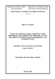
The recombinant proteins contained a His6 tag at their N-terminus, allowing purification by Ni2+-nitrilotriacetic acid affinity chromatography. Although one cycle of affinity chromatography substantially enriched the recombinant proteins from other bacterial proteins, a purification grade of > 95% was only achieved after a second chromato- graphic cycle as demonstrated by SDS/PAGE (Fig. 2).
Inhibitory activity of the recombinant proteins against HMW-uPA
used in a concentration of 400 UÆmL)1. Regeneration of the surface was achieved by injection of 10 mM HCl for 8 min. In order to check for reproducibility during the measure- ment, at first 80 lL of a 200 UÆmL)1 dilution of wild-type PAI-1 were injected for two subsequent experiments, followed by two subsequent measurements of 80 lL of a 200 UÆmL)1 dilution of a PAI-1 mutant. Then, wild-type PAI-1 was measured a third time in duplicate, followed by a measurement in duplicate of the next mutant and so on. Thus, for each PAI-1 variant at least two binding profiles were recorded. The kinetics obtained in the collagen type IV-coated flow cell were subtracted from the kinetics derived from the Vn-coated flow cell in order to obtain binding profiles without bulk effects.
R E S U L T S
Expression and purification of recombinant PAI-1, PAI-2, and PAI-1 variants
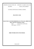
The coding sequences for wild-type PAI-1 and wild-type PAI-2, respectively, have previously been cloned by us in expression vector pQE-30 [21]. By reverse PCR, a series of PAI-1 variants was generated using pQE-30-wild-type PAI-1 as the template. Due to the fact that serpins have a very compact tertiary structure (Fig. 1A), large modifications of the molecule may result in misfolded, and therefore inactive, proteins. Because of this, we designed various strategies such as introduction of point mutations, substitution of few amino acids by alanine and glycine, substitution of selected epitopes by the homologue PAI-2 sequence, and short deletions (Fig. 1B). The generated PAI-1 variants are summarized in Table 1. IPTG-induced recombinant protein
PAI-1 is unique among serpins by its metastability that leads to a short half-life of (cid:25) 2 h under physiological conditions. it was not surprising that after purification Therefore, (performed at room temperature) most of the recombinant PAI-1 wild-type protein and variants were present in the inhibitory inactive latent conformation. However, denatur- ation and subsequent refolding by dialysis leads to the reactivation of latent PAI-1 [23]. An up to 87% inhibitory activity of wild-type PAI-1 and PAI-1 variants against HMW-uPA (100 000 UÆmg)1 defined as the maximum [24]) was reached after denaturation with 4 M guanidinium/HCl followed by dialysis in a buffer of high ionic strength at inhibitory activities of the protein pH 5.6. Moreover, preparations remained stable for more than one year in this buffer when stored at )80 (cid:176)C. The inhibitory activity of the generated PAI-1 mutants is summarized in Table 1. All inhibitory active mutants were metastable (half-life £ 2 h at 37 (cid:176)C) and the half-life was roughly doubled in the presence of Vn. Furthermore, inhibitory active variants formed SDS-stable complexes with HMW-uPA; the inactive
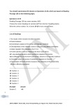
Fig. 1. Three-dimensional structure of active PAI-1. (A) Important structural elements of active PAI-1 (PDB 1B3K). The central b sheet A consisting of strands 1A, 2A, 3A, 5A, and 6A is indicated in yellow, helix D in green, and helix E (hE) in cyan blue. The P1-residue of PAI-1 (R346) is also indicated. (B) Location of amino acid alterations in some of the generated PAI-1 mutants. P73A, single amino acid exchange of P73 to alanine; A114–118, exchange of the sequence 114FRLFR118 to five alanines; D109–112, deletion of the four amino acids 109MPHF112; Q123K, single amino acid exchange of Q123 to lysine.
188 N. Arroyo De Prada et al. (Eur. J. Biochem. 269)
(cid:211) FEBS 2002
Table 1. PAI-1 variants and binding to Vn. PAI-1 mutants with corresponding specific inhibitory activity towards uPA and their ability to interact with Vn measured by use of Vn-coated microtiter plates (MP) and surface plasmon resonance (SPR). + Indicates binding to Vn; – indicates no binding to Vn. NT, not tested.
Binding to Vn
MP SPR PAI-1 variant Modificationa Inhibitory activity towards uPA (UÆmg)1)
Wild-type PAI-1 Wild-type PAI-2 Mutant 1 (D109-123) Mutant 2 (M2) Mutant 3 (M3) 87 000 90 000 Inactive Inactive Inactive + – – – – + NT NT NT –
Inactive 10 000 57 000 20 800 7 700 – + + + + – NT NT + + Mutant 4 (M4) Mutant 5 (D109-112) Mutant 6 (A114-115) Mutant 7 (A114–118) Mutant 8 (M8) Mutant 9 (M9)
a Numbering of PAI-1 according to Ny et al. [20]; b 141YIRLCQKYYSSEPQA155, and c 318YELRSILRSMG328, PAI-2 according to PIR protein sequence database: A32853.
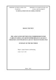
mutants did not (Fig. 3). PAI-2 was stable (no loss of inhibitory activity after incubation for 24 h at 37 (cid:176)C) and exerted an inhibitory activity against HMW-uPA of 90% (equivalent to 90 000 UÆmg)1) after the two-step affinity chromatography.
Interaction of (mutant) PAI-1 with Vn
All inhibitory active mutants but mutant Q123K [16] did interact with Vn as verified by measuring (mutant) PAI-1 binding to Vn-coated microtiter plates and by surface plasmon resonance analysis (Table 1). This binding was demonstrated to be highly specific, as binding of (mutant) PAI-1 was completely abolished by preincubation of recombinant PAI-1 with soluble Vn (10 lgÆmL)1) prior to adding it to the wells or before injection in a reproducible manner (data not shown). Furthermore, latent PAI-1 (data not shown) as well as heat-denatured PAI-1 (Fig. 4) did not bind to the immobilized Vn.
The
observed KD value
Mutant 10 (P73A) Mutant 11 (Q123K) D F109-Q123 F109-Q123 vs. AAGAGAA F109-Q123 vs. homologue PAI-2 sequenceb and E128G F109-Q123 vs. AAAA D F109-H112 F114A and R115A F114-R118 vs. AAAAA F114-R118 vs. AAAAA and D68G V284-G294 vs. homologue PAI-2 sequencec P73A Q123K Inactive 56 250 47 800 – + – – + –
for wild-type PAI-1 (KD (cid:136) 0.18 nM) as well as for P73A (KD (cid:136) 0.33 nM) is similar to that previously reported for the interaction of human PAI-1 recombinantly expressed in Chinese hamster ovary cells with vitronectin (KD (cid:136) 0.1 nM), also applying
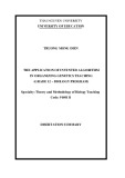
Fig. 2. Purification of recombinant wild-type PAI-1. Human recombi- nant wild-type PAI-1 equipped with an N-terminal His6 tag (wild-type PAI-1) was purified from an E. coli cell lysate by Ni2+-nitrilotriacetic acid a(cid:129)nity chromatography and analyzed by SDS/PAGE. M, marker; lane 1, E. coli lysate; lane 2, e(cid:130)uent of the first Ni2+-nitrilo- triacetic acid a(cid:129)nity chromatography cycle; lane 3, eluate of the first Ni2+-nitrilotriacetic acid a(cid:129)nity chromatography cycle; lane 4, eluate of the second Ni2+-nitrilotriacetic acid a(cid:129)nity chromatography cycle.
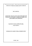
Fig. 3. Formation of SDS stable complexes. Wild-type PAI-1 or variants thereof in the presence (+ uPA) or absence (– uPA) of 100 U ((cid:25) 0.7 lg) HMW-uPA were incubated for 10 min at room temperature and then subjected to nonreducing SDS/PAGE. Subsequently, the gels were silver- stained. 200 U of PAI-1, 110 U of Q123K, 50 U of P73A and of A114–118 were applied; alternatively 1.4 lg of (inactive) M4 and 10 U of PAI-1 were used.
(cid:211) FEBS 2002
Interaction of PAI-1 variants with vitronectin (Eur. J. Biochem. 269) 189
M
M
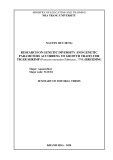
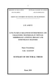
SPR analysis [22]. Mutant A114–118 displayed a slower association to (on-rate: 5.35 · 105 )1Æs)1) and a faster dissociation from Vn (off-rate: 1.0 · 10)3 s)1) compared to wild-type PAI-1 (on-rate: 1.0 · 106 )1Æs)1; off-rate: 1.9 · 10)4 s)1). Only a low amount of mutant Q123K associated to Vn and dissociated immediately after washing with buffer, indicating an unspecific binding to Vn (Fig. 4). Moreover, mutant Q123K did not show any change in binding to solid phase Vn in the presence or absence of soluble Vn (data not shown). All Vn-binding mutants dissociated immediately from Vn after uPA injection (Fig. 4). Thus, these results clearly demonstrate that (mutant) PAI-1, but not Q123K, specifically binds to Vn immobilized to the dextran matrix of the CM5 chip, and then still was able to form complexes with uPA.
Inhibition of thrombin by PAI-1 and binding to Hep
Fig. 4. Surface plasmon resonance: binding of (mutant) PAI-1 to VN. Approximately 2000 RU of collagen type IV or Vn were immobilized to a CM5 sensor chip inserted in the BIAcore 2000 system. Then, wild-type PAI-1 (200 UÆmL)1) was injected and allowed to bind to Vn, which was followed by a washing step with bu(cid:128)er. Finally, HMW-uPA (400 UÆmL)1) was injected. Two subsequent measurements of the binding kinetics of wild-type PAI-1 were followed by two independent measurements of a PAI-1 variant (200 UÆmL)1). After that, wild-type PAI-1 was measured a third time, followed by a measurement in duplicate of the next mutant and so on. Thus, for each PAI-1 variant at least two independent binding profiles were obtained. All experiments were performed at a flow rate of 20 lLÆmin)1; regeneration of the surface was achieved by treatment with 10 mM HCl for 8 min. The kinetics obtained in the collagen type IV-coated flow cell were subtracted from the kinetics derived from the Vn-coated flow cell in order to obtain binding profiles without bulk e(cid:128)ects. Heat denatured controls were measured to compare the binding signal with the unspecific binding to the identical surface conditions.
Binding to Vn provides wild-type PAI-1 with thrombin inhibitory properties [10]. Therefore, we tested whether the PAI-1 mutants generated were also able to inhibit thrombin in the presence of Vn. In line with our data obtained by SPR, the Vn-interacting mutants A114–118 and P73A, but not Q123K, inhibited thrombin in the presence of Vn to about 40%. Vn alone did not reduce thrombin activity significantly (Fig. 5).
seen in Western blots visualizing the formation of SDS stable complexes between thrombin and PAI-1 with or without Vn or Hep. Detection of a higher residual thrombin activity correlated with a lower amount of SDS stable complexes (data not shown).
The reduced capacity of A114–118 to inhibit thrombin with Hep as a cofactor is not related to an altered affinity to
We also tested the effects of Hep on the inhibitory activity of PAI-1 towards thrombin. All mutants tested but A114– 118 displayed similar properties as wild-type PAI-1 (> 90% inhibition; Fig. 5). A114–118 inhibited thrombin in the presence of Hep clearly less effectively (about 40%; Fig. 5). These results determined by amidolytic assays measuring (residual) thrombin activity were supported by the results
Fig. 5. Inhibition of thrombin. Fifty units of recombinant PAI-1 ((cid:25) 35 nM) in the presence or absence of 140 nM Vn or one UÆmL)1 Hep were incubated with 0.1 U of thrombin in a total volume of 130 lL of Tris/NaCl/Tween bu(cid:128)er at 37 (cid:176)C for 1 h. Then, 10 lL of chromogenic substrate were added and the thrombin activity measured monitoring the change of optical density at 405 nm. The activity of thrombin in the absence of inhibitor was set to 100%, the other activities were calculated accordingly. Data shown are from three independent experiments, each measured in duplicate ((cid:139) SD). As a control, the e(cid:128)ect of 140 nM Vn or 1 UÆmL)1 Hep without inhibitor was measured. Black bars, bu(cid:128)er, only; hatched bars, plus Vn (140 nM); grey bars, plus Hep (1 UÆmL)1).
190 N. Arroyo De Prada et al. (Eur. J. Biochem. 269)
(cid:211) FEBS 2002
(D109-123, M2, M3, M4, M9), which contained more than seven modified amino-acid positions did not display any inhibitory activity towards uPA, indicating that larger modifications at these positions are not tolerated and lead to a loss of activity most likely due to misfolding of the compact PAI-1 structure.
Hep, as in SPR we observed that A114–118 displayed similar binding to biotinylated Hep immobilized on a SA-5 sensor chip as wild-type PAI-1 (data not shown). Interest- ingly, Hep did not only provide A114–118 with thrombin inhibitory properties less efficiently than the other tested mutants, it also reduced the inhibitory property of A114– 118 towards uPA in a dose dependent manner. Hep had only marginal effects on the inhibition of uPA by other inhibitory active PAI-1 proteins (wild-type PAI-1, P73A, Q123K, data not shown).
D I S C U S S I O N
The active PAI-1 variants behaved similarly to wild-type PAI-1 with respect to their metastability and the stabiliza- tion of the active form by Vn. In contrast to wild-type PAI-1 and its active variants, recombinant PAI-2 did not show any significant loss of activity after purification under native conditions, underlining the uniqueness of the metastable PAI-1 among serpins.
Purification and inhibitory activity of recombinant PAI-1, PAI-2, and PAI-1 variants
Interaction of (mutant) PAI-1 with Vn
Recombinant expression in a bacterial system is an easy and quick method for the production of large amounts of human PAI-1 and PAI-2. It has been shown previously, that, although PAI-1 and extracellular PAI-2 are glycosy- lated proteins, expression of both proteins in a nonglycosy- lated form in prokaryotes does not affect production and inhibitory activity of these proteins [25]. We cloned the cDNA sequence of both wild-type PAI-1 and wild-type PAI-2 in an expression vector that provides an additional histidine6-sequence at the N-terminus of the recombinant proteins [21], thus allowing purification by Ni2+-nitrilotri- acetic acid affinity chromatography. The obtained specific activities of wild-type PAI-1 and wild-type PAI-2, respec- tively, strongly indicate that this modification does not have any significant effect on the inhibitory activity of the recombinant in comparison to the natural proteins. This is most likely due to the fact that the RCL of both serpins is located close to the C-terminus.
The recombinant proteins were purified under native conditions using a modified version of a buffer that had been described previously for the isolation of PAI-1 from conditioned medium of human endothelial cells [26]. This buffer is characterized by a low pH (5.6) as well as a relatively high ionic strength (1 M NaCl). These conditions resemble the inner milieu of the a-granula contained in thrombocytes, which are the main reservoir of PAI-1 in blood [27]. Sancho et al. [28] reported the isolation of significant amounts of active PAI-1 under native conditions by using such a buffer. However, we did not obtain such high amounts of active PAI-1 under similar conditions, but the inhibitory activity of PAI-1 was dramatically enhanced by denaturation, refolding, and storage in a buffer at pH 5.6 containing 1 M NaCl.
The generated PAI-1 variants did also not display any inhibitory activity after purification under native conditions. Again, the inhibitory activity of at least some of the PAI-1 mutants could be restored by denaturation and refolding. The achieved activities among the variants were highly related to the number of modifications introduced. The variants A114–115, P73A, and Q123K, in which only one or two amino-acid were exchanged, showed the highest inhibitory capacity against HMW-uPA among the PAI-1 variants ((cid:25) 50 000 UÆmg)1). Mutant A114–118 containing five amino-acid substitutions displayed 24%, D109–112 (four deleted amino acids) and M8 (six amino-acid substi- tutions) about 10% of the inhibitory activity of recombinant wild-type PAI-1, only. All of the other PAI-1 variants
Using Vn-coated microtiter plates and SPR, we analyzed specific binding of wild-type PAI-1 and PAI-1 variants to Vn. In general, SPR measurements are considered to be very quantitative and association/dissociation constants are normally easily analyzed by Langmuir binding isotherms to obtain the respective binding constants. However, in the case of PAI-1 and variants thereof, this kind of measure- ments may not be accurate for the following reasons: PAI-1 spontaneously converts from its active to a latent form. To determine the proportion of active PAI-1, we measured the specific inhibitory activity against uPA with a theoretical maximum defined as 100 000 UÆmg)1 [24]. This method can be used for wild-type PAI-1, but for the PAI-1 mutants a different maximum for the specific activity may exist. Furthermore, within the time frame of determination of the amount of active PAI-1 in a given preparation to the time- point when PAI-1 is used in the SPR analysis, another as yet unknown part of active PAI-1 converts to the latent form and thus cannot bind to Vn anymore. Therefore, the analysis of the interaction of PAI-1 and, especially, PAI-1 variants with Vn can only be semiquantitative. It has to be emphasized, however, that in the present study the main aim was to analyze whether mutants with variations in hE are still able to bind to Vn and not to compare the binding affinities of the various mutants in a quantitative manner. As only inhibitory active PAI-1 binds to Vn [8], conclusions about the Vn-binding site on PAI-1 can only be drawn from the mutants with inhibitory activity. All mutants with variations covering the whole area of hE (D109-123, M2, M3, M4) yielded inactive PAI-1 variants. However, D109-112, in which the N-terminal amino acids of hE were deleted, as well as A114–118, with changes in the C-terminal region of hE, still bound to Vn and inhibited uPA. Thus, mutations within hE of PAI-1 are tolerated to some extent. Our results from measuring binding of wild- type PAI-1 and its variants to Vn-coated microtiter plates were reproduced in SPR. Furthermore, as these results are in line with those obtained in thrombin inhibition experi- ments, it can be concluded that the mutants still interact functionally with Vn. Moreover, concerning Q123K and its dramatically reduced affinity to Vn, we reproduced the results of Lawrence et al. [16], which again underlines the functionality of our test systems. Lawrence et al. [16] also reported about another mutation (L116P) in hE that led to Vn-binding deficiency. In two of our mutants (A114–118, M8), L116, among other amino acid changes, was substi- tuted by alanine. The resulting mutant proteins, however,
(cid:211) FEBS 2002
Interaction of PAI-1 variants with vitronectin (Eur. J. Biochem. 269) 191
C O N C L U S I O N S
Comparison of the main protease targets of PAI-1, uPA and tPA, has previously shown that the proteolytic activity of these enzymes is not exclusively the relevant feature for cancer spread. Rather, it seems that further interactions of one of the proteases, uPA, with other molecules support tumor invasion and metastasis. Whereas tPA, at least in breast and ovarian cancer, does not play a major role in tumor cell invasion, uPA is an important, multifunctional component of the invasion machinery most likely due to effects excerted upon interaction with its specific receptor, uPAR [3]. Similarly, the additional binding partners of PAI- 1 (Vn, Hep, and fibrin) strongly differentiate it from PAI-2, which extracellularly targets serine proteases only. Especial- ly, interaction of PAI-1 with Vn is strongly related to the modulation of cancer cell adhesion and, thus, may facilitate tumor cell invasion via a balanced interference/induction of tumor cell attachment/migration [11,12,33,34].
still displayed Vn-binding activity. This may indicate that the rather conservative alteration of L116 to alanine (as compared to proline) may not be dramatic enough to eliminate the Vn-binding capacity of these PAI-1 variants. Padmanabhan and Sane [19] located the Vn-binding site on PAI-1 to amino acids 115–130 employing PAI-1/PAI-2 chimera and protease-digested PAI-1. This seems to be contradictory to our results, but one has to keep in mind that proteolytic treatment of PAI-1 most likely leads to an altered overall structure in the resulting fragments. Fur- thermore, all of the chimera that did not bind to Vn were not only modified around hE but additionally in the area around Q123 of PAI-1. Van Meijer et al. [17] localized the Vn-binding region of PAI-1 to amino acids 110–145 using epitope-mapped monoclonal antibodies which inhibited Vn/ PAI)1-interaction. The region encompassing amino acids 110–145 not only comprises hE but also the region of the strand 1 edge of b sheet A, where Q123 is located [29]. Cross- linking studies reported by Deng et al. [18] localized the Vn-binding region of PAI-1 to the same region. However, Sui and Wiman [30] did not report any changes in the Vn-binding behavior of mutants with single amino-acid substitutions in the region of F113 to D138, which is in line with our results. Summarizing these data, it is much likely that the importance of hE for Vn-binding has been overestimated previously. Although hE still plays a role for high affinity binding of PAI-1 to Vn as demonstrated in the altered association to and especially dissociation from Vn in the case of A114–118 in SPR, there seems to exist some cooperation of the region around strand 1 of b sheet A with hE in Vn-binding.
Inhibition of thrombin by PAI-1 and binding to Hep
Detailed knowledge of the structural region(s) of the PAI-1 molecule implicated in the PAI-1/Vn-interaction is the basis for the rational development of site-specific PAI-1 modu- lators [35]. Surface-exposed loop structures, such as hE in PAI-1, represent attractive targets for the development of such modulators because of their high accessibility. hE has been implicated in the binding of PAI-1 to Vn [16–19]. An hE-blocking compound may not block further PAI-1 activities (inhibition of serine proteases or binding to fibrin) and, thus, would not alter functions of PAI-1 important for physiological processes such as fibrinolysis. However, in the present paper, we demonstrate that the region around hE in PAI-1 is not a prerequisite for binding to Vn and, thus, may not be a target for the development of a therapeutically applicable PAI-1 modulator.
A C K N O W L E D G E M E N T S
The excellent technical assistance of S. Creutzburg is gratefully acknowledged. We thank J. Stu¨ rzebecher, J. Krol, and S. Sato for stimulating discussions. Part of this work was supported by grants of the Graduiertenkolleg 333, the Sonderforschungsbereich 469 (A4), and the Sonderforschungsbereich 456 (B9) of the Deutsche Forschungs- gemeinschaft.
R E F E R E N C E S
1. Andreasen, P.A., Kjøller, L., Christensen, L. & Du(cid:128)y, M.J. (1997) The urokinase-type plasminogen activator system in cancer metastasis: a review. Int. J. Cancer 72, 1–22.
2. Schmitt, M., Harbeck, N., Thomssen, C., Wilhelm, O., Magdolen, V., Reuning, U., Ulm, K., Ho¨ fler, H., Ja¨ nicke, F. & Grae(cid:128), H. (1997) Clinical impact of the plasminogen activation system in tumor invasion and metastasis: prognostic relevance and target for therapy. Thromb. Haemost. 78, 285–296.
3. Reuning, U., Magdolen, V., Wilhelm, O., Fischer, K., Lutz, V., Grae(cid:128), H. & Schmitt, M. (1998) Multifunctional potential of the plasminogen activation system in tumor invasion and metastasis. Int. J. Oncol. 13, 893–906.
[10] demonstrated that wild-type PAI-1 Ehrlich et al. inhibits thrombin in the presence of Vn. This is also true for all tested variants in the present study that did interact with Vn. Furthermore, in line with the results from Ehrlich et al. [31], 1 UÆmL)1 was determined as the ideal Hep concentration for inhibition of thrombin by wild-type PAI- 1. In addition to wild-type PAI-1, we also tested the mutants A114–118, Q123K, and P73A at this concentration. P73A did not show any differences in interaction with Hep, although this amino acid is located in helix D, which was previously reported to contribute to the Hep binding site [32]. As expected, Hep-bound Q123K inhibited thrombin exactly like wild-type PAI-1. A114–118 did not display a significantly altered affinity to Hep in SPR, although it was not able to inhibit thrombin as efficiently as wild-type PAI-1 together with Hep. This contrasts with the results of Sui and Wiman [30] who detected a change in affinity to Hep in their mutant R118D and proposed that mainly ionic interactions occur between PAI-1 and Hep. However, a change of R118 to alanine (as in the mutant A114–118) and not to aspartate (as in R118D) may not change the surface charge of this region significantly enough to reduce affinity to Hep. Surprisingly, the inhibitory activity of A114–118 towards uPA was reduced in a dose dependent manner by addition of Hep. This may suggest that upon Hep-binding to A114– 118 conformational changes occur, affecting the inhibition characteristics of this PAI-1 mutant towards both thrombin and uPA.
4. Andreasen, P.A., Egelund, R. & Petersen, H.H. (2000) The plasminogen activation system in tumor growth, invasion and metastasis. Cell. Mol. Life Sci. 57, 25–40. 5. Stratikos, E. & Gettins, P.G. (1999) Formation of
the covalent serpin-proteinase complex involves translocation of the proteinase by more than 70 A˚ and full insertion of the reactive
192 N. Arroyo De Prada et al. (Eur. J. Biochem. 269)
(cid:211) FEBS 2002
center loop into b-sheet A. Proc. Natl Acad. Sci. USA 96, 4808– 4813.
Kessler, H., Wilhelm, O.G. & Magdolen, V. (2000) Epitope mapping of monoclonal antibodies directed to PAI-1 using PAI-1/ PAI-2 chimera and PAI-1-derived synthetic peptides. Thromb. Res. 98, 73–81. 6. Gils, A. & Declerck, P.J. (1998) Structure-function relationships in serpins: current concepts and controversies. Thromb. Haemost. 80, 531–541.
22. Ehnebom, J., Bjo¨ rquist, P., Sigurdardottir, O. & Deinum, J. (2000) Characterization of the interaction of plasminogen activator inhibitor type 1 with vitronectin by surface plasmon resonance. Fibrinol. Proteol. 14, 47–57. 7. Mottonen, J., Strand, A., Symersky, J., Sweet, R.M., Danley, D.E., Geoghegan, K., Gerard, R.D. & Goldsmith, E.J. (1992) Structural basis of latency in plasminogen activator inhibitor-1. Nature 355, 270–273.
23. Hekman, C.M. & Loskuto(cid:128), D.J. (1985) Endothelial cells produce a latent inhibitor of plasminogen activators that can be activated by denaturants. J. Biol. Chem. 260, 11581–11587.
24. Ga(cid:128)ney, P.J. & Heath, A.B. (1990) A collaborative study to establish a standard for high molecular weight urinary-type plasminogen activator (HMW/u-PA). Thromb. Haemost. 64, 398– 401. 8. Lawrence, D.A., Palaniappan, S., Stefansson, S., Olson, S.T., Francis-Chmura, A.M., Shore, J.D. & Ginsburg, D. (1997) Characterization of the binding of di(cid:128)erent conformational forms of plasminogen activator inhibitor-1 to vitronectin. Implications for the regulation of pericellular proteolysis. J. Biol. Chem. 272, 7676–7680. 9. Loskuto(cid:128), D.J., Curriden, S.A., Hu, G. & Deng, G. (1999) Reg- ulation of cell adhesion by PAI-1. APMIS 107, 54–61.
25. Lawrence, D.A., Strandberg, L., Grundstro¨ m, T. & Ny, T. (1989) Purification of active human plasminogen activator inhibitor 1 from Escherichia coli. Comparison with natural and recombinant forms purified from eucaryotic cells. Eur. J. Biochem. 186, 523– 533. 10. Ehrlich, H.J., Gebbink, R.K., Keijer, J., Linders, M., Preissner, K.T. & Pannekoek, H. (1990) Alteration of serpin specificity by a protein cofactor. J. Biol. Chem. 265, 13029–13035.
26. Booth, N.A., MacGregor, I.R., Hunter, N.R. & Bennett, B. (1987) Plasminogen activator inhibitor from human endothelial cells. Purification and partial characterization. Eur. J. Biochem. 165, 595–600. 11. Deng, G., Curriden, S.A., Wang, S., Rosenberg, S. & Loskuto(cid:128), D.J. (1996) Is plasminogen activator inhibitor-1 the molecular switch that governs urokinase receptor-mediated cell adhesion and release? J. Cell. Biol. 134, 1563–1571. 27. Holmson, H. (1985) Platelet metabolism and activation. Semin. Hematol. 22, 219–240.
12. Bajou, K., Noe¨ l, A., Gerard, R.D., Masson, V., Bru¨ nner, N., Holst-Hansen, C., Skobe, M., Fusenig, N.E., Carmeliet, P., Collen, D. & Foidart, J.M. (1998) Absence of host plasminogen activator inhibitor 1 prevents cancer invasion and vascularization. Nat. Med. 4, 923–928. 28. Sancho, E., Tonge, D.W., Hockney, R.C. & Booth, N.A. (1994) Purification and characterization of active and stable recombinant plasminogen activator inhibitor accumulated at high levels in Escherichia coli. Eur. J. Biochem. 224, 125–134.
29. Sharp, A.M., Stein, P.E., Pannu, N.S., Carrell, R.W., Berkenpas, M.B., Ginsburg, D., Lawrence, D.A. & Read, R.J. (1999) The active conformation of plasminogen activator inhibitor 1, a target for drugs to control fibrinolysis and cell adhesion. Structure 7, 111–118. 13. Bajou, K., Masson, V., Gerard, R.D., Schmitt, P.M., Albert, V., Praus, M., Lund, L.R., Frandsen, T.L., Bru¨ nner, N., Danø, K., Fusenig, N.E., Weidle, U., Carmeliet, G., Loskuto(cid:128), D., Collen, D., Carmeliet, P., Foidart, J.M. & Noe¨ l, A. (2001) The plasmi- nogen activator inhibitor PAI-1 controls in vivo tumor vascular- ization by interaction with proteases, not vitronectin. Implications for antiangiogenic strategies. J. Cell Biol. 152, 777–784.
30. Sui, G. & Wiman, B. (1998) Functional e(cid:128)ects of single amino acid substitutions in the region of Phe113 to Asp138 in the plasminogen activator inhibitor 1 molecule. Biochem. J. 331, 409–415.
14. Hapke, S., Kessler, H., Arroyo De Prada, N., Benge, A., Schmitt, M., Lengyel, E. & Reuning, U. (2001) Integrin avb3/vitronectin- interaction e(cid:128)ects expression of the urokinase system in human ovarian cancer cells. J. Biol. Chem. 276, 26340–26348.
31. Ehrlich, H.J., Keijer, J., Preissner, K.T., Gebbink, R.K. & Pannekoek, H. (1991) Functional interaction of plasminogen activator inhibitor type 1 (PAI-1) and heparin. Biochemistry 30, 1021–1028.
15. Stefansson, S., Petitclerc, E., Wong, M.K., McMahon, G.A., Brooks, P.C. & Lawrence, D.A. (2001) Inhibition of angiogenesis in vivo by plasminogen activator inhibitor-1. J. Biol. Chem. 276, 8135–8141.
32. Ehrlich, H.J., Gebbink, R.K., Keijer, J. & Pannekoek, H. (1992) Elucidation of structural requirements on plasminogen activator inhibitor 1 for binding to heparin. J. Biol. Chem. 267, 11606– 11611.
33. Magdolen, V., Arroyo de Prada, N., Sperl, S., Muehlenweg, B., Luther, T., Wilhelm, O.G., Magdolen, U., Grae(cid:128), H., Reuning, U. & Schmitt, M. (2000) Natural and synthetic inhibitors of the tumor-associated serine protease urokinase-type plasminogen activator. Adv. Exp. Med. Biol. 477, 331–342.
16. Lawrence, D.A., Berkenpas, M.B., Palaniappan, S. & Ginsburg, D. (1994) Localization of vitronectin binding domain in plasmi- nogen activator inhibitor-1. J. Biol. Chem. 269, 15223–15228. 17. van Meijer, M., Gebbink, R.K., Preissner, K.T. & Pannekoek, H. (1994) Determination of the vitronectin binding site on plasmi- nogen activator inhibitor 1 (PAI-1). FEBS Lett. 352, 342–346. 18. Deng, G., Royle, G., Sei(cid:128)ert, D. & Loskuto(cid:128), D.J. (1995) The PAI-1/vitronectin interaction: two cats in a bag? Thromb. Hae- most. 74, 66–70.
34. Deng, G., Curriden, S.A., Hu, G., Czekay, R.P. & Loskuto(cid:128), D.J. (2001) Plasminogen activator inhibitor-1 regulates cell adhesion by binding to the somatomedin B domain of vitronectin. J. Cell. Physiol. 189, 23–33. 19. Padmanabhan, J. & Sane, D.C. (1995) Localization of a vitro- nectin binding region of plasminogen activator inhibitor-1. Thromb. Haemost. 73, 829–834.
1 20. Ny, T., Sawdey, M., Lawrence, D., Millan, J.L. & Loskuto(cid:128), D.J. (1986) Cloning and sequence of a cDNA coding for the human beta-migrating endothelial-cell-type plasminogen activator inhib- itor. Proc. Natl Acad. Sci. USA 83, 6776–6780. 35. Bijnens, A.P., Gils, A., Stassen, J.M., Komissarov, A.A., Knockaert, I., Brouwers, E., Shore, J.D. & Declerck, P.J. (2001) The distal hinge of the reactive site loop and its proximity: a target to modulate plasminogen activator inhibitor-1 activity. J. Biol. Chem. DOI: 10.1074/jbc.M103077200. [epub ahead of print].
21. Muehlenweg, B., Guthaus, E., Arroyo de Prada, N., Schmitt, M., Schmiedeberg, N., Kotzsch, M., Creutzburg, S., Kramer, M.D.,