Functional hierarchy of plasminogen kringles 1 and 4 in fibrinolysis and plasmin-induced cell detachment and apoptosis Benoıˆt Ho-Tin-Noe´ 1, Gertrudis Rojas2, Roger Vranckx1, H. Roger Lijnen3 and Eduardo Angle´ s-Cano1
1 INSERM U698, Centre Hospitalier Universitaire Bichat-Claude Bernard, Paris, France 2 Center for Genetic Engineering and Biotechnology, Havana, Cuba 3 Center for Molecular and Vascular Biology, Katholieke Universiteit Leuven, Campus Gasthuisberg, Leuven, Belgium
Keywords cell detachment-induced apoptosis; extracellular matrix proteolysis; fibrinolysis; lysine-binding site; plasminogen activation
Correspondence E. Angle´ s-Cano, INSERM U698, CHU Bichat-Claude Bernard, 46 rue Henri Huchard, F-75877-Cdx, Paris 18, France Fax: +33 1 40 25 86 10 Tel: +33 1 40 25 86 11 E-mail: angles@infobiogen.fr
(Received 30 March 2005, revised 5 May 2005, accepted 9 May 2005)
doi:10.1111/j.1742-4658.2005.04754.x
Plasmin(ogen) kringles 1 and 4 are involved in anchorage of plasmin(ogen) to fibrin and cells, an essential step in fibrinolysis and pericellular proteo- lysis. Their contribution to these processes was investigated by selective neutralization of their lysine-binding function. Blocking the kringle 1 lysine- binding site with monoclonal antibody 34D3 fully abolished binding and activation of Glu-plasminogen and prevented both fibrinolysis and plasmin- induced cell detachment-induced apoptosis. In contrast, blocking the kringle 4 lysine-binding site with monoclonal antibody A10.2 did not impair its activation although it partially inhibited plasmin(ogen) binding, fibrinolysis and cell detachment. This remarkable, biologically relevant, distinctive response was not observed for plasmin or Lys-plasminogen; each antibody inhibited their binding and activation of Lys-plasminogen to a limited extent, and full inhibition of fibrinolysis required simultaneous neutraliza- tion of both kringles. Thus, in Lys-plasminogen and plasmin, kringles 1 and 4 act as independent and complementary domains, both able to support binding and activation. We conclude that Glu- ⁄ Lys-plasminogen and plas- min conformations are associated with transitions in the lysine-binding function of kringles 1 and 4 that modulate fibrinolysis and pericellular proteolysis and may be of biological relevance during athero-thrombosis and inflammatory states. These findings constitute the first biological link between plasmin(ogen) transitions and functions.
Plasminogen, a 92-kDa single-chain zymogen, present in blood and most extravascular fluids, has a broad physiological and pathophysiological role; it is essen- tial for efficient fibrinolysis [1–3], facilitates cell migra- tion [4–7] and participates in vascular remodelling [8]. Recent in vitro studies indicate that it may be implica- ted in cell detachment-induced apoptosis [9–13]. These activities depend upon the ability of plasminogen and its activators to assemble onto macromolecules (such
as fibrin) or cellular receptors [14,15], where it becomes the two-chain serine protease plasmin following clea- vage of the Arg561–Val562 peptide bond. Plasmin for- mation is regulated in part by conformational changes due to interactions between plasminogen domains (reviewed in [16]). Glu-plasminogen (Glu-Pg) consists of an amino-terminal peptide Glu1–Lys77, five kringle domains at the N terminus and a serine-protease domain at the C-terminal end [17,18]. Cleavage of the
Abbreviations 6-Ahx, 6-aminohexanoic acid; ABTS, 2,2¢-azino-bis (3-ethylbenzthiazoline)-6-sulphonic acid; CBS0065, (methylmalonyl)-hydroxyprolylarginine- p-nitroanilide; CHO, Chinese hamster ovary; DAPI, 4¢, 6-diamino-2-phenylindole; D-Val-Phe-Lys-CH2Cl, D-valyl-L-phenylalanyl-L-lysine chloromethylketone; Glu-Pg, Glu-plasminogen; HRP, horseradish peroxydase; K, kringle; LBS, lysine-binding site; Lys-Pg, Lys-plasminogen; mAb, monoclonal antibody; MTT, 3-(4,5-dimethylthiazol-2-yl)-2,5-diphenyltetrazolium bromide; t-PA, tissue-type plasminogen activator; TUNEL, terminal deoxynucleotidyltransferase-mediated dUTP nick end labelling.
FEBS Journal 272 (2005) 3387–3400 ª 2005 FEBS
3387
B. Ho-Tin-Noe´ et al.
Plasminogen transitions and proteolysis
lysis [36,37] and cell detachment-induced apoptosis [9,10].
anchorage
and
N-terminal peptide at either Lys62, Arg68 or Lys77 by plasmin yields a truncated form collectively known as Lys-Pg. Kringles (K) are triple-loop structures of about 80 amino acids constrained by three disulfide bridges. Lysine-binding site (LBS) substructures pre- sent in K1 and K4 [19] display affinity for lysine resi- dues, physiologically relevant for binding to fibrin [20,21], to extracellular matrix components [22] and to cells [15,23]. K5 has some affinity for lysine analogues, but it is too weak to firmly anchor plasminogen to sur- face lysines [17,24,25].
We demonstrate that initial binding and activation of native Glu-Pg onto fibrin and cells is governed by K1-LBS, whereas K4-LBS contributes to reinforce plasmin(ogen) functioning. Both K1- and K4-LBS support Lys-plasmin(ogen) binding and activation. These structural–functional transitions are determinant in the initiation and development of both fibrinolysis and cell detachment-induced apopto- sis and may therefore be of relevance during athero- thrombosis and inflammatory states.
Results
Contribution of plasminogen K1 and K4 to surface plasminogen activation
In native Glu-Pg, the LBS in K1 is exposed and functional such that binding to targets probably occurs through this site and not through the LBS in K4, which is masked in Glu-Pg [26]. These differences in LBS availability are explained by several physicochem- ical studies indicating that native Glu-Pg is a compact molecule with inter-domain interactions between the N-terminal Glu1–Lys77 peptide and K5 in the C-ter- minal region [26,27] and between K3 and K4 [28], organized in a spiral-type domain conformation [26,29–32]. Rupture of these interactions by proteolytic removal of the N-terminal peptide (Glu- to Lys-Pg conversion) or upon occupation of a weak LBS (ligan- ded state) by 6-aminohexanoic acid (6-Ahx) results in transition to an extended open conformation. Thus, Lys-Pg has an open conformation similar to that of 6-Ahx-liganded Glu-Pg [33].
It has been previously shown that mAb A10.2 binds specifically to the LBS of immobilized plasminogen K4 with high affinity (Kd ¼ 0.25 nm) [35], whereas the high affinity (Kd ¼ 1.28 nm) binding of mAb 34D3 is localized to the K1-3 region of plasminogen [34]. In the present study we demonstrate that this interaction is K1-LBS-specific as indicated by its complete inhibi- tion [50% inhibitory concentration (IC50) ¼ 0.75 mm] with 6-Ahx. Because 6-Ahx also fully inhibited the interaction of the smaller (50 kDa) Fab fragments with plasminogen (data not shown), the possibility that antibody binding produces steric hindrance rather than specific LBS blockage was excluded.
The compact spiral-type conformation of Glu-Pg is resistant to activation, whereas a shift to the open extended form increases the accessibility of its activa- tion peptide bond to plasminogen activators and thereby accelerates the local rate of plasmin forma- tion [16,33]. Transitions in plasminogen conformation can be induced by interactions with macromolecules such as fibrin [30]. It has been suggested that the open form can be induced only if the LBS in K1 and K4 are both occupied in the same molecule [33]. However, the relative contribution of the LBS func- tion in each of these kringles to the regulation of plasminogen activation on fibrin and cell surfaces as well as its consequences on fibrinolysis and cell beha- viour (migration, survival) remains unknown. This information is important to assess the relevance of a functional hierarchy of plasminogen kringle inter- actions with cells in cell detachment-induced apoptosis. We have developed monoclonal antibodies (mAbs) that specifically inhibit the LBS function of either K1 [34] or K4 [35] and have used them to analyse, in functional isolation, the role of these LBS on plas- min(ogen) binding and activation. The effect of these mAbs on plasmin formation and function was further analysed by using well established models for fibrino-
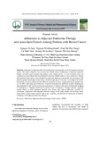
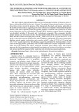
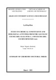
Glu-Pg interactions with fibrin surfaces and Chinese hamster ovary (CHO)-K1 cells were of similar affinity (Kd ¼ 0.9 ± 0.2 lm, Kd ¼ 1.05 ± 0.3 lm, respect- ively), in agreement with previous reports [37,38]. They were specifically inhibited by 6-Ahx that blocks the LBS of plasminogen K1 and K4 (IC50 ¼ 0.9 mm on fibrin, IC50 ¼ 1.7 mm on cells). Single LBS-targeted blockage of either K1 by mAb 34D3 or of K4 by mAb A10.2 prevented binding of Glu-Pg to both CHO-K1 cells (Fig. 1A) and fibrin (Fig. 1B) in a concentration- dependent manner. However, at saturating mAb con- centrations, mAb 34D3 fully inhibited the binding of Glu-Pg, whereas mAb A10.2 produced a partial inhibi- tory effect as indicated by the residual amount (< 5% vs. (cid:1) 60%, respectively) of plasminogen bound to cells inhibitory effect of and fibrin (Fig. 1). The partial mAb A10.2 is most probably related to the dynamic equilibrium between the open and compact conforma- tions of Glu-Pg in the unliganded state [16]. The lower immobilized mAb A10.2 for apparent affinity of Glu-Pg (Kd ¼ 1.64 nm ± 0.2 nm) as compare to its affinity for Lys-Pg (Kd ¼ 0.16 nm ± 0.06 nm) in which
FEBS Journal 272 (2005) 3387–3400 ª 2005 FEBS
3388
B. Ho-Tin-Noe´ et al.
Plasminogen transitions and proteolysis
Fig. 1. Inhibition of Glu-Pg binding to CHO-K1 cells and fibrin by anti-LBS A10.2 and 34D3 mAbs. CHO-K1 cells (A) or fibrin plates (B) were incubated with Glu-Pg (100 nM and 50 nM, respectively) supplemented with varying concentrations of A10.2 or ⁄ and 34D3 mAbs (0–1 lM). After two washes, bound plasminogen was detected using HRP-conjugated CPL-15 mAb. The inhibitory effect of the mAbs on plasminogen binding to fibrin and cells is expressed as a percentage (mean ± SD, n ¼ 3) relative to the amount of plasminogen bound in the absence of mAbs. The unre- lated mAb P6E9G11used as control did not affect plasminogen binding (data not shown). Data were fitted to a hyperbolic decay equation allowing calculation of mAb concentrations that produce 50% of the inhibitory effect, IC50, on, respectively, cells and fibrin: A10.2 ¼ 9.2 nM and 3 nM; 34D3 ¼ 13.1 nM and 5 nM; mAb mix ¼ 11.8 nM and 7 nM.
K4-LBS is permanently exposed, supports this hypo- thesis. Blockage by mAb A10.2 of K4-LBS exposed upon Glu-Pg binding to surfaces, may also contribute to this inhibitory effect.
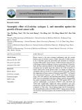
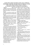
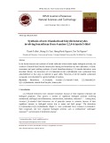
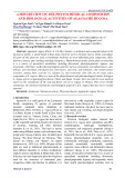
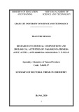
We then investigated the consequence of the inhibi- tion of plasminogen binding on plasmin formation by fibrin-bound tissue plasminogen activator (t-PA) or by CHO-K1 cells. For that purpose, t-PA was first bound to a fibrin surface, whereas no exogenous t-PA was added to CHO-K1 cells as they constitutively express t-PA at their membrane (detected by zymography as described [39]). CHO-K1 cells are indeed able to acti- vate plasminogen (Km ¼ 43.3 ± 3.2 nm, Fig. 2A) in an LBS-dependent manner as indicated by the com- plete inhibition of this activation by 6-Ahx (IC50 ¼ 5 mm, Fig. 2A, inset). By blocking the LBS of Glu-Pg K1, mAb 34D3 inhibits plasminogen binding (Fig. 1) and activation on both cells (IC50 ¼ 116 nm, Fig. 2B) and fibrin (IC50 ¼ 17 nm, Fig. 2B, inset). Interestingly, mAb A10.2 that blocks the LBS of plasminogen K4 and was shown to partially inhibit Glu-Pg binding (Fig. 1), does not impair its activation either on cells or on fibrin (Fig. 2B). These results were confirmed by Western blot analysis of cell-conditioned media from CHO-K1 cells treated with 0.5 lm Glu-Pg in the pres- ence or absence of 0.5 lm of mAb A10.2 or 34D3 (Fig. 2C). In the absence of mAbs, plasminogen was fully converted into plasmin by CHO-K1 cells (lane 4). Plasmin formation was not affected by mAb A10.2 (lane 5). However, the amount of plasmin activity that remains associated to cells and fibrin was progressively decreased by mAb A10.2 in a concentration-dependent manner, to 30% ± 1.4% cell-associated plasmin activ- the highest mAb concentration used (1 lm) ity at (Fig. 2D, to simplify the plot only results on cells are shown). In contrast, the conversion of plasminogen fully inhibited by mAb 34D3 into plasmin was the rate of plasmin formation lane 6): (Fig. 2C, (Fig. 2B) and the amount of cell-associated plasmin activity (Fig. 2D) decreased in parallel, reaching back- ground level (6.7% ± 0.9% maximum of cell-associ- ated plasmin activity) at 125 nm mAb concentration. This fully inhibitory dose of anti-K1-LBS mAb 34D3 is eightfold lower than the dose of anti-K4-LBS mAb A10.2 required to produce only a partial reduction in cell-associated plasmin. Because Glu-Pg is in a closed conformation, we further analysed the effect of mAb A10.2 on the activation of Lys-Pg that is known to be in an open conformation. In contrast to the full inhibi- tion of Glu-Pg activation by mAb 34D3 (Fig. 2B), blocking either the K4-LBS by mAb A10.2 or the K1-LBS by mAb 34D3 on Lys-Pg only partially inhib- its both its binding to fibrin (Fig. 3A, IC50 A10.2 ¼ IC50 34D3 ¼ 420 nm) and its activation 290 nm, (Fig. 3B, IC50 A10.2 ¼ 185 nm, IC50 34D3 ¼ 106 nm). A complete inhibition of Lys-Pg binding (Fig. 3A, IC50 mix ¼ 52 nm) and activation (Fig. 3B, IC50
FEBS Journal 272 (2005) 3387–3400 ª 2005 FEBS
3389
B. Ho-Tin-Noe´ et al.
Plasminogen transitions and proteolysis
A
B
C
D
Fig. 2. Effect of anti-LBS A10.2 and 34D3 mAbs on Glu-plasminogen activation by CHO-K1 cells and fibrin-bound t-PA. (A) CHO-K1 cells were incubated with varying concentrations of Glu-Pg (0–2 lM) and 0.75 mM CBS0065. Kinetics of plasmin formation was followed for 16 h. Data were fitted according to the Michaelis–Menten equation. Inset: inhibition of Glu-Pg activation (200 nM) by varying concentrations of 6-Ahx (0–100 mM). Results are expressed as a percentage (mean ± SD, n ¼ 3) relative to the maximal rate of plasmin formation in the absence of 6-Ahx. Data were fitted as indicated in Fig. 1 (IC50 ¼ 5 mM). (B) Kinetics of plasmin formation performed on cells (main figure) and fibrin (inset) in the presence of varying concentrations of mAb A10.2 or mAb 34D3 (0–1 lM for cells and 0–0.25 lM for fibrin) and fixed concentrations of Glu-Pg (200 nM for cells and 50 nM for fibrin) and CBS0065 (0.75 mM). Results are expressed as a percentage (mean ± SD, n ¼ 3) relative to the rate of plasmin formation in the absence of mAbs. (C) CHO-K1 cells were incubated for 16 h with 0.5 lM Glu-Pg in the presence or absence of 0.5 lM of mAb A10.2 or mAb 34D3. Cell conditioned media were analysed by SDS ⁄ PAGE (7.5% acryl- amide, nonreducing conditions) and western blotting using HRP-conjugated mAb CPL-15. Lane 1, Control Glu-Pg; lane 2, control plasmin; lane 3, untreated control cells; lane 4, Glu-Pg-treated cells; lane 5, Glu-Pg- and mAb A10.2-treated cells; lane 6, Glu-Pg- and mAb 34D3-trea- ted cells. Bands in lanes 1 and 2 correspond to forms I and II of plasmin(ogen). High molecular weight bands in lanes 5 and 6 correspond to mAb A10.2- and mAb 34D3-plasminogen complexes. (D) Cells were treated as in (B), washed twice with NaCl ⁄ Pi and residual bound plas- min was detected by adding CBS0065 at 0.75 mM. Results are expressed as a percentage (mean ± SD, n ¼ 3) relative to the amount of cell-associated plasmin activity detected in the absence of mAbs. *P < 0.0001 vs. control.
mix ¼ 15 nm) was obtained only when the two mAbs thus indicating an additive effect. were combined, Experiments were performed to exclude nonspecific effects of intact mAbs, including the reproduction of
similar results with their Fab fragments and the absence of effect on plasminogen binding and activa- tion in the presence of the unrelated anti-D-dimers mAb P6E9G11 (data not shown).
FEBS Journal 272 (2005) 3387–3400 ª 2005 FEBS
3390
B. Ho-Tin-Noe´ et al.
Plasminogen transitions and proteolysis
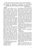
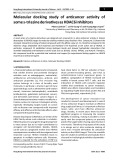
manner (Fig. 2A). The plasmin generated in situ indu- ces cell detachment (Fig. 4A). An inverse correlation (r ¼ 0.89, P ¼ 0.0013) was found between the amount of cell-associated plasmin and the percentage of resid- ual adherent cells as assessed by the 3-(4,5-dimethyl- thiazol-2-yl)-2,5-diphenyltetrazolium bromide (MTT) test. Plasmin(ogen)-induced cell detachment was pre- vented in a dose-dependent manner by mAb 34D3 or mAb A10.2; however, at similar concentrations, the effect of mAb 34D3 was more pronounced (Fig. 4B). Interestingly, a synergistic effect was observed by combining single doses of mAb 34D3 and mAb A10.2 that have little or no protective effect on cell detach- ment induced by 500 nm of plasminogen (Fig. 4C). Cell anchorage was indeed fully preserved by a com- bination of 75 nm doses of these mAbs (Fig. 4C). In a similar fashion, the concomitant blocking of plasmi- nogen K1-LBS and K4-LBS by 6-Ahx fully prevented cell detachment (data not shown). This protective effect of the two anti-LBS mAbs 34D3 and A10.2 (Fig. 4B,C) was parallel to a decrease in cell-associ- ated plasmin activity (Fig. 2D). An inverse correlation (r ¼ 0.94, P ¼ 0.005 for mAb A10.2, r ¼ 0.94, P ¼ 0.017 for mAb 34D3) was indeed found between the amount of cell-associated plasmin and the percentage of residual adherent cells. The decrease in cell-associ- ated plasmin (Fig. 2D) can be due to a decrease in plasmin formation, to a decrease in plasmin binding or to a combination of the two. We demonstrate that both mAb 34D3 and mAb A10.2 inhibit plasmin binding to cells (Fig. 5A) and fibrin (Fig. 5B) in a concentration-dependent manner. When combined, these mAbs produced an additive effect for the inhibi- tion of plasmin binding to cells (Fig. 5A) and fibrin (Fig. 5B).
Fig. 3. Effect of anti-LBS A10.2 and 34D3 mAbs on Lys-Pg binding and activation by fibrin-bound t-PA. (A) Fibrin surfaces were incuba- ted with a fixed concentration of Lys-Pg (100 nM) supplemented with varying concentrations of A10.2 or ⁄ and 34D3 mAbs (0–1 lM). After two washes, bound plasminogen was detected with HRP-con- jugated CPL-15 mAb as indicated in Fig. 1. Results are expressed as a percentage relative to the amount of Lys-Pg bound to fibrin in the absence of mAbs. Data were fitted as indicated in Fig. 2. (B) Fibrin surfaces with bound t-PA (10 IUÆmL)1) were incubated with different concentrations of A10.2 or ⁄ and 34D3 mAbs (0–1 lM) in the presence of fixed concentrations of Lys-Pg (50 nM) and CBS0065 (0.75 mM). Kinetics of plasmin formation was followed for 3 h. Results are expressed as a percentage (mean ± SD, n ¼ 3) rel- ative to the rate of plasmin formation in the absence of mAbs.
residual adherent cells
Plasmin-detached cells undergo apoptosis as indica- ted by the terminal deoxynucleotidyltransferase-medi- ated dUTP nick end labelling (TUNEL) assay (Fig. 6). The apoptotic index of the Glu-Pg treated cells reached 56.6 ± 6.7% in detached cells (Fig. 6C) and 37.1 ± 9.2% in the residual adherent cells (Fig. 6B) as compared to 0.9 ± 0.5% in untreated control cells the antiplasminogen (Fig. 6A). In the presence of K4-LBS mAb A10.2 the apoptotic index fall to (Fig. 6E) and to 7.9 ± 5.7% for detached cells 7.6 ± 4.3% for (Fig. 6D). Blocking of Glu-Pg K1-LBS by mAb 34D3 fully pre- vented loss of cell adhesion and apoptosis (apoptotic index 0.4 ± 0.2%, Fig. 6F).
Plasmin-induced cell detachment and apoptosis are impaired by selectively blocking the LBS of plasminogen K1 or K4
The incubation of CHO-K1 cells with plasminogen leads to plasmin formation in a saturable and specific
The higher protective effect on cell detachment (Fig. 4B) and apoptosis (Fig. 6F) produced by mAb 34D3 as compared to mAb A10.2 reflects its ability to prevent both plasmin formation (Fig. 2B) and binding
FEBS Journal 272 (2005) 3387–3400 ª 2005 FEBS
3391
B. Ho-Tin-Noe´ et al.
Plasminogen transitions and proteolysis
A
Fig. 4. Anti-LBS A10.2 and 34D3 mAbs protect cells from plasmino- gen activation-induced cell detachment. CHO-K1 cells were incuba- ted with Glu-Pg at varying concentrations (0–2 lM) or at a fixed concentration (0.5 lM) supplemented with varying concentrations of mAb A10.2 or 34D3 (0–1 lM). After 16 h of incubation, nonad- herent CHO-K1 cells were removed by washing with NaCl ⁄ Pi and residual adherent cells were detected with the colorimetric MTT test. (A) Bars represent the percentage of residual adherent cells as a function of Glu-Pg concentration. (B) Residual adherent cells at varying concentrations of mAb A10.2 or mAb 34D3 (0–1 lM) and a fixed concentration of Glu-Pg (0.5 lM) (mean ± SD, n ¼ 3). (C) Residual adherent cells at single 75 nM dose of mAb A10.2 or mAb 34D3 and their combination (mix) for a fixed concentration of Glu-Pg (0.5 lM) (mean ± SD, n ¼ 3). Results are expressed as a percentage relative to the amount of adherent untreated-control cells incubated with Ham-F12 medium alone. *P < 0.0001 vs. control.
B
to cells (Fig. 5A) whereas mAb A10.2 prevents only plasmin binding to cells (Figs 2D and 5A).
Anti-LBS mAbs 34D3 and A10.2 inhibit fibrinolysis
C
As fibrinolysis is the main physiological function of fibrin-bound plasmin, we explored the effect of the two anti-LBS mAbs on this process. Both anti-LBS mAbs decreased fibrinolysis in a dose-dependent manner (Fig. 7A). The decrease in fibrinolysis was directly rela- ted to the amount of fibrin-associated plasmin (r ¼ 0.94, P ¼ 0.0002, Fig. 7C). No effect on fibrinolysis could be detected in the presence of the unrelated anti- D-dimers mAb P6E9G11 (data not shown). As for Glu-Pg binding (Fig. 1) and activation (Fig. 2B), the inhibitory effect on fibrinolysis was more pronounced with the anti-K1-LBS mAb 34D3 than with the anti- K4-LBS mAb A10.2 (Fig. 7A). However, with Lys-Pg and plasmin, the antifibrinolytic effect of the two anti- LBS mAbs was comparable (39 ± 5% maximum of fibrinolysis for mAb A10.2 and 49 ± 4% for mAb 34D3) and a strong additive effect was observed when combined doses of mAbs were used (Fig. 7B, to sim- plify the plot only Lys-Pg data are shown).
Discussion
Cell detachment-induced apoptosis [9,10] and throm- bus lysis [14] are distinct biological processes that share plasmin(ogen) dependence. In vitro and ex vivo studies have recently shown that degradation of extra- cellular matrix proteins (fibronectin, laminin) by cell- bound plasmin plays a key role in triggering apoptosis following cell detachment [9,10]. These biological pro- cesses require LBS-dependent plasminogen binding to
FEBS Journal 272 (2005) 3387–3400 ª 2005 FEBS
3392
B. Ho-Tin-Noe´ et al.
Plasminogen transitions and proteolysis
tion into plasmin in situ. However, data linking plasminogen conformations and LBS accessibility to its functional properties such as binding to fibrin and cells, activation on these surfaces and the effect of plasmin proteolytic activity on fibrinolysis and extra- cellular proteolysis, are lacking. Furthermore, the relative importance of K1 and K4 in mediating inter- actions of plasminogen with fibrin and cells remains unsolved. This information could not be obtained with plasminogen fragments or with lysine analogues, as plasminogen fragments directly block binding sites on fibrin and cells [23] and the LBS of both K1 and K4 simultaneously occupied by lysine analogues are [41,42]. However, by nature of their conservative amino acids substitutions at contributing aromatic sites of the LBS [19], K1 and K4 have different abilities to lysine-Sepharose [40] bind lysine analogues [41–43], and most probably proteins in which lysine residues are accessible [16,20–23]. As the LBS are not structur- ally identical [19], we hypothesized that this problem can be studied with the use of specific LBS-targeted immunological probes. It has indeed been possible to raise polyclonal antibodies [44,45] and mAbs that spe- cifically differentiate the LBS of plasminogen K1 and K4 [34,35,46,47].
In this study, using two mAbs that allow direct and selective blockage of the LBS function of either K1 (mAb 34D3) [34] or K4 (mAb A10.2) [35], we were able to evaluate their respective contribution to Glu- and Lys-Pg binding and activation onto fibrin and cells, as well as their effects on fibrinolysis and on plas- min-induced cell apoptosis. The LBS specificity of these mAbs is based on their absence of interaction with 6-Ahx-blocked plasmin(ogen) and its isolated K4 and K1-3 fragments [35]. As expected, simultaneous inhibition with both mAbs resulted in a comple- mentary blocking effect of plasmin biological activities (Figs 4C and 7). These data indicate that the observed biological effects of these mAbs are strictly dependent on LBS interactions and reasonably exclude the possi- bility of steric hindrance. The reproduction of similar results with Fab fragments is in agreement with this concept.
Fig. 5. Inhibition of plasmin binding to CHO-K1 cells and fibrin by the anti-LBS A10.2 and 34D3 mAbs. CHO-K1 cells (A) or fibrin (B) were incubated with a constant concentration of D-Val-Phe-Lys- CH2Cl-inactivated plasmin (50 nM for cells, 5 nM for fibrin) supple- mented with varying concentrations of A10.2 or ⁄ and 34D3 mAbs (0–0.5 lM for cells, 0–50 nM for fibrin). After two washes with NaCl ⁄ Pi, bound plasmin was detected by incubating the cells with HRP-conjugated CPL-15 mAb. Results are expressed as a percent- age (mean ± SD, n ¼ 3) relative to the amount of plasmin bound in the absence of mAbs.
[28,40], previous
significantly affect
fibrin and cell surfaces [21,37,38]. Functional LBS in plasminogen have been identified in K1 and K4. As K4 in native Glu-Pg is most probably masked by inter-domain interactions studies using lysine analogues or plasminogen fragments have indirectly suggested the role of K1 in mediating inter- actions with cells [23] and fibrin [21] for its transforma-
We demonstrate that the selective neutralization of the K1-LBS with mAb 34D3 completely abolished the specific binding of Glu-Pg to fibrin and cells (Fig. 1). As a consequence, a dose-dependent inhibition of plas- min formation was observed (Fig. 2B), fibrinolysis was impaired (Fig. 7), and plasmin-induced cell detachment (Fig. 4B,C) and apoptosis (Fig. 6) were fully preven- the anti-K4-LBS mAb A10.2 did ted. In contrast, not the generation of plasmin (Fig. 2B,C), although it decreased the amount of
FEBS Journal 272 (2005) 3387–3400 ª 2005 FEBS
3393
B. Ho-Tin-Noe´ et al.
Plasminogen transitions and proteolysis
Fig. 6. Anti-LBS A10.2 and 34D3 mAbs protect cells from plasminogen activation-induced apoptosis. CHO-K1 cells were incubated for 36 h without (control) or with 0.5 lM of Glu-Pg in the presence or absence of 0.5 lM mAb A10.2 or mAb 34D3. The TUNEL reaction was then used to visualize DNA fragmentation (green fluorescence) in adherent cells and detached cytospun cells. Counterstaining with DAPI (blue fluorescence) was performed to visualize all nuclei. The apoptotic index (bars graph) was calculated as the percentage of TUNEL-positive nuclei relative to total DAPI-stained nuclei (bars represent mean ± SD, n ¼ 5). (A) Untreated control cells. (B, C) Cells incubated with Glu-Pg: (B) residual adherent cells; (C) detached cytospun cells. (D, E) Cells incubated with Glu-Pg and 0.5 lM of mAb A10.2: (D) residual adherent cells; (E) detached cytospun cells. (F) Cells incubated with Glu-Pg and 0.5 lM of mAb 34D3. No cells were recovered after cytospining the supernatant of control and Glu-Pg ⁄ mAb 34D3-treated cells. *P < 0.0001 vs. control.
the U-shaped open form of Lys-Pg, occurs upon occu- pancy of an LBS with the lysine analogue 6-Ahx. It has therefore been predicted that, as for the 6-Ahx- liganded form, Glu-Pg would adopt, upon binding to its lysine targets on fibrin and cells, an open conforma- tion in which K4 is exposed [16]. An increase in affin- ity of K4 for 6-Ahx upon transformation of Glu-Pg (Kd ¼ 5 mm) to Lys-Pg (Kd ¼ 36 lm) has indeed been documented [40,41].
cell-bound plasmin (Fig. 2D). This remarkable differ- ence in plasmin formation directly shows that in the native compact form of Glu-Pg, the LBS of K1 is readily exposed and plays a primary role in its initial binding to fibrin and cells. The subsequent accessibility of the K4-LBS is necessary to reinforce plasmin(ogen) anchorage as its occupancy by mAb A10.2 weakly inhibits Glu-Pg binding (Fig. 1), decreases fibrinolysis and plasmin-induced cell detachment, thus suggesting that this interaction may be of biological relevance.
These
results are
This mechanism was further explored by measuring the effect of the mAbs on the interaction of purified plasmin and Lys-Pg with fibrin and cells. The binding of Lys-Pg was partially inhibited by both 34D3 and A10.2 mAbs to a similar degree (Fig. 3A). The anti- K1-LBS mAb 34D3 was indeed unable to induce an inhibition of Lys-Pg binding (Fig. 3A) as marked as for the Glu-Pg form (Fig. 1). A complete inhibition of
in agreement with previous physicochemical studies [30,31,48–50]. It has thus been established that in the native spiral-like Glu-Pg form the K4-LBS is masked by inter-domain interactions [27,28] and that conformational changes in Glu-Pg are induced by ligand binding. A conformational shift to the more expanded form, roughly similar to that of
FEBS Journal 272 (2005) 3387–3400 ª 2005 FEBS
3394
B. Ho-Tin-Noe´ et al.
Plasminogen transitions and proteolysis
A
(C)
Fig. 7. Anti-LBS A10.2 and 34D3 mAbs decrease fibrinolysis. Fibrin surfaces were incubated with 10 IUÆmL)1 human t-PA. The plates were then washed to eliminate unbound proteins and the reaction was started by adding 50 nM of either (A) Glu-plasminogen or (B) Lys-Pg in the presence or in the absence of varying concentrations (0–1 lM) of A10-2 and ⁄ or 34D3 mAbs. The generated plasmin was eluted with 100 mM 6-Ahx containing 20 lM D-Val-Phe-Lys-CH2Cl in Tris ⁄ HCl pH 5 and the degree of fibrinolysis was estimated by detecting the plasmin-generated fibrin fragment E with mAb FDP-14 and an HRP-conjugated goat anti-mouse IgG. Results are expressed as a percentage relative to the extent of fibrin degrada- tion in the absence of mAbs (bars represent mean ± SD, n ¼ 3). *P < 0.0001 vs. control. In parallel experiments, fibrin-bound plasmin was not eluted but detected with HRP-conjugated CPl-15 mAb as indicated. The graph represents the correlation between the amount of fibrin-associated plasmin activity and the degree of fibrinolysis (r ¼ 0.94, P ¼ 0.0002).
B
resulted in the
that
C
Lys-Pg binding was obtained only when the LBS of both K1 and K4 were blocked simultaneously (Fig. 3A). When the binding of purified active site- inhibited plasmin was tested directly, similar partial and complete inhibitory effects were observed (Fig. 5) thus indicating accessibility and participation of both K1 and K4 in firmly anchoring plasmin to fibrin and cell surfaces. Because the expanded Lys-form of plasminogen may be an intermediary in plasmin for- mation [51,52], we have also evaluated its mechanism of activation by direct inhibition with the mAbs. The the mAbs on Lys-Pg binding observed effects of corresponding parallel (Fig. 3A) decrease in plasmin formation: it was completely abol- ished when both antibodies were added at concentra- tions if used singly produce only a partial inhibition (Fig. 3B). These results indicate that in con- trast to the selective K1-dependent binding of Glu-Pg to fibrin and cells, in Lys-Pg either the K1- or the K4-LBS can mediate initial binding for its activation. These studies provide biological support to data indi- cating that in the Lys-Pg open conformation K1 and K4 are equally exposed [33].
that
this
Furthermore, we add new data about their respect- ive role in the binding ⁄ activation mechanism by link- functions of ing them directly to the biological plasmin. In particular, we demonstrate that these inter- actions are determinant for the activities of plasmin that we have evaluated, namely: fibrinolysis, cell in agreement detachment and apoptosis. We show, with previous work [9,10], that uncontrolled plasmin formation by adherent cells leads to cell detachment (Fig. 4A) and subsequent apoptosis (Fig. 6B,C). We plasmin(ogen)-dependent demonstrate sequence of events can be prevented to different the initial degrees by selectively inhibiting either
FEBS Journal 272 (2005) 3387–3400 ª 2005 FEBS
3395
B. Ho-Tin-Noe´ et al.
Plasminogen transitions and proteolysis
Cell culture
benzthiazoline)-6-sulphonic acid (ABTS) was from Roche (Mannheim, Germany). Human t-PA was from Biopool (Uppsala, Sweden). Fibrin and partially degraded fibrin surfaces were prepared and characterized as described pre- viously [37].
Plasminogen and monoclonal antibodies
binding and activation of native Glu-plasminogen with mAb 34D3 or the anchoring of plasmin with mAb A10.2. A complete inhibition of plasmin formation (Fig. 2B), prevention of fibrinolysis (Fig. 7), protection from cell detachment (Fig. 4B) and apoptosis (Fig. 6F) were readily obtained by blocking the K1-LBS of Glu-Pg with mAb 34D3. Blockage of K4-LBS with mAb A10.2 did not significantly affect plasmin forma- tion (Fig. 2B) but produced a dose-dependent partial reduction in cell-bound plasmin that progressively pro- tected cells from detachment (Fig. 4B) and significantly limited the rate of apoptosis (Fig. 6D,E). Furthermore, to prevent plas- these mAbs at doses insufficient min(ogen)-dependent cell detachment, were able to fully preserve cell adhesion if added in combination (Fig. 4C). Because cell detachment-induced apoptosis may be implicated in the development of several pathologies in which the fibrinolytic system is activated [53–55], these mAbs represent new tools that could be the basis for the development of pharmacological agents that would prevent this phenomenon in vivo.
CHO-K1 cells (American Type Culture Collection CCL-61) were seeded in 96-well plates and cultured at 37 (cid:1)C in a humidified atmosphere of 5% (v ⁄ v) CO2 using Ham-F12 medium supplemented with 2 mm glutamine and 10% (v ⁄ v) fetal bovine serum. The cells at near confluence were starved from serum for 6 h before experiments.
In conclusion, we demonstrate that
initial binding
Human Glu- and Lys-Pg were purified as described [37,56] and were considered to be > 99% pure as assessed by SDS ⁄ PAGE and by N-terminal sequence analysis. Plasmin was prepared by activation of Glu-plasminogen with immo- bilized urokinase as described previously [57].
mAb A10.2 was raised in mice immunized with human recombinant apolipoprotein(a), cloned using classical hybri- doma procedures and purified as described [35]. mAb A10.2 is an IgG1 that specifically binds and masks the LBS of apolipoprotein(a) KIV-10 and of plasminogen K4, with- out cross-reaction with the LBS of plasminogen K1 [58].
in Glu-Pg, K1-LBS mediates and activation whereas K4-LBS is secondarily required to reinforce plasmin(ogen) anchorage. In contrast, in Lys-Pg as in plasmin, K1- and K4-LBS act as independent and complementary domains both able to support plasmi- nogen binding and activation, and both required to ensure plasmin anchorage and subsequent proteolytic transitions activity. These structural and functional are determinant in the initiation and acceleration of fibrinolysis and cell detachment and may be of biologi- cal relevance in inflammatory states. As plasminogen activation is involved in several physiological and pathological conditions, the effects of the anti-LBS mAbs on fibrinolysis and plasmin-induced apoptosis may also have a pharmacological interest; they may serve for future strategies in the development of poten- tial therapeutic agents in cardiovascular diseases where uncontrolled plasmin formation results in unwanted cell death.
mAb 34D3 was raised in mice immunized with human plasmin-a2-antiplasmin complex, cloned and purified as described [34]. This mAb reacts with plasminogen frag- ment K1+2+3 and shows no cross-reaction with plasmi- nogen K4 [34]. The specificity of this mAb for the LBS of plasminogen K1 was determined as follows. Glutaralde- hyde-immobilized mAb 34D3 was incubated with a fixed concentration of plasminogen (25 nm) supplemented with varying concentrations of 6-Ahx (0–200 mm) in binding buffer (50 mm sodium phosphate pH 6.8, 80 mm NaCl, 4 mgÆmL)1 BSA, 2 mm EDTA, 0.01% Tween 20, 0.01% azide). Bound plasminogen was detected as indicated in the following section.
Experimental procedures
Reagents
FEBS Journal 272 (2005) 3387–3400 ª 2005 FEBS
3396
mAb CPL-15 is an IgG1 specifically directed against an epitope of plasminogen K1 not overlapping with the LBS. It was produced and characterized previously [59]. Horse- radish peroxidase (HRP)-conjugated CPL-15 mAb was obtained using a peroxydase labelling kit according to the manufacturer’s instructions (Roche, Mannheim, Germany). mAb FDP-14, an IgG1 that reacts with a neo-epitope exposed in the Bb chain stretch 54–118 of the fibrin degra- dation products X, Y and E but not in fibrin [60], was kindly provided by W. Nieuwenhuizen (Gaubius Laborat- ory, Leiden, the Netherlands). chloromethylketone mAb P6E9G11 directed against fibrin d-dimers (provided by Biomerieux France) was used as a control. The chromogenic substrate selective for plasmin (methyl- malonyl)-hydroxyprolylarginine-p-nitroanilide (CBS0065) was from Stago (Asnie` res, France). The lysine analogue 6-Ahx was from Sigma (St-Louis, MO, USA). d-Valyl-l- (d-Val-Phe-Lys- phenylalanyl-l-lysine CH2Cl), a plasmin inhibitor was from Calbiochem (La Jolla, CA, USA). Peroxydase substrate 2,2¢-azino-bis (3-ethyl-
B. Ho-Tin-Noe´ et al.
Plasminogen transitions and proteolysis
Binding of plasmin(ogen) to fibrin and cells
Effect of anti-LBS mAbs on plasminogen activation
Inhibition of plasminogen activation by 6-Ahx
To determine the effect of the mAbs 34D3 and A10.2 on plasminogen activation, the fibrin plates and the cells were incubated with different concentrations of these mAbs in the presence of fixed concentrations of plasminogen and CBS0065. mAb P6E9G11 was used as a plasmin(ogen)- unrelated control. We previously determined that the amidolytic activity of plasmin was not modified in the pres- ence of the mAbs.
CHO-K1 cells and partially degraded fibrin surfaces were incubated for 1 h at 37 (cid:1)C with different concentrations of plasminogen or d-Val-Phe-Lys-CH2Cl-inactivated plasmin in binding buffer (for binding to fibrin) or in Ham-F12 medium supplemented with 1% (w ⁄ v) BSA (for binding to cells). After two washes with NaCl ⁄ Pi, bound proteins were detected by incubation for 1 h at 37 (cid:1)C with HRP- conjugated mAb CPL-15. After washing with NaCl ⁄ Pi, 1 mgÆmL)1 ABTS was used as a substrate for colour devel- opment and the absorbance at 405 nm was measured in a multiwell plate reader.
CHO-K1 cells were incubated with varying concentrations of 6-Ahx (0–100 mm) and fixed concentrations of plasmino- gen (200 nm) and CBS0065 (0.75 mm) in a total volume of 100 lL of Ham-F12 medium per well. Kinetics of plasmin formation was followed for 16 h as indicated.
Western blot analysis of plasminogen activation
To determine the effect of 34D3 and A10.2 mAbs on plasmin(ogen) binding to fibrin and CHO-K1 cells, fibrin plates and cells were incubated with a fixed concentration of plasmin(ogen) supplemented with varying concentrations of mAbs. Bound plasmin(ogen) was detected as indicated above. mAb P6E9G11 directed against fibrin d-dimers was used as a plasmin(ogen)-unrelated control.
incubating CHO-K1 cells
Data were fitted according to the Langmuir equation for single site interactions and to a hyperbolic decay equation that allows calculation of the concentration of mAb that produces 50% of the inhibitory effect (IC50) [61]. cell
Effect of anti-LBS mAbs on the activation of plasminogen by fibrin- and cell-bound t-PA
Plasminogen activation on fibrin
Effects of anti-LBS mAbs on matrix degradation and cell survival
for 16 h with 0.5 lm After Glu-Pg in the presence or absence of 0.5 lm of mAb A10.2 or 34D3, conditioned media were analysed by SDS ⁄ PAGE (7.5% acrylamide, nonreducing conditions) and western blotting using HRP-conjugated mAb CPL-15.
Cell detachment assay
After the plasminogen activation experiments described above, nonadherent CHO-K1 cells were removed by wash- ing with NaCl ⁄ Pi and the following analyses were per- formed.
Plasminogen activation on cells
The activation of plasminogen by fibrin-bound t-PA was studied as described elsewhere [36,62]. Briefly, a 96-well plate coated with fibrin was incubated for 1 h at 37 (cid:1)C with 10 IUÆmL)1 human t-PA in binding buffer (50 lLÆ well)1). The plate was then washed twice to eliminate unbound proteins, and the reaction was started by adding 50 lL per well of plasminogen and of CBS0065 in the same buffer. Kinetics of plasmin formation was followed for 5 h by measuring the release of p-nitroaniline, detected as a change in absorbance (DA405(cid:2) (cid:3)1 min ), using a multiwell plate reader (MX5000, Dynex) held at 37 (cid:1)C. Rates of plasmin production were calculated from the slopes of curves of A405 vs. time and transformed into plasmin con- centration as described [62].
Terminal deoxynucleotidyltransferase-mediated TUNEL and 4¢, 6-diamino-2-phenylindole (DAPI) staining
The residual adherent cells were incubated for 1 h at 37 (cid:1)C with 0.5 mgÆmL)1 of the tetrazolium salt MTT in NaCl ⁄ Pi. Remaining living adherent cells form formazan crystals that are dissolved in dimethylsulfoxide and colorimetrically detected at A550 using a multiwell plate reader. Absorbance readings are proportional to the number of living cells. Results are expressed as a percentage relative to the amount of residual adherent cells obtained for control cells incuba- ted with Ham-F12 medium.
CHO-K1 cells were starved of serum and then incubated with varying concentrations of Glu-Pg (0–2 lm) and a fixed concentration (0.75 mm final concentration) of CBS0065 in a total volume of 100 lL of Ham-F12 medium per well. Kinetics of plasmin formation was followed for 16 h as indicated above.
FEBS Journal 272 (2005) 3387–3400 ª 2005 FEBS
3397
After the indicated experiments, cells grown on eight-cham- bered slides (Labtek) or detached cytospun cells were fixed room in 3.7% (v ⁄ v) paraformaldehyde for 30 min at After the activation reaction on both cells and fibrin, two washes in NaCl ⁄ Pi were performed and the residual bound plasmin was detected by adding CBS0065 at 0.75 mm.
B. Ho-Tin-Noe´ et al.
Plasminogen transitions and proteolysis
disruption of the plasminogen gene on thrombosis, growth, and health in mice. Circulation 92, 2585–2593. 3 Lijnen HR, Carmeliet P, Bouche A, Moons L, Ploplis
VA, Plow EF & Collen D (1996) Restoration of thrombolytic potential in plasminogen-deficient mice by bolus administration of plasminogen. Blood 88, 870–876.
Effect of anti-LBS mAbs on fibrinolysis
temperature and then permeabilized in 0.1% (v ⁄ v) Triton X-100, 0.1% (w ⁄ v) sodium citrate for 2 min on ice. TU- NEL (Roche Applied Science) used to visualize DNA frag- mentation and 4¢, 6-diamino-2-phenylindole (DAPI; Sigma) nuclear counterstaining to visualize all nuclei were per- formed according to the manufacturer’s instructions. After washing, the slides were mounted with Fluoprep (DAKO, Carpinteria, CA, USA) and observed under an epifluores- cence microscope. The apoptotic index was calculated as the percentage of TUNEL-positive nuclei relative to total DAPI-stained nuclei. 4 Carmeliet P, Moons L, Ploplis V, Plow E & Collen D (1997) Impaired arterial neointima formation in mice with disruption of the plasminogen gene. J Clin Invest 99, 200–208.
5 Ploplis VA, French EL, Carmeliet P, Collen D & Plow EF (1998) Plasminogen deficiency differentially affects recruitment of inflammatory cell populations in mice. Blood 91, 2005–2009.
6 Plow EF, Carmeliet P & Collen D (1999) Plasminogen and cell migration in vivo. Fibrinolysis Proteolysis 13, 49–53.
7 Creemers E, Cleutjens J, Smits J, Heymans S, Moons L, Collen D, Daemen M & Carmeliet P (2000) Disruption of the plasminogen gene in mice abolishes wound healing after myocardial infarction. Am J Pathol 156, 1865–1873. secondary antibody
8 Lijnen HR (2001) Plasmin and matrix metalloprotein- ases in vascular remodeling. Thromb Haemost 86, 324–333. 9 Meilhac O, Ho-Tin-Noe B, Houard X, Philippe M,
Statistical analysis
Michel JB & Angles-Cano E (2003) Pericellular plasmin induces smooth muscle cell anoikis. FASEB J 17, 1301–1303. The activation of 50 nm of either Glu- or Lys-Pg by fibrin- bound t-PA was performed as indicated above in the pres- ence or in the absence of mAbs A10.2 or ⁄ and mAb 34D3 (0–1 lm). The generated plasmin was eluted with 100 mm 6-Ahx, 20 lm d-Val-Phe-Lys-CH2Cl, in Tris ⁄ HCl buffer pH 5, and the degree of fibrinolysis was estimated by detecting the plasmin-generated fibrin fragment E. For this purpose the specific antifragment E mAb FDP-14 was used at 200 ngÆmL)1 followed by an HRP-conjugated goat anti- mouse (DAKO AS, Glostrup, Denmark). After washing with binding buffer, 1 mgÆmL)1 ABTS was used as a substrate for colour development and the absorbance at 405 nm was measured in a multiwell plate reader. In parallel experiments, fibrin-bound plasmin was not eluted but detected with the HRP-conjugated CPL-15 at 250 ngÆmL)1 as indicated above.
10 Rossignol P, Ho-Tin-Noe B, Vranckx R, Bouton MC, Meilhac O, Lijnen HR, Guillin MC, Michel JB & Angles-Cano E (2004) Protease-nexin-1 inhibits plasmi- nogen activation-induced apoptosis of adherent cells. J Biol Chem 279, 10346–10356.
Statistical analysis was performed using statview 5.0 soft- ware. Results are expressed as means ± SD (at least three independent experiments performed in triplicate). Compari- sons used one-way analysis of variance with Scheffe’s F-test. Statistical significance was set at P < 0.05.
11 Reijerkerk A, Mosnier LO, Kranenburg O, Bouma BN, Carmeliet P, Drixler T, Meijers JC, Voest EE & Geb- bink MF (2003) Amyloid endostatin induces endothelial cell detachment by stimulation of the plasminogen acti- vation system. Mol Cancer Res 1, 561–568.
Acknowledgements
This study was founded by the INSERM and grants Adrienne et Pierre Sommer from the Fondation de France to E. AC. and Interuniversity Attraction Poles (P5 ⁄ o2) to H.R.L.
12 Zhang X, Chaudhry A & Chintala SK (2003) Inhibition of plasminogen activation protects against ganglion cell loss in a mouse model of retinal damage. Mol Vis 9, 238–248.
References
13 Davis J, Wagner MR, Zhang W, Xu F & Van Nostrand WE (2003) Amyloid beta-protein stimulates the expres- sion of urokinase-type plasminogen activator (uPA) and its receptor (uPAR) in human cerebrovascular smooth muscle cells. J Biol Chem 278, 19054–19061. 1 Bugge TH, Flick MJ, Daugherty CC & Degen JL 14 Wiman B & Collen D (1978) Molecular mechanism of physiological fibrinolysis. Nature 272, 549–550.
(1995) Plasminogen deficiency causes severe thrombosis but is compatible with development and reproduction. Genes Dev 9, 794–807.
FEBS Journal 272 (2005) 3387–3400 ª 2005 FEBS
3398
2 Ploplis VA, Carmeliet P, Vazirzadeh S, Van Vlaenderen I, Moons L, Plow EF & Collen D (1995) Effects of 15 Plow EF, Freaney DE, Plescia J & Miles LA (1986) The plasminogen system and cell surfaces: evidence for plas- minogen and urokinase receptors on the same cell type. J Cell Biol 103, 2411–2420.
B. Ho-Tin-Noe´ et al.
Plasminogen transitions and proteolysis
16 Markus G (1996) Conformational changes in plasmino- gen, their effect on activation, and the agents that modulate activation rates – a review. Fibrinolysis 10, 75–85. 30 Weisel JW, Nagaswami C, Korsholm B, Petersen LC & Suenson E (1994) Interactions of plasminogen with polymerizing fibrin and its derivatives, monitored with a photoaffinity cross-linker and electron microscopy. J Mol Biol 235, 1117–1135.
31 Mangel WF, Lin BH & Ramakrishnan V (1990) Char- acterization of an extremely large, ligand-induced con- formational change in plasminogen. Science 248, 69–73.
17 Sottrup-Jensen L, Claeys H, Zajdel M, Petersen TE & Magnusson S (1978) The primary structure of human plamsinogen: isolation of two lysine-binding fragments and one ‘mini’-plasminogen (MW, 38,000) by elastase- catalyzed-specific limited proteolysis. Prog Chem Fibri- nol Thrombol 3, 191–209. 18 Petersen TE, Martzen MR, Ichinose A & Davie EW
32 Ponting CP, Holland SK, Cederholm-Williams SA, Marshall JM, Brown AJ, Spraggon G & Blake CC (1992) The compact domain conformation of human Glu-plasminogen in solution. Biochim Biophys Acta 1159, 155–161. (1990) Characterization of the gene for human plasmi- nogen, a key proenzyme in the fibrinolytic system. J Biol Chem 265, 6104–6111.
33 Ramakrishnan V, Patthy L & Mangel WF (1991) Con- formation of Lys-plasminogen and the kringle 1–3 frag- ment of plasminogen analyzed by small-angle neutron scattering. Biochemistry 30, 3963–3969. 34 Lijnen HR, Bloemmen F, Vereecken A & Collen D
19 Tulinsky A, Park CH, Mao B & Llinas M (1988) Lysi- ne ⁄ fibrin binding sites of kringles modeled after the structure of kringle 1 of prothrombin. Proteins 3, 85–96. 20 Thorsen S (1975) Differences in the binding to fibrin of native plasminogen and plasminogen modified by pro- teolytic degradation. Influence of omega-aminocarboxy- lic acids Biochim Biophys Acta 393, 55–65. (2001) Enzyme-linked immunosorbent assay for the spe- cific detection of angiostatin-like plasminogen moieties in biological samples. Thromb Res 102, 53–59.
21 Lucas MA, Fretto LJ & McKee PA (1983) The binding of human plasminogen to fibrin and fibrinogen. J Biol Chem 258, 4249–4256.
22 Knudsen BS, Silverstein RL, Leung LL, Harpel PC & Nachman RL (1986) Binding of plasminogen to extra- cellular matrix. J Biol Chem 261, 10765–10771.
23 Miles LA, Dahlberg CM & Plow EF (1988) The cell- binding domains of plasminogen and their function in plasma. J Biol Chem 263, 11928–11934. 35 Dominguez M, Rojas G, Loyau S, Bazurco M, Sorell L & Angles-Cano E (2001) Kringles of the plasminogen – prothrombin gene family share conformational epitopes with recombinant apolipoprotein (a): specificity of the fibrin-binding site. Biochim Biophys Acta 1548, 72–80. 36 Rouy D, Grailhe P, Nigon F, Chapman J & Angles- Cano E (1991) Lipoprotein (a) impairs generation of plasmin by fibrin-bound tissue-type plasminogen activa- tor. In vitro studies in a plasma milieu. Arterioscler Thromb 11, 629–638.
24 Thewes T, Constantine K, Byeon IJ & Llinas M (1990) Ligand interactions with the kringle 5 domain of plas- minogen. A study by 1H NMR spectroscopy. J Biol Chem 265, 3906–3915. 37 Fleury V & Angles-Cano E (1991) Characterization of the binding of plasminogen to fibrin surfaces: the role of carboxy-terminal lysines. Biochemistry 30, 7630–7638. 38 Plow EF, Herren T, Redlitz A, Miles LA & Hoover-
Plow JL (1995) The cell biology of the plasminogen sys- tem. FASEB J 9, 939–945. 25 Castellino FJ, Ploplis VA, Powell JR & Strickland DK (1981) The existence of independent domain structures in human Lys77-plasminogen. J Biol Chem 256, 4778–4782. 39 Gaussem P, Grailhe P & Angles-Cano E (1993) Sodium 26 Banyai L & Patthy L (1984) Importance of intramole-
cular interactions in the control of the fibrin affinity and activation of human plasminogen. J Biol Chem 259, 6466–6471.
dodecyl sulfate-induced dissociation of complexes between human tissue plasminogen activator and its specific inhibitor. J Biol Chem 268, 12150–12155. 40 Vali Z & Patthy L (1982) Location of the intermediate and high affinity omega-aminocarboxylic acid-binding sites in human plasminogen. J Biol Chem 257, 2104–2110.
27 Cockell CS, Marshall JM, Dawson KM, Cederholm- Williams SA & Ponting CP (1998) Evidence that the conformation of unliganded human plasminogen is maintained via an intramolecular interaction between the lysine-binding site of kringle 5 and the N-terminal peptide. Biochem J 333, 99–105. 41 Markus G, DePasquale JL & Wissler FC (1978) Quanti- tative determination of the binding of epsilon-amino- caproic acid to native plasminogen. J Biol Chem 253, 727–732.
28 Marshall JM, Brown AJ & Ponting CP (1994) Confor- mational studies of human plasminogen and plasmino- gen fragments: evidence for a novel third conformation of plasminogen. Biochemistry 33, 3599–3606. 29 Tranqui L, Prandini M-H & Chapel A (1979) The 42 Markus G, Priore RL & Wissler FC (1979) The binding of tranexamic acid to native (Glu) and modified (Lys) human plasminogen and its effect on conformation. J Biol Chem 254, 1211–1216.
FEBS Journal 272 (2005) 3387–3400 ª 2005 FEBS
3399
43 De Marco A, Petros AM, Laursen RA & Llinas M (1987) Analysis of ligand-binding to the kringle 4 Structure of Plasminogen Studied by Electron Micro- scopy. Biologie Cellulaire 34, 39–42.
B. Ho-Tin-Noe´ et al.
Plasminogen transitions and proteolysis
fragment from human plasminogen. Eur Biophys J 14, 359–368.
54 Johnsen M, Lund LR, Romer J, Almholt K & Dano K (1998) Cancer invasion and tissue remodeling: common themes in proteolytic matrix degradation. Curr Opin Cell Biol 10, 667–671. 44 Plow EF & Collen D (1981) Immunochemical character- ization of a low affinity lysine binding site within plas- minogen. J Biol Chem 256, 10864–10869. 45 Hochschwender SM & Laursen RA (1981) Immuno-
55 Bauriedel G, Hutter R, Welsch U, Bach R, Sievert H & Luderitz B (1999) Role of smooth muscle cell death in advanced coronary primary lesions: implications for pla- que instability. Cardiovasc Res 41, 480–488. chemical characterization of the kringle 4 fragment of human plasminogen. J Biol Chem 256, 11166–11171. 46 Cummings HS & Castellino FJ (1985) A monoclonal
56 Holvoet P, Lijnen HR & Collen D (1985) A monoclonal antibody specific for Lys-plasminogen. Application to the study of the activation pathways of plasminogen in vivo. J Biol Chem 260, 12106–12111. antibody to the epsilon-aminocaproic acid binding site on the kringle 4 region of human plasminogen that accelerates the activation of Glu1-plasminogen by urokinase. Arch Biochem Biophys 236, 612–618.
57 Wiman B & Wallen P (1973) Activation of human plas- minogen by an insoluble derivative of urokinase. Struc- tural changes of plasminogen in the course of activation to plasmin and demonstration of a possible intermediate compound. Eur J Biochem 36, 25–31.
58 Dominguez M, Montes R, Paramo JA & Angles-Cano E (2002) Bivalency of plasminogen monoclonal antibo- dies is required for plasminogen bridging to fibrin and enhanced plasmin formation. Biochim Biophys Acta 1598, 165–176. 59 Montes R, Paramo JA, Angles-Cano E & Rocha E
47 Holvoet P, Lijnen HR & Collen D (1986) A monoclonal antibody directed against the high-affinity lysine-binding site (LBS) of human plasminogen. Role of LBS in the regulation of fibrinolysis. Eur J Biochem 157, 65–69. 48 Brockway WJ & Castellino FJ (1972) Measurement of the binding of antifibrinolytic amino acids to various plasminogens. Arch Biochem Biophys 151, 194–199. 49 Castellino FJ, Brockway WJ, Thomas JK, Liano HT & Rawitch AB (1973) Rotational diffusion analysis of the conformational alterations produced in plasminogen by certain antifibrinolytic amino acids. Biochemistry 12, 2787–2791. (1996) Development and clinical application of a new ELISA assay to determine plasmin-alpha2-antiplasmin complexes in plasma. Br J Haematol 92, 979–985.
50 Sjoholm I (1973) Studies on the conformational changes of plasminogen induced during activation to plasmin and by 6-aminohexanoic acid. Eur J Biochem 39, 471–479. 51 Hajjar KA & Nachman RL (1988) Endothelial cell-
60 Koppert PW, Koopman J, Haverkate F & Nieuwenhui- zen W (1986) Production and characterization of a monoclonal antibody reactive with a specific neoanti- genic determinant (comprising B beta 54–118) in degra- dation products of fibrin and of fibrinogen. Blood 68, 437–441. 61 Adamson AW (1992) Physical Chemistry of Surfaces. mediated conversion of Glu-plasminogen to Lys-plasmi- nogen. Further evidence for assembly of the fibrinolytic system on the endothelial cell surface. J Clin Invest 82, 1769–1778. Wiley-Interscience Publication, New York.
62 Fleury V, Lijnen HR & Angles-Cano E (1993) Mechan- ism of the enhanced intrinsic activity of single-chain urokinase-type plasminogen activator during ongoing fibrinolysis. J Biol Chem 268, 18554–18559. 52 Gong Y, Kim SO, Felez J, Grella DK, Castellino FJ & Miles LA (2001) Conversion of Glu-plasminogen to Lys-plasminogen is necessary for optimal stimulation of plasminogen activation on the endothelial cell surface. J Biol Chem 276, 19078–19083.
FEBS Journal 272 (2005) 3387–3400 ª 2005 FEBS
3400
53 Chen ZL & Strickland S (1997) Neuronal death in the hippocampus is promoted by plasmin-catalyzed degra- dation of laminin. Cell 91, 917–925.