M I N I R E V I E W
Post-ischemic brain damage: targeting PARP-1 within the ischemic neurovascular units as a realistic avenue to stroke treatment Flavio Moroni and Alberto Chiarugi
Department of Preclinical and Clinical Pharmacology, University of Florence, Italy
Keywords blood brain barrier; endothelium; inflammation; ischemia; microglia; neuroprotection; neurovascular unit; PARP-1; pericytes; stroke
Correspondence F. Moroni, Dipartimento di Farmacologia, Viale Pieraccini 6, 50139 Firenze, Italy Fax: +39 055 4271226 Tel: +39 055 4271280 E-mail: flavio.moroni@unifi.it
Stroke is the third leading cause of death in industrialized countries but efficacious stroke treatment is still an unmet need. Preclinical research indi- cates that different molecules afford protection from ischemic neurodegen- eration, but all clinical trials conducted so far have inexorably failed. Critical re-evaluation of experimental data shows that all the components of the neurovascular unit, such as neurons, glia, endothelia and basal mem- branes, must be protected during the ischemic insult to obtain substantial and long-lasting neuroprotection. Here, we propose the nuclear enzyme poly(ADP-ribose) polymerase (PARP-1) as a key effector of cell death in the various elements of the neurovascular units, and assert that drugs inhibiting PARP-1 may therefore represent valuable tools for pharmacolog- ical treatment of stroke patients.
(Received 3 July 2008, revised 11 September 2008, accepted 14 October 2008)
doi:10.1111/j.1742-4658.2008.06768.x
research for
and industrial
neurologic disability by administering recombinant tis- sue plasminogen activator within 3 h from when the stroke symptoms first start. Conversely, the cellular approach has been so far clinically unsuccessful, and none of the numerous neuroprotective strategies that have been tested in clinical trials have reached the clinical arena [3,4].
Exciting, radical, suicidal and inflamed – the many pathways of ischemic brain injury
clinically
vascular
therapy
led
to
The enormous body of information on ischemic neuro- degeneration in different experimental stroke models has shed light on the complex signaling pathways and responsible for neuronal damage molecular events
Therapeutic strategies aimed at reducing brain dam- age after ischemic stroke have been a major focus of academic the past 30 years. Two primary therapeutic approaches have been intensively studied: the first can be defined as the ‘vascular approach’ and its main goal is the rapid re-opening of occluded blood vessels so that oxygen and nutrients may return to the ischemic region. The second may be defined as the ‘cellular approach’ and is based on the possibility of interfering with the sig- naling pathways, leading to loss of neurons and dam- age of other cellular elements present in the affected brain region [1,2]. Efforts directed at developing effec- useful tive procedures and have clearly demonstrated that it is selectively, brain damage and possible to reduce,
Abbreviations AIF, apoptosis-inducing factor; BBB, blood–brain barrier; HMGB1, high-mobility-group protein box 1; IL, interleukin; MMP, matrix metalloproteinase; NMDA, N-methyl-D-aspartate; PARG, poly(ADP-ribose) glycohydrolase; PARP, poly(ADP-ribose) polymerase; PARP-1, poly(ADP-ribose) polymerase 1; ROS, reactive oxygen species; TNF-a, tumor necrosis factor-a.
FEBS Journal 276 (2009) 36–45 ª 2008 The Authors Journal compilation ª 2008 FEBS
36
F. Moroni and A. Chiarugi
PARP-1 and the ischemic neurovascular unit
the main target
the
Another event widely recognized to be of key patho- genetic relevance to post-ischemic brain damage is immune activation of resident glial cells and leukocytes infiltrating from blood vessels [16,17]. In this regard, several therapeutic approaches aimed at counteracting post-ischemic immune activation and infiltration have been tested in clinical trials. Some, such as the anti-leu- kocyte adhesion molecules enlimonab and HU23F2G, proved inefficacious and harmful, respectively. Others, such as the interleukin (IL)-1 receptor antagonist, pro- vided inconclusive results. Failure might be a result of the fact that both protective as well as detrimental effects of the inflammatory response during ischemic neurodegeneration have been reported [18].
Critical re-evaluation of drug development in stroke
when blood flow to a brain region drops below a criti- cal threshold and when it returns because of vessel re-opening and tissue reperfusion. In the past, particu- lar attention was directed to derangement of excitatory amino acid-mediated neurotransmission that became, for neuroprotection. for years, Hypoxia ⁄ ischemia increases concentrations of extracellular glutamate [5,6] with excessive stimulation of ionotropic and metabotropic glutamate receptors, which initiates a chain of events leading to excitotoxic neuronal death [7,8]. This concept is strongly sup- ported by the observation that, in a number of in vitro and in vivo experimental models of ischemia, glutamate receptor antagonists, acting either on ionotropic [N-methyl-d-aspartate (NMDA) or Gk alpha-amino-3- hydroxy-5-methyl-4-isoxazolone propinate] or on group I metabotropic receptors, are effective neuroprotective agents [9–13]. Unfortunately, however, none of the glutamate receptor antagonists tested in clinical trials showed positive results or had an acceptable benefit ⁄ side effects ratio.
scavengers
Triggered by the excitotoxic events as well as by impairment of mitochondrial respiration, a burst of reactive oxygen species (ROS) and reactive nitrogen species typically occurs within the ischemic brain tis- sue. Again, inhibition of radical formation as well as of radical scavengers provides significant neuroprotec- tion in animal stroke models. Agents acting as free- radical therefore have been repeatedly proposed as useful drugs for stroke therapy, but most were rapidly discarded because of cardiovascular toxic- ity. Recently, however, the spin-trap nitrone NXY-059 from AstraZeneca reached the clinical arena with some success [14]. The putative neuroprotectant is probably n-t-butyl hydroxylamine and ⁄ or its parent spin-trap 2-methyl-2-nitrosopropane, produced by hydrolysis of NXY-059. Unfortunately, the positive outcome of the first clinical trial was not confirmed in a second clinical trial, and NXY-059 development was dropped, leaving widespread scepticism in the field regarding the possi- bility of obtaining ischemic neuroprotection in humans [15].
stroke treatment
for efficacious
Preclinical studies clearly show that it is feasible to protect the brain from ischemic injury by means of pharmacological or genetic approaches aimed at tar- geting the molecular mechanisms involved in ischemic neurodegeneration. Hence, because there are no appar- ent reasons why these strategies should not be effective in humans, it is reasonable to predict that effective neuroprotective strategies identified at the preclinical level also reach clinical practice. Then, the question is why has this not yet happened? An increasing body of literature is accumulating on this subject, and several critical points that have been identified are the past and, unfortunately, present criteria and methodologies used for drug development in the stroke field [3,4,19]. To summarize, it is now clear that animal models should closely reproduce the complex cardiovascular and cerebral pathophysiology of stroke patients, and neuroprotection should be evaluated on a long-lasting and functional basis, rather than on an acute and his- tological basis. Also, careful and rigorous selection of patients with salvageable tissue [evidenced using mag- netic resonance imaging as the presence of an area of hypoperfusion larger than that of altered water diffu- sion (the latter is an index of necrosis), the so-called ‘Perfusion ⁄ Diffusion (PWI ⁄ DWI) mismatch’] should be conducted before treating them with an anti-stroke drug candidate [4]. Finally, the concepts of ‘pleiotypic drugs’ (i.e. drugs with several mechanisms of action) and ‘synergistic combinatorial drug therapy’ emerge as key requisites [4]. Indeed, one of the possible reasons for the lack of clin- ical efficacy of drugs tested in clinical trials for brain ischemia is their selective mechanism of action. For instance, glutamate antagonists act exclusively (or pre- dominantly) on neurons. So, even if neurons are the
Apoptotic mechanisms also contribute to ischemic neuronal demise. This suicidal form of neurodegenera- tion seems to occur mainly in specific types of brain ischemia, including the global type of brain ischemia. Also, activation of the apoptotic program typically occurs in a delayed manner in brain regions present in the surroundings of the ischemic core (the so-called ‘penumbra’, see below) and is thought to be a key com- ponent of time-dependent brain infarct evolution [1]. Yet, strategies aimed at inhibiting the several apoptotic effectors have not been exploited at the clinical level.
FEBS Journal 276 (2009) 36–45 ª 2008 The Authors Journal compilation ª 2008 FEBS
37
F. Moroni and A. Chiarugi
PARP-1 and the ischemic neurovascular unit
scepticism permeated the field. As above, general outlined below, we claim that poly(ADP-ribose) poly- inhibitors are among the most merase 1 (PARP-1) efficacious protectants of the neurovascular unit currently available.
PARP-1 activation and cell death in the neurovascular unit
localized within the
first cell type to lose their function when blood supply is insufficient, the other cell types present in the ner- vous tissue are of the utmost importance to support neuronal functioning. When capillaries and glia are damaged, neurons cannot survive in spite of protection from excitotoxic insults. Similarly, selective blocking of apoptosis or inflammation within the ischemic tissue cannot provide protection when the other detrimental events are unrestricted. As a whole, efficacious stroke treatment needs concomitant targeting of the various pathogenetic events actively contributing to neurode- generation in cells ischemic penumbra.
Penumbra and the neurovascular unit
is now clear that
Poly(ADP-ribose) polymerases (PARPs) are NAD- dependent enzymes that are able to catalyse the trans- fer of ADP-ribose units from NAD to substrate proteins, thereby contributing to the control of geno- mic integrity, cell cycle and gene expression [23]. Among PARPs, nuclear PARP-1 is a DNA damage- activated enzyme of 113 kDa molecular mass and is the most abundant and commonly studied member of the family. Its enzymatic activity leads to poly(ADP ribose) formation, and it was first described over 40 years ago in liver cell nuclei incubated with NAD and ATP in Paul Mandel’s laboratory in Strasburg [24]. Although this seminal observation was made in a neuroscience laboratory, for the following 30 years, research on PARP-1 was exclusively carried out by researchers mainly involved in genome stability, DNA community repair and cancer. The neuroscience ignored PARP-1 until the early 1990s when it was shown that it mediates glutamate-induced and nitric oxide-induced neuronal death [25,26]. Excellent work carried out in the following years uncovered several molecular events causally linking PARP-1 activation to ischemic cell death [27]. As for the triggers of PARP-1 hyperactivity during ischemia, ROS-dependent DNA damage is thought to play a major role. However, Ca2+-dependent and kinase-dependent PARP-1 activa- tion might also contribute [28–30]. Ambiguity also exists regarding the molecular mechanisms underlying the detrimental role of the enzyme in ischemic brain injury [31,32]. Indeed, although we know in part the mechanisms activated by PARP-1 and triggering neurotoxicity, which of these is causally involved in PARP-1-dependent ischemic neurodegeneration still needs to be elucidated.
the components of
targeting of all
Experimental data demonstrate that, upon different stresses, activation of PARP-1 can exert detrimental effects in every cell type of the neurovascular unit the ischemic challenge mimics (Fig. 1). Given that these stresses, we reason that during brain ischemia PARP-1-dependent cytotoxicity occurs in all the com- ponents of the neurovascular unit. It is obvious that time courses and final effects of PARP-1 triggers, activation in endothelial, muscle and glial cells, as well as in infiltrating leukocytes, are different from those
The ischemic brain region may be divided into a zone in which blood flow is completely absent (‘ischemic core’) and a peripheral zone in which collateral ves- sels supply only a fraction of the oxygen and glucose required for the normal activity of neural cells (‘ische- mic penumbra’) [2,20]. While all cell types in the core region undergo typical necrotic features and die form- ing an infarct zone, the ischemic penumbra may initially retain its morphological integrity, even if its functions (i.e. electrical activity, synthetic processes, bioenergetic functions, etc.) are temporally lost. How- ever, if sufficient blood flow eventually returns to the ischemic region within a reasonable time (hours) it is possible to rescue this area, thus limiting the neuro- logical damage. It in order to obtain full functional recovery, not only neurons, but all cell types (i.e. astrocytes, microglia, oligodendro- cytes, endothelial cells, muscle cells, pericytes) and structures (mainly basal membranes) present in the ‘penumbra area’ should be rescued [21,22]. Thus, ischemic neuroprotection can be achieved only if the is classic, oversimplified strategy ‘save the neurons’ changed into ‘save neural and stromal cells’. Overall, neural and stromal cells are grouped into a functional entity: the so-called ‘neurovascular unit’. Operatively, the latter is a very complex network of functions brought about by different cells and aimed at main- taining the homeostatic milieu necessary for normal brain activities. Protection of the components of the neurovascular unit seems therefore essential to reduce brain damage and neurological deficits after a stroke. To achieve this, different strategies have been pro- posed and evaluated in preclinical settings. Yet, con- comitant the neurovascular units adds substantial complexity to the feasibility of obtaining ischemic neuroprotection by pharmacological approaches and, as mentioned
FEBS Journal 276 (2009) 36–45 ª 2008 The Authors Journal compilation ª 2008 FEBS
38
F. Moroni and A. Chiarugi
PARP-1 and the ischemic neurovascular unit
etycortsA
1-PRAP
norueN
yrotammalfnI srotaidem
FIA
a
1-PRAP
P
M
a l l a m i n
M
s
1-PRAP
a
B
1BGMH
muilehtodnE
P
M
X
M
T R P M 2
aC +2
P
X
M
M
GB1
H M
yrotammalfnI srotaidem
FIA
X
yrotammalfnI srotaidem
nemuL
X
1-PRAP
1-PRAP
1-PRAP
etycokueL
glia Micro
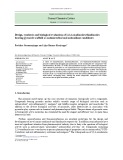
Fig. 1. The role of PARP-1 within the ischemic neurovascular units. PARP-1 exerts its detrimental role within the ischemic neurovascular unit by promoting necrosis and AIF-dependent apoptosis in neurons, astrocytes and endothelial cells. PARP-1 also plays a key role in immune activation and migration of microglial cells upon different noxious stimuli to the central nervous system. The expression of adhesion molecules by endothelial cells is also promoted by PARP-1-dependent transcriptional activation, thereby promoting leukocyte recruitment within the ischemic brain tissue and their detrimental effects on ischemic injury. Hence, the pharmacological inhibition of the enzyme exerts ischemic neuroprotection by targeting several pleiotypic events of pathogenetic relevance to post-ischemic brain damage. X, adhesion mole- cules. ADP-ribose monomers are depicted as black circles binding to the transient receptor potential melastatin-2 receptor.
occurring in neurons. Regardless, the hyperactivation of PARP-1 in each single component of the neurovas- cular unit triggers dysfunction ⁄ cytotoxicity and, indi- rectly, severely affects the functioning of neighbouring neurons. As a whole, PARP-1-dependent derangement of the integrity of the neurovascular unit is caused by the enzyme’s ability to prompt an increase of blood– brain barrier (BBB) permeability, the release of pro- inflammatory mediators, mitochondrial dysfunction and bioenergetic failure, as well as the activation of specific apoptotic pathways.
PARP-1, endothelia and post-ischemic BBB breakdown
[35–37]. Accordingly,
Ischemia causes rapid structural changes and break- down of the BBB, allowing plasma exudation and immune cell infiltration, which contribute to ischemic brain damage [22]. Very early after the onset of brain ischemia, and especially after a reperfusion period, abundant free radicals are generated in macrophages, endothelial cells, perycites, astrocytes, microglia and
neurons, causing significant damage to brain capillaries and disruption of the BBB [33]. Free radicals formed both inside and outside the vessels prompt genotoxic stress and activate PARP-1 in endothelial cells. Under conditions of chronic hypoxia, PARP-1 activation within endothelia triggers cell proliferation and slowly developing brain damage. The molecular mechanisms of cell proliferation include the generation and release of ROS from NADPH oxidase and mitochondria, sus- tained increase of the cytosolic Ca2+ concentration and finally nuclear translocation of mitogen-activated protein kinase kinase ⁄ extracellular regulated protein kinase with cell cycle activation [34]. Conversely, during ischemia, PARP-1 hyperactivation causes endo- thelial cell death. The latter occurs because of cellular the PARP-1 product poly(ADP- accumulation of ribose), which causes translocation of apoptosis-induc- ing factor (AIF) from mitochondria to the nucleus and activation of a caspase-independent programmed cell- the potent death pathway PARP-1 inhibitor, PJ34, administered to rats with transient focal brain ischemia, preserves the integrity
FEBS Journal 276 (2009) 36–45 ª 2008 The Authors Journal compilation ª 2008 FEBS
39
F. Moroni and A. Chiarugi
PARP-1 and the ischemic neurovascular unit
products,
death
and
cell
capillary basal lamina, opening of the BBB and infil- tration of blood-borne leukocytes. This prompts a vicious circle comprising waves of release of cytotoxic inflammatory recruit- ment ⁄ activation of blood or bystander immune cells. Eventually, the neuroimmune response causes collapse of the structures and functions of the neurovascular unit [16,17,45].
inducible nitric oxide
factor-alfa
(TNF-a)
of endothelial tight junctions and decreases the expres- sion of the adhesion molecule intercellular adhesion molecule-1, thus limiting leukocyte infiltration and the subsequent inflammatory damage to the ischemic brain [35,38]. It has also been proposed that post-ischemic PARP-1 activation contributes to increased expression of matrix metalloproteinases (MMPs), a group of zinc- containing proteases with key roles in matrix degrada- tion and disruption of capillary permeability during stoke [39]. Indeed, pharmacological PARP-1 inhibition reduces MMP-9 expression levels in plasma and brain [40], prevents brain matrix degradation, reduces delayed increase of BBB permeability and edema for- mation, preserves endothelial tight junction proteins and decreases delayed infiltration of leukocytes into the brain of rats with middle cerebral artery occlusion [41]. The key role of PARP-1 hyperactivation in endo- thelial dysfunction in experimental models of diabetes underscores the pathogenetic relevance of the enzyme to disorders of this key component of the neurovascu- lar unit [42]. Accordingly, gene array studies have demonstrated that upregulation of inflammatory genes is hampered in PARP-1) ⁄ ) endothelial cells exposed to tumor necrosis [43]. Taken together, these findings point to basal PARP-1 activity to homeostatic regulation of endothelial as central function, whereas its hyperactivation appears causal to BBB damage and immune cell infiltration during ischemia.
activated T-cells
are positively
PARP-1, glia and post-ischemic inflammatory events
Again, PARP-1 plays a key role in this scenario. Indeed, numerous reports demonstrate that PARP-1 activity promotes the neuroimmune response thanks to its ability to assist transcriptional activation and epigenetic remodeling in immune cells. In this light, it has been speculated that ischemic neuroprotection afforded by PARP inhibitors is at least partially med- iated by their anti-inflammatory properties [46]. expression of Indeed, PARP inhibitors decrease such as CD11b, inflammatory markers ⁄ mediators cyclooxygenase-2, synthase, TNF-a, IL-1b, IL-6, intracellular adhesion molecule-1, interferon-gamma and E-selectin in different models these of neurodegeneration [40,47–55]. Remarkably, molecules actively contribute to ischemic neurodegen- eration. A key role for PARP-1 in microglia activa- tion and migration towards injured neurons has also been reported [56]. Reduced expression of pro-inflam- matory mediators is probably a result of the fact that inflammatory transcription factors such as nuclear factor-kappaB, activator protein-1 and nuclear factor regulated by of PARP-1. PARP-1 protein per se, as well as its enzy- matic activity, promote transcription factor binding to DNA as well as supramolecular complex formation transcription-regulating proteins containing several and RNA polymerase II [23,53,57]. These findings taken together may explain why post-treatment with PARP-1 inhibitors reduces the neuroimmune response in different stroke models [58–60].
Recently,
to reduce
strategies able
[62].
Interestingly,
impairment it has
Activation of resident immune cells as well as infiltra- tion of leukocytes within the ischemic area lead to excessive release of inflammatory mediators and ensu- ing worsening of brain damage. In keeping with this, astrocytes, microglia and blood-derived leukocytes contribute to ischemic neurodegeneration, whereas immunosuppressant the inflammatory response decrease infarct volumes in dif- ferent stroke models [16,17]. Microglial cells are resi- dent brain macrophages displaying a ‘resting’ highly ramified phenotype. Upon ischemic challenge, before neuronal damage can be morphologically detected [44], microglia assume amoeboid morphology and acquire phagocytic activity, producing ROS and other inflam- matory ⁄ cytotoxic factors such as nitric oxide, prosta- noids, TNF-a, IL-1b and MMPs. Astrocytes and infiltrating leukocytes within the ischemic brain tissue also contribute to the synthesis and release of pro- inflammatory mediators [17]. It is now widely accepted that the latter are responsible for disruption of the
the tetracycline, minocycline, has been proposed as a clinically relevant tool to limit post- ischemic brain damage because of its ability to inhibit microglia activation. Minocycline is indeed able to reduce brain infarct volumes in preclinical models [61], as well as neurological in stroke recently been patients reported that minocycline is a powerful inhibitor of PARP-1 [63]. Whether PARP-1 inhibition underpins the drug’s neuroprotective effects in stroke patients is currently unknown. Yet, given that minocycline has been largely used without significant side effects, these observations indicate that acute inhibition of PARP-1 in vivo might be a rather safe procedure and could be the ischemic proposed to preserve the integrity of
FEBS Journal 276 (2009) 36–45 ª 2008 The Authors Journal compilation ª 2008 FEBS
40
F. Moroni and A. Chiarugi
PARP-1 and the ischemic neurovascular unit
to the hypothesis
neurovascular unit and limit post-ischemic brain damage in humans.
PARP-1 and post-ischemic death in neurons
that PARP-1 apparent contrast worsens ischemic neurodegeneration by reducing ATP levels within the injured tissue, however, ischemia- induced energy derangement is similar in the affected brain areas of PARP-1+ ⁄ + and PARP-1) ⁄ ) mice, despite the latter showing significant reduction of ischemic volumes [73].
In this
intracellular [Ca2+]
(but also see
[83]
reduce
Controversy still exists on the molecular mecha- nisms involved in PARP-1-dependent neuronal death during ischemia. regard it has been very recently reported that exposure of cultured neurons to poly (ADP-ribose) is sufficient to trigger nuclear translocation of mitochondrial AIF and cell demise [74]. The poly(ADP-ribose)-degrading enzyme, poly (ADP-ribose) glycohydrolase (PARG), should be, in principle, a neuroprotective agent [75]. Consistently, PARG-110 kDa) ⁄ ) or PARG+ ⁄ ) mice show increased sensitivity to brain ischemia [36,76]. Also, PARP-1 activity seems to be essential for AIF release within neurons of the infarct area, and AIF-deficient (Harle- quin) mice are less sensitive to post-ischemic brain damage [77]. Data therefore point to PARP-1 activ- ity-dependent AIF release from mitochondria as a key molecular event underlying ischemic neuronal death. Interestingly, the ADP-ribose monomers origi- nating from the polymer degradation through PARG might also contribute to neuronal demise by activat- ing transient receptor potential melastatin-2 receptors and massive Ca2+ influx [78,79]. Finally, the finding that, when released in the extracellular space, high- mobility-group protein box 1 (HMGB1) promotes the neuroinflammatory response and worsens brain ische- mia [80–82], along with evidence that PARP-1 pro- [82]), motes HMGB1 release indicate that HMGB1 may mediate, in part, the toxic effect of PARP-1 hyperactivation within the ischemic brain tissue. Overall, a wealth of evidence points to the synthesis of poly (ADP-ribose) within ischemic neurons as a crucial event contributing to derange- ment of the neurovascular unit.
Conclusion
for pharmacological
Excitotoxicity and PARP-1 activation have been caus- ally linked since 1994 when it was reported that gluta- mate increases poly(ADP-ribose) synthesis and causes a type of cell death that is prevented by both NMDA antagonists and PARP-1 inhibitors [25,26]. The pro- posed molecular events underlying these observations include: overactivation of NMDA glutamate receptors with consequent intracellular Ca2+ influx; and subse- quent ROS production mainly caused by neuronal nitric oxide synthase activity, which, in turn, triggers DNA damage-dependent hyperactivation of PARP-1, depletion of intracellular NAD and ATP stores, and neuronal death [26]. PARP-1 activation may also occur in neurons without NMDA receptor activation, as triggered by K+- increases of induced depolarization or inositol 3-phosphate-recep- tor activation are sufficient to trigger poly(ADP-ribose) formation [28,64]. In keeping with this toxic cascade of events, neurons obtained from PARP-1-deficient mice are resistant to NMDA toxicity and to oxygen and glucose deprivation [65]. It was also shown that NMDA-induced overload of cytosolic Ca2+ not only activates neuronal nitric oxide synthase in the cytosol, but is also responsible for mitochondrial ROS produc- tion [66], which contributes to DNA damage and fur- ther activation of PARP-1 [67,68]. Substantial DNA damage, evaluated by means of the comet assay, is present in cells isolated from the rat ischemic cortex or caudate. NMDA receptor antagonists the extent of the damage and provide ischemic neuropro- tection, while PARP inhibitors decrease infarct vol- umes without affecting the severity of DNA damage [69]. These observations suggest that NMDA receptor channel openings, ROS formation, DNA damage and PARP activation are sequential crucial steps in the process leading to neuronal death. They also indicate that stroke protection can be achieved without reduc- ing DNA damage. Energy failure following PARP-1 activation is not only caused by NAD resynthesis but also by glycolysis block because of NAD depletion, which results in reduced synthesis of both glycolysis- derived ATP and mitochondrial energetic substrates [70]. Accordingly, tricarboxylic acid cycle substrates or extracellular NAD supplementation protect neurons activation [71], whereas from excessive PARP-1 PARP-1 inhibitors prevent ischemia-induced NAD+ depletion and reduce ischemic brain injury [72]. In
To reduce brain damage after stroke it is not sufficient to protect neurons from excitotoxic insults, but it is mandatory to rescue all cellular and structural compo- nents of the neurovascular unit. As outlined above, PARP-1 activation during brain ischemia plays a detri- mental role in all cell types of the neurovascular unit. Inhibitors of PARP-1 might therefore represent a class of ‘pleiotypic drugs’, which are considered the most promising tools treatment of stroke. Also, the different temporal kinetics of PARP-1
FEBS Journal 276 (2009) 36–45 ª 2008 The Authors Journal compilation ª 2008 FEBS
41
F. Moroni and A. Chiarugi
PARP-1 and the ischemic neurovascular unit
11 Moroni F, Lombardi G, Thomsen C, Leonardi P,
Attucci S, Peruginelli F, Albani Torregrossa S, Pelleg- rini-Giampietro DE, Luneia R & Pellicciari R (1997) Pharmacological characterization of 1-aminoindan-1,5- dicarboxylic acid (AIDA), a potent mGluR1 antagonist. J Pharmacol Exp Ther 281, 721–729.
activation within the components of the neurovascular unit would warrant a significant ‘window of opportu- nity’ to be harnessed for the treatment of stroke patients. Remarkably, the clinical relevance of PARP-1 inhibitors in stroke treatment is emphasized by the fact that these drugs are well tolerated by patients enrolled in clinical trials for treatment of tumor malignancies or coronary bypass, and that, theoretically, anti-stroke treatment with PARP-1 inhibitors would require an acute, 4–6-day treatment. This, of course, would reduce the risk of side effects. The latter might be fur- ther reduced by the forthcoming development of PARP isoform-specific inhibitors [84]. In conclusion, preclinical and clinical data indicate that PARP-1 is a very promising target for ischemic neuroprotection, and PARP-1 inhibitors represent a realistic new avenue to stroke treatment.
12 Pellegrini-Giampietro DE, Peruginelli F, Meli E, Cozzi A, Albani Torregrossa S, Pellicciari R & Moroni F (1999) Protection with metabotropic glutamate 1 receptor antagonists in models of ischemic neuronal death: time course and mechanisms. Neuropharmacology 38, 1607–1621.
13 Moroni F, Attucci S, Cozzi A, Meli E, Picca R, Schei- deler MA, Pellicciari R, Noe C, Sarichelou I & Pelleg- rini-Giampietro DE (2002) The novel and systemically active metabotropic glutamate 1 (mGlu1) receptor antagonist 3-MATIDA reduces post-ischemic neuronal death. Neuropharmacology 42, 741–751.
References
1 Lo EH, Dalkara T & Moskowitz MA (2003) Mecha- 14 Lees KR, Zivin JA, Ashwood T, Davalos A, Davis SM, Diener HC, Grotta J, Lyden P, Shuaib A, Hardemark HG et al. (2006) NXY-059 for acute ischemic stroke. N Engl J Med 354, 588–600. 15 Garber K (2007) Stroke treatment–light at the end of nisms, challenges and opportunities in stroke. Nat Rev Neurosci 4, 399–415. the tunnel? Nat Biotechnol 25, 838–840. 16 Barone FC & Feuerstain GZ (1999) Inflammatory
2 Dirnagl U, Iadecola C & Moskowitz MA (1999) Patho- biology of ischaemic stroke: an integrated view. Trends Neurosci 22, 391–397.
mediators and stroke: novel opportunities for novel therapeutics. J Cereb Blood Flow Metab 19, 819–834. 17 Iadecola C & Alexander M (2001) Cerebral ischemia and inflammation. Curr Opin Neurol 14, 89–94. 18 Wang X & Feuerstein GZ (2004) The Janus face of 3 O’Collins VE, Macleod MR, Donnan GA, Horky LL, van der Worp BH & Howells DW (2006) 1,026 experi- mental treatments in acute stroke. Ann Neurol 59, 467–477. inflammation in ischemic brain injury. Acta Neurochir Suppl 89, 49–54. 19 Feuerstein GZ, Zaleska MM, Krams M, Wang X, 4 Savitz SI & Fisher M (2007) Future of neuroprotection for acute stroke: in the aftermath of the SAINT trials. Ann Neurol 61, 396–402. 5 Benveniste H, Drejer J, Schousboe A & Diemer N
(1984) Elevation of extracellular concentrations of glu- tamate and aspartate in rat hippocampus during tran- sient cerebral ischemia monitored by intracerebral microdialysis. J Neurochem 43, 1369–1374. Day M, Rutkowski JL, Finklestein SP, Pangalos MN, Poole M, Stiles GL et al. (2008) Missing steps in the STAIR case: a Translational Medicine perspective on the development of NXY-059 for treatment of acute ischemic stroke. J Cereb Blood Flow Metab 28, 217– 219. 6 Lombardi G & Moroni F (1992) GM1 ganglioside
20 Astrup J, Siesjo¨ BK & Symon L (1981) Thresholds in cerebral ischemia: the ischemic penumbra. Stroke 12, 723–725. reduces ischemia-induced excitatory amino acid output: a microdialysis study in the gerbil hippocampus. Neuro- sci Lett 134, 171–174. 21 Iadecola C, Goldman SS, Harder DR, Heistad DD, 7 Choi DW (1992) Excitotoxic cell death. J Neurobiol 23, 1261–1276. 8 Lipton SA & Rosenberg PA (1994) Excitatory amino
acids as a final common pathway for neurological disor- ders. N Engl J Med 330, 613–622. Katusic ZS, Moskowitz MA, Simard JM, Sloan MA, Traystman RJ & Velletri PA (2006) Recommendations of the National Heart, Lung, and Blood Institute work- ing group on cerebrovascular biology and disease. Stroke 37, 1578–1581.
9 Meldrum B & Garthwaite J (1990) Excitatory amino acid neurotoxicity and neurodegenerative disease. Trends Pharmacol Sci 11, 379–387. 22 del Zoppo GJ & Mabuchi T (2003) Cerebral microves- sel responses to focal ischemia. J Cereb Blood Flow Metab 23, 879–894. 10 Nicoletti F, Bruno VM, Copani A, Casabona G &
FEBS Journal 276 (2009) 36–45 ª 2008 The Authors Journal compilation ª 2008 FEBS
42
Knopfel T (1996) Metabotropic glutamate receptors: a new target for the therapy of neurodegenerative disor- ders? Trends Neurosci 19, 267–271. 23 Hassa PO, Haenni SS, Elser M & Hottiger MO (2006) Nuclear ADP-ribosylation reactions in mammalian cells: where are we today and where are we going? Microbiol Mol Biol Rev 70, 789–829.
F. Moroni and A. Chiarugi
PARP-1 and the ischemic neurovascular unit
24 Chambon P, Weill JD & Mandel P (1963) Nicotinamide mononucleotide activation of new DNA-dependent polyadenylic acid synthesizing nuclear enzyme. Biochem Biophys Res Commun 11, 39–43. 38 Zhang Y, Park TS & Gidday JM (2007) Hypoxic pre- conditioning protects human brain endothelium from ischemic apoptosis by Akt-dependent survivin activa- tion. Am J Physiol Heart Circ Physiol 292, H2573– H2581.
39 Rosell A & Lo EH (2008) Multiphasic roles for matrix metalloproteinases after stroke. Curr Opin Pharmacol 8, 82–89. 25 Cosi C, Suzuki H, Milani D, Facci L, Menegazzi M, Vantini G, Kanai Y & Skaper SD (1994) Poly(ADP- ribose)polymerase: early involvement in glutamate- induced neurotoxicity in cultured cerebellar granule cells. J Neurosci Res 39, 38–46.
26 Zhang J, Dawson VL, Dawson T & Snyder SH (1994) Nitric oxide activation of poly(ADP-ribose) synthetase in neurotoxicity. Science 263, 687–689. 40 Koh SH, Chang DI, Kim HT, Kim J, Kim MH, Kim KS, Bae I, Kim H, Kim DW & Kim SH (2005) Effect of 3-aminobenzamide, PARP inhibitor, on matrix me- talloproteinase-9 level in plasma and brain of ischemic stroke model. Toxicology 214, 131–139. 41 Lenzser G, Kis B, Snipes JA, Gaspar T, Sandor P, 27 Szabo C & Dawson VL (1998) Role of poly(ADP- ribose) synthetase in inflammation and ischaemia- reperfusion. Trends Pharmacol Sci 19, 287–298.
Komjati K, Szabo C & Busija DW (2007) Contribution of poly(ADP-ribose) polymerase to postischemic blood-brain barrier damage in rats. J Cereb Blood Flow Metab 27, 1318–1326.
28 Homburg S, Visochek L, Moran N, Dantzer F, Priel E, Asculai E, Schwartz D, Rotter V, dekel N & Cohen- Armon M (2000) A fast signal induced activation of poly(ADP-ribose)polymerase: a downstream target of phospholipase C. J Cell Biol 150, 293–307.
42 Pacher P, Liaudet L, Oriano FG, Abley JG, Czabo E & Czabo C (2002) The role of poly(ADP-ribose) polymer- ase activation in the development of myocardial and endothelial dysfunction in diabetes. Diabetes 51, 514– 521. 29 Szabo C, Pacher P & Swanson RA (2006) Novel modu- lators of poly(ADP-ribose) polymerase. Trends Pharma- col Sci 27, 626–630. 30 Alano CC & Swanson RA (2006) Players in the PARP-1
cell-death pathway: JNK1 joins the cast. Trends Biochem Sci 31, 309–311. 31 Chiarugi A (2005) Poly(ADP-ribosyl)ation and stroke. 43 Carrillo A, Monreal Y, Ramirez P, Marin L, Parrilla P, Oliver FJ & Yelamos J (2004) Transcription regulation of TNF-alpha-early response genes by poly(ADP- ribose) polymerase-1 in murine heart endothelial cells. Nucleic Acids Res 32, 757–766. Pharmacol Res 52, 15–24. 44 Jorgensen MB, Finsen BR, Jensen MB, Castellano B, 32 Chiarugi A (2005) Intrinsic mechanisms of poly(ADP-
ribose) neurotoxicity: three hypotheses. Neurotoxicology 26, 847–855.
Diemer NH & Zimmer J (1993) Microglial and astrogli- al reactions to ischemic and kainic acid-induced lesions of the adult rat hippocampus. Exp Neurol 120, 70–88. 45 del Zoppo GJ (2006) Stroke and neurovascular protec- tion. N Engl J Med 354, 553–555.
46 Chiarugi A (2002) PARP-1: killer or conspirator? The suicide hypothesis revisited. Trends Pharmacol Sci 23, 122–129.
33 Gursoy-Ozdemir Y, Can A & Dalkara T (2004) Reperfu- sion-induced oxidative ⁄ nitrative injury to neurovascular unit after focal cerebral ischemia. Stroke 35, 1449–1453. 34 Abdallah Y, Gligorievski D, Kasseckert SA, Dieterich L, Schafer M, Kuhlmann CR, Noll T, Sauer H, Piper HM & Schafer C (2007) The role of poly(ADP-ribose) polymerase (PARP) in the autonomous proliferative response of endothelial cells to hypoxia. Cardiovasc Res 73, 568–574. 35 Zhang Y, Zhang X, Park TS & Gidday JM (2005) 47 Scott GS, Hake P, Kean RB, Virag L, Szabo C & Hoo- per DC (2001) Role of poly(ADP-ribose) synthetase activation in the development of experimental allergic encephalomyelitis. J Neuroimm 117, 78–86.
Cerebral endothelial cell apoptosis after ischemia-reper- fusion: role of PARP activation and AIF translocation. J Cereb Blood Flow Metab 25, 868–877. 36 Yu SW, Andrabi SA, Wang H, Kim NS, Poirier GG, 48 Chiarugi A (2002) Inhibitors of poly(ADP-ribose) poly- merase-1 suppress transcriptional activation in lympho- cytes and ameliorate autoimmune encephalomyelitis in rats. Br J Pharmacol 137, 761–770. 49 Ha HC, Hester LD & Snyder SH (2002) Poly(ADP-
Dawson TM & Dawson VL (2006) Apoptosis-inducing factor mediates poly(ADP-ribose) (PAR) polymer- induced cell death. Proc Natl Acad Sci U S A 103, 18314–18319. ribose) polymerase-1 dependence of stress-induced tran- scription factors and associated gene expression in glia. Proc Natl Acad Sci U S A 99, 3270–3275. 37 Moubarak RS, Yuste VJ, Artus C, Bouharrour A,
50 Ha HC, Hester LD & Snyder SH (2002) Poly(ADP- ribose) polymerase-1 dependence of stress-induced transcription factors and associated gene expression in glia. Proc Natl Acad Sci U S A 99, 3270–3275.
FEBS Journal 276 (2009) 36–45 ª 2008 The Authors Journal compilation ª 2008 FEBS
43
51 Chiarugi A & Moskowitz MA (2003) Poly(ADP-ribose) polymerase-1 activity promotes NF-kappaB-driven tran- Greer PA, Menissier-de Murcia J & Susin SA (2007) Sequential activation of poly(ADP-ribose) polymerase 1, calpains, and Bax is essential in apoptosis-inducing factor-mediated programmed necrosis. Mol Cell Biol 27, 4844–4862.
F. Moroni and A. Chiarugi
PARP-1 and the ischemic neurovascular unit
glutamate receptors stimulate the activity of poly(ADP- ribose) polymerase in mammalian mGlu1-transfected cells and in cortical cell cultures. Neuropharmacology 49(Suppl 1), 80–88.
scription and microglial activation: implication for neu- rodegenerative disorders. J Neurochem 85, 306–317. 52 Koh SH, Park Y, Song CW, Kim JG, Kim K, Kim J, Kim MH, Lee SR, Kim DW, Yu HJ et al. (2004) The effect of PARP inhibitor on ischaemic cell death, its related inflammation and survival signals. Eur J Neuro- sci 20, 1461–1472. 65 Pieper A, Verma A, Zhang J & Snyder SH (1999) Poly (ADP-ribose)polymerase, nitric oxide and cell death. Trends Pharmacol Sci 20, 171–181.
66 Reynolds IJ & Hastings TG (1995) Glutamate induces the production of reactive oxygen species in cultured forebrain neurons following NMDA receptor activa- tion. J Neurosci 15, 3318–3327.
53 Nakajima H, Nagaso H, Kakui N, Ishikawa M, Hira- numa T & Hoshiko S (2004) Critical role of the auto- modification of poly(ADP-ribose) polymerase-1 in nuclear factor-kappaB-dependent gene expression in pri- mary cultured mouse glial cells. J Biol Chem 279, 42774–42786.
67 Mandir AS, Poitras MF, Berliner AR, Herring WJ, Guastella DB, Feldman A, Poirier GG, Wang ZQ, Dawson TM & Dawson VL (2000) NMDA but not non-NMDA excitotoxicity is mediated by Poly(ADP- ribose) polymerase. J Neurosci 20, 8005–8011.
54 Haddad M, Rhinn H, Bloquel C, Coqueran B, Szabo C, Plotkine M, Scherman D & Margaill I (2006) Anti- inflammatory effects of PJ34, a poly(ADP-ribose) poly- merase inhibitor, in transient focal cerebral ischemia in mice. Br J Pharmacol 149, 23–30.
68 Duan Y, Gross RA & Sheu SS (2007) Ca2 + -depen- dent generation of mitochondrial reactive oxygen spe- cies serves as a signal for poly(ADP-ribose) polymerase- 1 activation during glutamate excitotoxicity. J Physiol 585, 741–758.
55 Lee JH, Park SY, Shin HK, Kim CD, Lee WS & Hong KW (2007) Poly(ADP-ribose) polymerase inhibition by cilostazol is implicated in the neuroprotective effect against focal cerebral ischemic infarct in rat. Brain Res 1152, 182–190. 56 Ullrich O, Diestel A, Eyupoglu IY & Nitsch R (2001)
69 Giovannelli L, Cozzi A, Guarnieri I, Dolara P & Mor- oni F (2002) Comet assay as a novel approach for studying DNA damage in focal cerebral ischemia: dif- ferential effects of NMDA receptor antagonists and poly(ADP-ribose) polymerase inhibitors. J Cereb Blood Flow Metab 22, 697–704. 70 Ying W, Alano CC, Garnier P & Swanson RA (2005)
Regulation of microglial expression of integrins by poly (ADP-ribose) polymerase-1. Nat Cell Biol 3, 1035–1042. 57 Kraus WL (2008) Transcriptional control by PARP-1: chromatin modulation, enhancer-binding, coregulation, and insulation. Curr Opin Cell Biol 20, 294–302. 58 Ducrocq S, Benjelloun N, Plotkine M, Ben-Ari Y & NAD+ as a metabolic link between DNA damage and cell death. J Neurosci Res 79, 216–223.
Charriaut-Marlangue C (2000) Poly(ADP-ribose) Syn- thethase inhibition reduces ischemic injury and inflamma- tion in neonatal rat brain. J Neurochem 74, 2504–2511. 59 Strosznajder RP, Jesko H & Zambrzycka A (2005) 71 Ying W, Chen Y, Alano CC & Swanson RA (2002) Tri- carboxylic acid cycle substrates prevent PARP-mediated death of neurons and astrocytes. J Cereb Blood Flow Metab 22, 774–779.
Poly(ADP-ribose) polymerase: the nuclear target in sig- nal transduction and its role in brain ischemia-reperfu- sion injury. Mol Neurobiol 31, 149–167. 72 Endres M, Wang ZQ, Namura S, Waeber C & Mosko- witz MA (1997) Ischemic brain injury is mediated by the activation of poly(ADP-ribose)polymerase. J Cereb Blood Flow Metab 17, 1143–1151.
60 Hamby AM, Suh SW, Kauppinen TM & Swanson RA (2007) Use of a poly(ADP-ribose) polymerase inhibitor to suppress inflammation and neuronal death after cere- bral ischemia-reperfusion. Stroke 38, 632–636. 61 Yrjanheikki J, Keinanen R, Pellikka M, Hokfelt T & 73 Goto S, Xue R, Sugo N, Sawada M, Blizzard KK, Poi- tras MF, Johns DC, Dawson TM, Dawson VL, Crain BJ et al. (2002) Poly(ADP-ribose) polymerase impairs early and long-term experimental stroke recovery. Stroke 33, 1101–1106. 74 Andrabi SA, Kim NS, Yu SW, Wang H, Koh DW, Koistinaho J (1998) Tetracyclines inhibit microglial acti- vation and are neuroprotective in global brain ischemia. Proc Natl Acad Sci U S A 95, 15769–15774.
Sasaki M, Klaus JA, Otsuka T, Zhang Z, Koehler RC et al. (2006) Poly(ADP-ribose) (PAR) polymer is a death signal. Proc Natl Acad Sci U S A 103, 18308– 18313.
75 Koh DW, Dawson VL & Dawson TM (2005) The road to survival goes through PARG. Cell Cycle 4, 397–399.
FEBS Journal 276 (2009) 36–45 ª 2008 The Authors Journal compilation ª 2008 FEBS
44
62 Lampl Y, Boaz M, Gilad R, Lorberboym M, Dabby R, Rapoport A, Anca-Hershkowitz M & Sadeh M (2007) Minocycline treatment in acute stroke: an open-label, evaluator-blinded study. Neurology 69, 1404–1410. 63 Alano CC, Kauppinen TM, Valls AV & Swanson RA (2006) Minocycline inhibits poly(ADP-ribose) polymer- ase-1 at nanomolar concentrations. Proc Natl Acad Sci U S A 103, 9685–9690. 64 Meli E, Baronti R, Pangallo M, Picca R, Moroni F & 76 Cozzi A, Cipriani G, Fossati S, Faraco G, Formentini L, Min W, Cortes U, Wang ZQ, Moroni F & Chiarugi A (2006) Poly(ADP-ribose) accumulation and enhance- ment of postischemic brain damage in 110-kDa poly Pellegrini-Giampietro DE (2005) Group I metabotropic
F. Moroni and A. Chiarugi
PARP-1 and the ischemic neurovascular unit
(ADP-ribose) glycohydrolase null mice. J Cereb Blood Flow Metab 26, 684–695.
81 Faraco G, Fossati S, Bianchi ME, Patrone M, Pedrazzi M, Sparatore B, Moroni F & Chiarugi A (2007) High mobility group box 1 protein is released by neural cells upon different stresses and worsens ischemic neuro- degeneration in vitro and in vivo. J Neurochem 103, 590–603.
77 Culmsee C, Zhu C, Landshamer S, Becattini B, Wagner E, Pellechia M, Blomgren K & Plesnila N (2005) Apop- tosis-inducing factor triggered by poly(ADP-Ribose) polymerase and bid mediates neuronal cell death after oxygen-glucose deprivation and focal cerebral ischemia. J Neurosci 25, 10262–10272.
78 Fonfria E, Marshall IC, Benham CD, Boyfield I, Brown JD, Hill K, Hughes JP, Skaper SD & McNulty S (2004) TRPM2 channel opening in response to oxidative stress is dependent on activation of poly(ADP-ribose) poly- merase. Br J Pharmacol 143, 186–192. 82 Qiu J, Nishimura M, Wang Y, Sims JR, Qiu S, Savitz SI, Salomone S & Moskowitz MA (2008) Early release of HMGB-1 from neurons after the onset of brain ischemia. J Cereb Blood Flow Metab 28, 927–938. 83 Ditsworth D, Zong WX & Thompson CB (2007) Activa- tion of poly(ADP)-ribose polymerase (PARP-1) induces release of the pro-inflammatory mediator HMGB1 from the nucleus. J Biol Chem 282, 17845–17854.
FEBS Journal 276 (2009) 36–45 ª 2008 The Authors Journal compilation ª 2008 FEBS
45
79 McNulty S & Fonfria E (2005) The role of TRPM channels in cell death. Pflugers Arch 451, 235–242. 80 Kim JB, Sig CJ, Yu YM, Nam K, Piao CS, Kim SW, Lee MH, Han PL, Park JS & Lee JK (2006) HMGB1, a novel cytokine-like mediator linking acute neuronal death and delayed neuroinflammation in the post- ischemic brain. J Neurosci 26, 6413–6421. 84 Pellicciari R, Camaioni E, Costantino G, Formentini L, Sabbatini P, Venturoni F, Eren G, Bellocchi D, Chia- rugi A & Moroni F (2008) On the way to selective PARP-2 inhibitors. Design, synthesis, and preliminary evaluation of a series of isoquinolinone derivatives. ChemMedChem 3, 914–923.