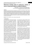
Repeats of LacdiNAc and fucosylated LacdiNAc on N-glycans of the human parasite Schistosoma mansoni Manfred Wuhrer, Carolien A. M. Koeleman, Andre´ M. Deelder and Cornelis H. Hokke
Biomolecular Mass Spectrometry Unit, Department of Parasitology, Center of Infectious Diseases, Leiden University Medical Center, The Netherlands
Keywords mass spectrometry; parasite; terminal N-acetylgalactosamine; trematode
Correspondence M. Wuhrer, Department of Parasitology, Leiden University Medical Center, P.O. Box 9600, 2300 RC Leiden, the Netherlands Fax: +31 71 526 6907 Tel: +31 71 526 5077 E-mail: m.wuhrer@lumc.nl
(Received 15 September 2005, revised 11 November 2005, accepted 21 November 2005)
doi:10.1111/j.1742-4658.2005.05068.x
N-Glycans from glycoproteins of the worm stage of the human parasite Schistosoma mansoni were enzymatically released, fluorescently labelled and analysed using various mass spectrometric and chromatographic methods. A family of 28 mainly core-a1–6-fucosylated, diantennary N-glycans of composition Hex3)4HexNAc6)12Fuc1)6 was found to carry dimers of N,N¢- diacetyllactosediamine [LacdiNAc or LDN; GalNAc(b1–4)GlcNAc(b1-] with or without fucose a1–3-linked to the N-acetylglucosamine residues in the antennae {GalNAc(b1–4)[±Fuc(a1–3)]GlcNAc(b1–3)GalNAc(b1– 4)[±Fuc(a1–3)]GlcNAc(b1-}. To date, oligomeric LDN and oligomeric fucosylated LDN (LDNF) have been found only on N-glycans from mammalian cells engineered to express Caenorhabditis elegans b4-GalNAc transferase and human a3-fucosyltransferase IX [Z. S. Kawar et al. (2005) J Biol Chem 280, 12810–12819]. It now appears that LDN(F) repeats can also occur in a natural system such as the schistosome parasite. Like monomeric LDN and LDNF, the dimeric LDN(F) moieties found here are expected to be targets of humoral and cellular immune responses during schistosome infection.
expressed by various pathogens, including the human parasite Schistosoma mansoni [2]. Glycans with LDN and LDNF {GalNAc(b1–4)[Fuc(a1–3)]GlcNAc(b1-} expressed by S. mansoni are targets of the humoral immune responses of the host [19], and LDN-contain- ing glycoconjugates of schistosomes may be ligands for galectin-3-mediated immune recognition [7].
The most common complex-type N-glycans of mam- mals contain N-acetyllactosamine-type antennae [Lac- NAc; Gal(b1–4)GlcNAc(b1-], which can be substituted in various positions by fucoses, sialic acids, sulfate, glucuronic acid, etc. LacNAc-type antennae are also seen in other eukaryotes [1–5]. The arising terminal structures can be antigenic [2,6] or may act as ligands of lectins [7,8]. They may modify cell–cell interaction, for example in development [9–12] and cancer [13–15]. An alternative antennary structure is N,N¢-diacetyl- [LacdiNAc or LDN; GalNAc(b1– lactosediamine 4)GlcNAc(b1-], which may likewise be modified by various substituents such as fucose, sulfate or sialic acid. LDN-based motifs have been found on N- or O-glycans of various mammalian glycoproteins inclu- ding seminal plasma glycodelin [16], thyrotropin [17] and tissue factor pathway inhibitor [18], but are also
In humans, biosynthesis of LDN can occur by two different b1–4-N-acetylgalactosaminyltransferases: b4GalNAc-T3, which is expressed in stomach, colon and testes at high levels [20]; and b4GalNAc-T4, which is transcribed in ovaries and brain tissues [21]. From the nematode Caenorhabditis elegans, a b1–4-galactos- aminyltransferase involved in LDN has likewise been identified [22]. The expression of this enzyme in Chi- nese hamster ovary (CHO) Lec8 cells has led to the production of N-glycans with LDN repeats, which
Abbreviations 2AB, 2-aminobenzamide; CHO, Chinese hamster ovary cells; F, deoxyhexose; H, hexose; IT, ion-trap; LacNAc, Gal(b1–4)GlcNAcb1-; LC, liquid chromatography; LDN or LacdiNAc, N,N¢-diacetyllactosediamine; LDNF, GalNAc(b1–4)[±Fuc(a1–3)]GlcNAc(b1-; N, N-acetylhexosamine; PNGase F, peptide N-glycosidase F.
FEBS Journal 273 (2006) 347–361 ª 2005 The Authors Journal compilation ª 2005 FEBS
347
M. Wuhrer et al.
S. mansoni N-glycans with dimeric LacdiNAc
A
B
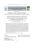
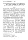
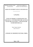
Fig. 1. MALDI-TOF-MS of N-glycans released from S. mansoni adult worms. (A) Low mass range; (B) high mass range; ·8, intensities eight times enlarged; F, deoxyhexose; H, hexose; N, N-acetylhexosamine. N-glycan species containing eight or more HexNAc residues are labelled in bold-type.
were converted to LDNF repeats by coexpression of human a1–3-fucosyltransferase IX [23]. The mam- malian b1–3-N-acetylglucosaminyltransferase(s) which contribute(s) to the synthesis of this alternating chain have yet to be identified [23].
Whereas LDN repeats have thus been registered after expressing a b4-GalNAc transferase in a hetero- logous system, oligo- or poly LDN units have hitherto not been described for natural sources. Here, we des- cribe the expression of dimeric, in part fucosylated LDN {GalNAc(b1–4)[±Fuc(a1–3)]GlcNAc(b1–3)Gal- NAc(b1–4)[±Fuc(a1–3)]GlcNAc(b1-} on N-glycans of S. mansoni adults.
Results
Evidence for N-glycans with chains of four HexNAc residues
N-Glycans were released from a total (glyco)protein preparation of S. mansoni adult worms using peptide
N-glycosidase F (PNGase F) treatment and analysed by MALDI-TOF-MS (Fig. 1). Almost all groups of glycans thus characterized had compositions in accord- ance with the published structures of N-glycans from S. mansoni adult worms [2–5] (Fig. 1). Glycans of com- position H2)10N2 were interpreted as being oligoman- nosidic structures with an additional terminal glucose residue for the H10N2 species. H2)4N2F1 represents a group of core-a1–6-fucosylated, paucimannosidic N-glycans. H3N4F0)2 and H3N6F1)3 are in agreement with complex-type structures containing one or two LDN or fucosylated LDN (GalNAc(b1–4)[Fuc(a1– 3)]GlcNAc(b1-; LDNF) antennae. Glycans with com- positions of H4N6F1, H4N5F0)3, H5N6F1)4, H4N4F1)2, H5N5F0)2 and H3N5F1 were interpreted as containing two or more LDN and ⁄ or partially truncated LacNAc antennae. H4N3F1)2, H5N4F0)3, H6N5F0)4, H7N6F1 most likely correspond to hybrid-type structures with 1, 2, 3 or 4 LacNAc and ⁄ or Lewis X antennae, although there are no detailed data in the literature to substantiate this.
FEBS Journal 273 (2006) 347–361 ª 2005 The Authors Journal compilation ª 2005 FEBS
348
M. Wuhrer et al.
S. mansoni N-glycans with dimeric LacdiNAc
A
y t i s n e t n I
CPB 7101CIE
236)SM/SM(CIE
8
21
61
02
)nim(emiT
#
SM 2
H)7101(
3N6F1A
B
3
3
2
3
3
1 . 6 1 9
4 . 4 1 8
3
5 . 7 1 0 1
A 1 F 4 N H
N
N
A 1 F 2 N H
A 1 F 3 N H
A 1 F 6 N H
A 1 F 2 N
3 . 2 3 6
4 . 4 4 9
2 . 9 2 4
5 . 5 0 6 1
4 . 9 9 1 1
5 . 2 0 4 1
4 . 3 1 7
9 . 2 4 8
4
5
3
y t i s n e t n I
3
3
3
A 1 F 1 N
1
N H
9 . 1 4 7
N H
N H
5 . 8 0 0 1
A N
9 . 6 0 9
2 . 0 1 5
4 . 1 2 3 1
2 . 4 6 3
5 . 4 2 5 1
4 . 8 1 1 1
4 . 6 5 2 1
5 . 0 6 4 1
004
006
008
0001
0021
0041
0061
z/m
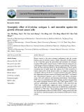
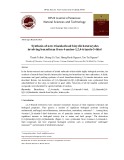
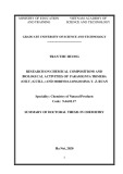
Fig. 2. RP-nano-LC-MS ⁄ MS of 2-aminobenzamide-labelled N-glycans from adult S. mansoni. (A) Chromatogram indicating the presence of H3N6F1A N-glycan species (double sodiated; EIC 1017) leading to a fragment ion at m ⁄ z 632 which corresponds to sodiated N3. (B) Fragment ion spectrum of the H3N6F1A N-glycan species. A, 2-aminobenzamide; BPC, base peak chromatogram; EIC, extracted ion chromatogram; F, deoxyhexose; H, hexose; N, N-acetylhexosamine; double-headed arrow with continuous line, N-acetylhexosamine; double headed arrow with dashed line, deoxyhexose; #, double-sodiated species.
N-glycans of this composition contained N-acetylhexo- samine chains of three or more residues.
One particular group of glycans, however, which showed compositions of H3N8)12F1)5 (Fig. 1), attrac- ted our attention by its high N-acetylhexosamine con- tent which led to the hypothesis that they contain elongated N-acetylhexosamine stretches on their anten- nae.
To test
corresponding
to
this hypothesis, RP-nano-LC-ESI-IT-MS ⁄ MS was performed on the N-glycans of S. mansoni adult worms after labelling with 2-aminobenzamide (2AB; Fig. 2). MS ⁄ MS data, obtained in the automatic mode, were screened for fragment ions that indicate oligo-N-acetylhexosamine stretches. A fragment ion corresponding to three N-acetylhexosamine residues (m ⁄ z 632) was found for the precursor at m ⁄ z 1017 ([M + 2Na]2+) the N-glycan H3N6F1A (A ¼ 2-aminobenzamide). The fragment ion spectrum (Fig. 2B) indicated that at least part of the
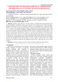
Because the MALDI-TOF-MS profile indicated that many of the N-glycans with high N-acetylhexosamine content were of low abundance, we chose a two- dimensional HPLC approach to allow MS ⁄ MS analy- sis of most of the species. In the first dimension, the 2AB-labelled N-glycans were separated by normal- phase HPLC on an amide column (Fig. 3). Individual peak fractions were analysed by MALDI-TOF-MS and separated in the second dimension by RP-nano- LC-IT-MS with automatic acquisition of MS ⁄ MS data. In order to obtain extensive MS ⁄ MS data of both sodium and proton adducts, each peak fraction was analysed by nano-LC-MS both with and with- out addition of sodium hydroxide to the running solvents, resulting in the registration ⁄ fragmentation of
FEBS Journal 273 (2006) 347–361 ª 2005 The Authors Journal compilation ª 2005 FEBS
349
M. Wuhrer et al.
S. mansoni N-glycans with dimeric LacdiNAc
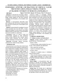
Fig. 3. Normal-phase HPLC separation of 2-aminobenzamide-labelled N-glycans from adult S. mansoni. Peaks are labelled with fraction num- bers and glycan compositions of the major 2-aminobenzamide-labelled species detected. Retention times of 2-aminobenzamide-labelled par- tial hydrolysate of dextran are indicated in italics in the upper part of the figure. All fractions were subjected to RP-nano-LC-MS ⁄ MS. Fractions containing species with a fragment ion at m ⁄ z 632 corresponding to sodiated N3 and ⁄ or an ion at m ⁄ z 813 corresponding to proto- nated N4 are labelled with (+). An overview of the N-glycan species giving rise to these characteristic ions is given in Table 1. F, deoxy- hexose; H, hexose; N, N-acetylhexosamine.
predominantly sodium adducts or proton adducts, respectively.
1
A
least this
42.rF
y t i s n e t n I
3
A
A
2
62.rF
51
02
)nim(emiT
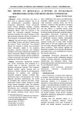
For glycans of composition H3N6F1A, which con- in part, N-acetylhexosamine stretches tain, at (Fig. 2), two-dimensional HPLC approach resolved three isomers eluting in normal-phase frac- tions 24 and 26 (Fig. 4). The MS ⁄ MS data obtained for these isobaric N-glycans (Fig. 5) indicated that iso- mers 1 and 2 were diantennary N-glycans with two LDN antennae differing only in the fucose attachment i.e. core-(a1–6)-fucosylation for isomer 1 and site, antenna-fucosylation for isomer 2 (Figs 4 and 5). Iso- mer 3, however, exhibited a stretch of four N-acetyl- hexosamine residues, which was tentatively interpreted [GalNAc(b1–4)GlcNAc(b1–3)Gal- as a diLDN unit NAc(b1–4)GlcNAc(b1-].
Fig. 4. RP-nano-LC-MS ⁄ MS of normal-phase HPLC fractions 24 and 26. Extracted ion chromatograms of m ⁄ z 1017 displayed for frac- tions 24 and 26 indicated three isomers separated by the two- dimensional HPLC system. The assigned structures are based on the MS ⁄ MS spectra shown on Fig. 5. Yellow square, N-acetylgal- actosamine; blue square, N-acetylglucosamine; green circle, man- nose; red triangle, fucose; A, 2-aminobenzamide.
Overall screening of the two-dimensional LC-MS ⁄ MS data set revealed a large group of N-glycans exhib- iting fragment ions indicative of HexNAc chains, as summarized in Table 1. For many of the N-glycan spe- cies, MS ⁄ MS data were obtained for sodium adducts
FEBS Journal 273 (2006) 347–361 ª 2005 The Authors Journal compilation ª 2005 FEBS
350
M. Wuhrer et al.
S. mansoni N-glycans with dimeric LacdiNAc
A
1remosi
A
A
# 3 . 6 1 9
A
# 3 . 4 1 8
A
A
# 2 . 3 1 7
1 . 4 6 3
4 . 2 0 4 1
5 . 5 0 6 1
3 . 9 9 1 1
A
# 0 . 3 4 8
# 6 . 9 3 6
# 6 . 1 4 7
# 3 . 4 4 9
1 . 9 2 4
A
2 . 3 5 0 1
3 . 6 5 2 1
5 . 9 5 4 1
3 . 8 1 1 1
3 . 0 1 2 1
2 . 0 1 5
4 . 1 2 3 1
# 9 . 8 0 0 1
1 . 6 4 3
4 . 7 8 5 1
9 . 7 9 6 1
2 . 0 9 5
4 . 4 2 5 1
B
2remosi
A
# 8 . 2 4 8
# 3 . 4 4 9
A
A
# 7 . 9 3 6
4 . 9 5 4 1
# 2 . 1 4 7
# 3 . 6 1 9
4 . 8 1 1 1
1 . 9 2 4
A
y t i s n e t n I
5 . 6 5 2 1
A
A
# 8 . 4 1 8
1 . 4 6 3
3 . 3 5 0 # 1 4 . 8 0 0 1
2 . 7 6 5
5 . 5 0 6 5 1 . 4 2 5 1
C
3remosi
A
A
A
# 3 . 5 3 8
1 . 9 2 4
2 . 2 3 6
5 . 2 0 4 1
# 2 . 6 1 9
5 . 5 0 6 1
# 1 . 3 1 7
4 . 0 0 2 1
# 6 . 4 1 8
# 7 . 2 4 8
5 . 0 6 4 1
A
4 . 6 5 2 1
A
# 3 . 4 4 9
5 . 3 5 0 # 1 9 . 8 0 0 1
2 . 0 1 5
3 . 4 6 3
1 . 7 6 5
5 . 4 2 5 1
4 . 1 7 6
2 . 4 4 3 1
3 . 9 4 6 1
3 . 1 2 3 1
3 . 8 1 1 1
006
0061
004
008
0021
0041
0001
z/m
Fig. 5. Fragment ion spectra (nano-LC-ESI-IT-MS ⁄ MS) of the three isomers displayed in Fig. 4. The deduced structures are boxed. Yellow square, N-acetylgalactosamine; blue square, N-acetylglucosamine; green circle, mannose; red triangle, fucose; A, 2-aminobenzamide; double- headed arrow with continuous line, N-acetylhexosamine; double-headed arrow with dashed line, deoxyhexose; #, double-sodiated fragment.
HexNAc chains seemed to be fucosylated as indicated by the corresponding reporter ions (e.g. m ⁄ z 778 for sodiated N3F1). In all cases the length of the HexNAc chain was deduced to be four HexNAc residues, and none of the fragment ion spectra indicated species with HexNAc chains of three or five HexNAc residues.
Detailed characterization of N-glycans
as well as proton adducts, with both fragment ion spectra revealing valuable structural information inclu- ding the HexNAc chain lengths (exemplified in Fig. 6). In the case of the protonated species, the length of the HexNAc chain was clearly indicated by the intense protonated HexNAc4 (m ⁄ z 813), whereas the protonat- ed HexNAc3 fragment was less abundant (m ⁄ z 610; Fig. 6A), indicating a particular lability of the Hex- NAc-Hex linkage. Although the fragment ion spectrum of the sodiated species likewise exhibited HexNAc3 and HexNAc4 ions (m ⁄ z 632 and 835), the intensity ratios were the reversed. The innermost linkage of the antenna HexNAc chain appeared to be particularly labile, resulting in an intense HexNAc3 peak and the HexNAc4 sodium a low-abundance signal for adduct (Fig. 6B). For some species, at least part of the
Few of the identified N-glycans with chains of four HexNAc were obtained in sufficient quantities and purity to allow more detailed structural characteriza- tion: the glycan species H3N8F4A detected in fraction 39 (Table 1) was purified by preparative RP-HPLC (not shown) and subjected to permethylation. Frag- ment ion analysis of permethylated H3N8F4A using
FEBS Journal 273 (2006) 347–361 ª 2005 The Authors Journal compilation ª 2005 FEBS
351
M. Wuhrer et al.
S. mansoni N-glycans with dimeric LacdiNAc
Table 1. Analytical data for S. mansoni adult worm N-glycans exhibiting partially fucosylated diLDN antennae. Compositions are given in terms of hexose (H), N-acetylhexosamine (N), and deoxyhexose (fucose; F). Fragment ions were determined by nano-LC-ESI-IT-MS ⁄ MS selecting sodiated and ⁄ or protonated precursors. n. d., not done.
Composition HPLC fractions Proposed structural features Ions registered by nano-LC-ESI-IT-MS (m ⁄ z) Characteristic fragmentions (m ⁄ z)
core-(a1–6)-fucosylation, LDN-LDN- antenna no core-fucosylation, LDNF-LDNF- antenna 26 37 1893.1b [M + Na]+; 1017.4 [M +2Na]2+ 2038.9b [M + Na]+; 1068.6 [M +2H]2+ H3N6F1 H3N6F2
39 core-(a1–6)- fucosylation, LDNF-LDNF- antenna H3N6F3 2185.2b [M + Na]+; 1141.6 [M +2H]2+; 1163.4 [M +2Na]2+
27 H3N7F1 2096.1b [M + Na]+; 1196.9 [M +2H]2+; 1218.9 [M +2Na]2+
40 2696.0b [M + Na]+; 932.1 [M +3H]3+ H4N7F4
30 Fig. 5C 407 (N2)a, 553 (N2F1)a, 813 (N4)a, 778 (N3F1) 510.2 (N1F1A), 778 (N3F1), 553 (N2F1)a, 610 (N3)a, 699 (N2F2)a, 756 (N3F1)a, 813 (N4)a, 959 (N4F1)a, 1106 (N4F2)a 488.1 (N1F1A)a, 813 (N4)a, 632 (N3), 835 (N4), 1218.4 (H2N3F1A)a 407 (N2)a, 512 (H1N1F1)a, 813 (N4)a, 959 (N4F1)a Fig. 6 H3N8F1
37 n.d. H3N8F2
37 H3N8F3 core-(a1–6)- fucosylation, LDN- LDN- antenna, 1 antenna of a single HexNAc residue (truncated) core-(a1–6)- fucosylation, 1 LDNF-LDNF- antenna, 1 Lewis X- antenna core-(a1–6)- fucosylation, LDN- LDN- antenna, LDN- antenna 1 LDN-LDN- antenna, 1 LDN- antenna, both possibly fucosylated core-(a1–6)- fucosylation, 1 LDNF- LDNF- antenna, 1 LDN- antenna 2299.0b [M + Na]+; 1198.5 [M +2H]2+; 1220.5 [M +2Na]2+ 2445.1b [M + Na]+; 1271.5 [M +2H]2+; 1293.5 [M +2Na]2+ 2591.6b [M + Na]+; 896.9 [M +3H]3+; 918.8 [M +3Na]3+
38–40 H3N8F4 2737.5b [M + Na]+; 945.6 [M +3H]3+; 967.4 [M +3Na]3+; 1439.6 [M +2Na]2+ core-(a1–6)- fucosylation, 1 LDNF- LDNF- antenna, 1 LDNF- antenna
37 510 (N1F1A), 575 (N2F1), 778 (N3F1), 989 [M +2Na]2+ (H3N5F2A), 1549 (H3N3F2A) 496 (N1F2)a, 699 (N2F2)a, 813 (N4)a, 1106 (N4F2)a, 510 (N1F1A), 632 (N3), 778 (N3F1), 1062 [M +2Na]2+ (H3N5F3A), 1164 [M +2Na]2+ (H3N6F3A), 1338 [M +2Na]2+ (H3N7F4A), 1810 (H3N4F2A), 1900 (H3N4F3A); see also Fig. 7 n.d. H3N9F1
37 H3N9F2
39 H3N9F3 1 LDN-LDN- antenna, 1 LDN- antenna, 1 antenna of a single HexNAc residue (truncated) core-(a1–6)- fucosylation, 1 LDNF-LDN- antenna, 1 antenna of a single HexNAc residue (truncated) core-(a1–6)-fucosylation, 1 LDNF-LDNF- antenna, 1 antenna of a single HexNAc residue (truncated) 2502.2b [M + Na]+; 1300.0 [M +2H]2+; 1321.9 [M +2Na]2+ 2648.4b [M + Na]+; 916.0 [M +3H]3+; 937.7 [M +3Na]3+ 2795.0b [M + Na]+; 964.6 [M +3H]3+; 986.4 [M +3Na]3+
38 38 2705.7b [M + Na]+; 1423.5 [M +2Na]2+; 2851.5b; 1005.4 [M +3Na]3+ H3N10F1 H3N10F2 2 LDN-LDN antennae, possibly fucosylated core-(a1–6)-fucosylation, 2 LDN-LDN- antennae, one of them singly fucosylated
40 2996.5b [M + Na]+; 1032.3 [M +3H]3+ core-(a1–6)-fucosylation, 2 LDN-LDN- antennae, H3N10F3
40–42 H3N10F4 core-(a1–6)-fucosylation, 1 LDNF-LDNF- antenna, 1 LDN-LDNF- antenna 3143.9b [M + Na]+; 1081.2 [M +3H]3+; 1102.8 [M +3Na]3+
FEBS Journal 273 (2006) 347–361 ª 2005 The Authors Journal compilation ª 2005 FEBS
352
813 (N4)a, 959 (N4F1)a, 510 (N1F1A), 778 (N3F1) 510 (N1F1A), 699 (N2F2)a, 813 (N4)a, 575 (N2F1), 632 (N3), 778 (N3F1), 981 (N4F1), 1090 [M +2Na]2+ (H3N6F2A), 1810 (H3N5F1A) n.d. 510.2 (N1F1A), 632 (N3), 778 (N3F1), 981 (N4F1), 1192 [M +2Na]2+ (H3N7F1A), 1753 (H3N4F2A), 1809 (H3N5F1A), 488 (N1F1A)a, 813 (N4)a, 959 (N4F1)a 510 (N1F1A), 813 (N4)a, 699 (N2F2)a, 1252 (N4F3)a, 575 (N2F1), 778 (N3F1), 1753 (H3N4F2A), 1899 (H3N4F3A)
M. Wuhrer et al.
S. mansoni N-glycans with dimeric LacdiNAc
Table 1. (Continued).
Composition HPLC fractions Proposed structural features Ions registered by nano-LC-ESI-IT-MS (m ⁄ z) Characteristic fragment ions (m ⁄ z)
42, 43 H3N10F5 3289.5b [M + Na]+; 1129.8 [M +3H]3+; 1151.5 [M +3Na]3+ core-(a1–6)-fucosylation, 2 LDNF-LDNF- antennae
37 H3N11F1 2909.0b [M + Na]+; 1002.3 [M +3H]3+; 1024.4 [M +3Na]3+ core-(a1–6)-fucosylation, 2 LDN-LDN- antennae, 1 antenna of a single HexNAc residue (truncated)
39, 40 1051.4 [M +3H]3+; 1072.8 [M +3Na]3+ H3N11F2
core-(a1–6)-fucosylation, 2 partially fucosylated LDN-LDN- antennae, 1 antenna of a single HexNAc residue (truncated) core-(a1–6)-fucosylation, 2 partially fucosylated LDN-LDN- antennae,
40, 41 1099.9 [M +3H]3+; 1121.8 [M +3Na]3+ H3N11F3
1 antenna of a single HexNAc residue (truncated) core-(a1–6)-fucosylation, 2 partly fucosylated LDN-LDN- antennae,
42, 43 1148.6 [M +3H]3+; 1170.5 [M +3Na]3+ H3N11F4 1 antenna of a single HexNAc residue (truncated)
43 1219.2 [M +3Na]3+ H3N11F5
39 40 H3N12F1 H3N12F2 core-(a1–6)-fucosylation, 2 LDNF-LDNF- antennae, 1 antenna of a single HexNAc residue (truncated) – core-(a1–6)-fucosylation, 1 LDN-LDN- antenna, 1 LDNF-LDN- antenna, 1 LDN- antenna 3111.8b [M + Na]+; 1092.1 [M +3Na]3+; 3257.8b [M + Na]+; 1118.8 [M +3H]3+; 1140.8 [M +3Na]3+
41, 42 H3N12F3 core-(a1–6)-fucosylation, 1 LDNF-LDN antenna, 1 LDN-LDN- antenna, 1 LDNF- antenna 3403.7b [M + Na]+; 1167.6 [M +3H]3+; 1189.5 [M +3Na]3+
43 1238.1 [M +3Na]3+ H3N12F4 core-(a1–6)-fucosylation, 2 partially fucosylated LDN-LDN- antennae, 1 LDN(F)- antenna
1265.1 [M +3H]3+; 1286.9 [M +3Na]3+ 43 H3N12F5 no core-fucosylation, 2 LDNF-LDNF- antennae, 1 LDNF- antenna
a Protonated fragment. b Sodium adducts of the native glycans were registered by MALDI-TOF-MS (average masses).
FEBS Journal 273 (2006) 347–361 ª 2005 The Authors Journal compilation ª 2005 FEBS
353
43 1335.6 [M +3Na]3+ H3N12F6 core-(a1–6)-fucosylation, 2 LDNF-LDNF- antennae, 1 LDNF- antenna 813 (N4)a, 902 (N3F2)a, 1106 (N4F2)a, 510 (N1F1A), 575 (N2F1), 778 (N3F1), 1897 (H3N4F3A) 813 (N4)a; 1380 (H3N3F1A)a; 510 (N1F1A), 429 (N2), 632 (N3), 1119 [M +2Na]2+ (H3N8F1A), 1221 [M +2Na]2+ (H3N9F1A), 1322 [M +2Na]2+ (H3N10F1A), 1809 (H3N5F1A), 1931 (H3N7) 488 (N1F1A)a, 553 (N2)a, 813 (N4)a, 1170 [M +2H]2+ (H3N7F2A)a, 1301 [M +2H]2+ (H3N9F1A)a, 1374 [M +2H]2+ (H3N9F2A)a, 1731 (H3N4F2A)a, 1294 [M +2Na]2+ (H3N8F2A), 1090 [M +2Na]2+ (H3N6F2A), 1323 [M +2Na]2+ (H3N9F1A), 1810 (H3N5F1A), 1955 (H3N5F2A), 1752 (H3N4F2A) 813 (N4)a, 959 (N4F1)a, 510 (N1F1A), 575 (N2F1), 778 (N3F1), 1293 [M +2Na]2+ (H3N8F2A), 1954 (H3N5F2A) 813 (N4)a, 699 (N2F2)a, 1252 (N4F3)a, 510 (N1F1A), 575 (N2F1), 778 (N3F1), 1955 (H3N5F2A) 510 (N1F1A), 429 (N2), 713 (N2F1A), 778 (N3F1), 2101 (H3N5F3A) n.d. 510 (N1F1A), 813 (N4)a, 959 (N4F1)a, 1120 [M +2H]2+ (H3N8F1A)a, 1177 (H3N2F1A)a, 1273 [M +2H]2+ (H3N8F2A)a, 1584 (H3N4F1A)1 510 (N1F1A), 575 (N2F1), 632 (N3),778 (N3F1), 2159 (H3N6F2A) 510 (N1F1A), 575 (N2F1), 713 (N2F1A), 778 (N3F1), 2158 (H3N6F2A), 2304 (H3N6F3A) 813 (N4)a, 699 (N2F2)a, 575 (N2F1), 778 (N3F1), 2304 (H3N6F3A) 510 (N1F1A), 575 (N2F1), 778 (N3F1), 2451 (H3N6F4A)
M. Wuhrer et al.
S. mansoni N-glycans with dimeric LacdiNAc
A
+2
A
z/m
(SM/SM
]H2+M[)8911
A
A
4 . 3 1 8
6 . 4 8 5 1
A
1 . 7 0 4
# 2 . 4 1 9
7 . 0 9 9 1
# 1 . 6 9 9
# 3 . 7 9 0 1
8 . 6 8 7 1
3 . 0 1 6
1 . 1 8 3 1
6 . 3 9 1 2
8 . 5 2 2 1
4 . 2 3 7
6 . 9 7 1 1
5 . 0 0 3 1
004
006
008
0001
0021
0041
0061
0081
0002
0022
+2
B
(SM/SM
z/m
]aN2+M[)0221
y t i s n e t n I
# 4
.
.
A
A
2 2 3 6
9 1 1 1
6
.
.
.
.
2 5 3 8
2 9 2 4
8 0 8 1
# 4 7 1 0 1
.
A
.
.
5
A
.
8
# 3
.
.
.
.
5
.
3
.
5
.
.
.
.
.
.
.
.
# 7 7 4 1 1
3 0 1 5
.
.
2 0 4 1
# 7 5 1 9
1 1 2 1
6 4 0 1
2 4 6 3
7 2 7 1
0 5 9 9 1
# 8 2 4 68 5 0 8
9 5 4 1
5 5 0 6 5 1 4 2 5 1
2 3 1 7
7 0 3 9 1
2 6 6 1
4 6 5 2 1
6 3 9 5
2 3 5 7
004
006
008
0001
0021
0041
0061
0081
0002
z/m
Fig. 6. Fragment ion spectra of the double-protonated (A) and double-sodiated (B) N-glycan species H3N8F1A. Spectra where obtained by RP-nano-LC-ESI-IT-MS ⁄ MS of the 2-aminobenzamide-labelled glycans of fraction 30. The deduced structure is boxed. Yellow square, N-ace- tylgalactosamine; blue square, N-acetylglucosamine; green circle, mannose; red triangle, fucose; A, 2-aminobenzamide; double-headed arrow with dashed line, deoxyhexose; #, double-sodiated fragment.
terminal Fuc as well as the following N-acetylhexosa- mine variants: terminal GalNAc, 3-substituted Gal- NAc and 3,4-disubstituted GlcNAc (Fig. 8). Notably, terminal GlcNAc was not detected. In conclusion, link- age analysis data were in line with the postulated LDNF and diLDNF antennae (Figs 7 and 9).
Furthermore, in order to obtain detailed information about the attachment of diLDN to upper and ⁄ or lower branch antennae for the above-mentioned isomer 3 (Figs 4 and 5C), fraction 26 was subjected to a-man- nosidase treatment, and partial removal of 1 hexose from the H3N6F1A isomer 1 was indicated by MALDI-TOF-MS (not shown). In order to determine the substitution position of the b-linked core mannose in isomer 3 after removal of the terminal mannose, the sample was subjected to permethylation and linkage
nano-LC-MS ⁄ MS indicated the attachment of one the subterminal HexNAc with a second fucose at fucose linked to the fourth HexNAc of the chain (Fig. 7). These data are in agreement with a fucosylat- ed version of the diLDN structure, namely a diLDNF {GalNAc(b1–4)[Fuc(a1–3)]GlcNAc(b1–3)Gal- motif NAc(b1–4)[Fuc(a1–3)]GlcNAc(b1-} as the antenna structure. In order to corroborate these findings, the permethylated N-glycan species H3N8F4A was subjec- ted to linkage analysis, i.e. hydrolysis, reduction and peracetylation, followed by GC-MS analysis of the obtained partially methylated alditol acetates using electron-impact In addition to ionization (Fig. 8). 2-substituted mannose, 3,6-disubstituted mannose and 4-substituted GlcNAc, which are in accordance with the trimannosyl core structure, linkage analysis revealed
FEBS Journal 273 (2006) 347–361 ª 2005 The Authors Journal compilation ª 2005 FEBS
354
M. Wuhrer et al.
S. mansoni N-glycans with dimeric LacdiNAc
N
-
A
SM 2
)2811(
3
A
A
dna
3 . 6 9 0 1
2
F 2 N
A
-
4 . 1 0 7
3
1 . 2 4 4
2 F 4 N
A
3 . 3 2 4 1
4 . 6 3 6
-
2
4 . 6 5 9
N
-
A F N
2
-
2
5 . 6 4 9
5 . 0 9 0 1
8 . 3 1 1 1
9 . 2 3 6 1
4 . 7 8 6
7 . 5 6 3 1
5 . 5 5 4 1
6 . 7 2 0 1
1 . 0 1 0 1
2 . 2 7 1 1
2 . 3 0 5 1
1 . 0 0 9 1
7 . 0 0 3 1 1 . 7 9 1 1
6 . 0 0 4 1
8 . 6 7 6 1
9 . 4 6 9 1
2 . 0 5 2
002
006
008
0001
0041
0061
0081
0002
0022
004
0021
→
B
SM 3
2811(
)3241
y t i s n e t n I
A
A
A
A
2
2
3 . 6 3 6
1 . 9 9 8 1
4 . 1 0 7
5 . 6 1 1 1
A
1 . 3 9 2 1
2 . 4 4 1 2
9 . 9 7 4 1
H
4 . 7 8 6
-
4 . 6 8 2 1
0 . 7 6 7
2
0 . 0 1 2 2
6 . 6 4 8 9 . 7 5 8
7 . 3 2 4 1
7 . 7 9 1 1
9 . 0 5 9 1
1 . 1 2 3 1
6 . 2 8 0 51 . 4 5 0 1
3 . 1 6 1 1
0002
005
0001
0051
z/m
Fig. 7. Fragment ion analysis of the triple-sodiated, permethylated N-glycan H3N8F4A. (A) Fragment ion spectrum obtained by RP-nano-LC- ESI-IT-MS ⁄ MS of the permethylated 2-aminobenzamide-labelled glycan from subfraction 39–4. (B) MS3 of the double-charged precursor at m ⁄ z 1423. The deduced structure is boxed. Yellow square, N-acetylgalactosamine; blue square, N-acetylglucosamine; green circle, mannose; red triangle, fucose; A, 2-aminobenzamide.
accordance with the postulated antennae structures of isomer 2 and isomer 3 occurring as a mixture in frac- tion 26 (Figs 4 and 5B,C).
Proposed structures
3-substituted GalNAc
Whereas the two N-glycans of composition H3N6F1 (isomer 3) and H3N8F4 (see above) were studied in detail revealing diLDN antennae and diLDNF anten- nae, respectively, the structural data obtained for the other N-glycans listed in Table 1 comprised mainly nano-LC-MS ⁄ MS analyses. Because the whole group of N-glycans exhibited consistent patterns of character-
analysis (not shown). In addition to 3,6-disubstituted mannose and 2-substituted mannose, which belong to the conventional trimannosyl core, 3-subsituted man- nose and 6-substituted mannose were detected in a ratio of (cid:1) 1 : 2. These monosaccharide derivatives arise from isomer 3 after removal of the terminal a-linked mannose. This would indicate that isomer 3 is a mix- ture of monoantennary structures carrying the diLDN motif on the lower branch (a1–3-linked mannose; (cid:1) 30%) and on the upper branch (a1–6-linked man- nose; (cid:1) 70%). Furthermore, terminal GalNAc, 4-sub- stituted GlcNAc, and 3,4-disubstituted GlcNAc were registered, which is in
FEBS Journal 273 (2006) 347–361 ª 2005 The Authors Journal compilation ª 2005 FEBS
355
M. Wuhrer et al.
S. mansoni N-glycans with dimeric LacdiNAc
A
detutitsbusid-4,3 cANclG
lanimret cANlaG
lanimret cANclG
detutitsbus-4 cANclG
detutitsbus-3 cANlaG
*
* *
*
04
54
)nim(emiT
Fig. 8. Linkage analysis of the N-glycan H3N8F4A. The extracted-ion chromatogram of m ⁄ z 158 indicates the HexNAc species, which were identified based on retention times and electron-impact fragmentation patterns. The elution position of terminal GlcNAc as determined with an authentic standard is indicated by an arrow. The 2AB-labelled, innermost GlcNAc is not detected in linkage analysis. Yellow square, N-ace- tylgalactosamine; blue square, N-acetylglucosamine; green circle, mannose; red triangle, fucose; A, 2-aminobenzamide; *, contaminant.
istic fragment ions revealing chains of four HexNAc residues as antenna structures carrying up to two fu- cose residues per antenna (Table 1), we postulate that these glycans also have antennae based on the diLDN motif. Structures are proposed as shown in Fig. 9 based on both the data summarized in Table 1, and the detailed analysis of compounds H3N6F1 (isomer 3) and H3N8F4.
Discussion
length might be due to the acceptor specificities of the involved b1–3-N-acetylglucosaminyltransferases, which have yet to be characterized in the case of schisto- somes as well as the CHO cells used for the expression the C. elegans GalNAc T [23]. The occurrence of of LDN(F) repeats parallels the poly(LacNAc) and poly(Lewis X) chains found on N-glycans from total S. mansoni worm glycoproteins [2–5,24], O-glycans of the circulating cathodic antigen of S. mansoni worms [2–5,25] as well as on glycoproteins and glycolipids of granulocytes [26,27]. The structural similarities of par- tially fucosylated LDN repeats and LacNAc repeats might be paralleled by the enzymatic repertoire recrui- ted for these biosyntheses: both the b3-GlcNAc trans- ferase and the a3-Fuc transferase are likely to be shared by the two biosynthetic pathways.
as
Many carbohydrate epitopes FLDN(F)
in schistosomiasis
response
We describe a very heterogeneous group of N-glycans expressed by adult worms of the human parasite S. mansoni, which feature repeats of LDN units. These units can also carry a fucose in the 3-position of N-acetylglucosamine residues, resulting in repetitive LDNF units. Repetitive LDN and LDNF structures have not previously been described in natural sources. Indications that such structures might occur in C. ele- gans were given by the heterologous expression of a C. elegans b1–4-N-acetylgalactosaminyltransferase in CHO Lec 8 cells leading to the production of oligo- LDN chains and, upon coexpression of an a1–3-fuco- syltransferase, oligo-LDNF chains [23]. Whereas Kawar et al. [23] found up to seven LDN units and up to five LDNF units per N-glycan chain, we found up to five LDN(F) units on schistosome N-glycans, with a maximum of four HexNAc residues in a row [di- LDN(F) antenna structures]. The differences in chain
from schistosomes, such {Fuc(a1–3)GalNAc(b1–4)[± Fuc(a1–3)]GlcNAc(b1-)}, LDN(F), and Lewis X, have been shown to be antigenic and several also appear to be targets for the innate immune system through recognition by human lectins [2–5,7,8,19,28–31]. In particular, LDN(F) units, repeats of which are des- cribed here, have been shown to be target of the host immune [19,29,38,39]. Despite the pronounced structural similarity between epitopes such as Lewis X and LDNF, the N-acetyl substitution at the 2-position of the Gal residue being the only difference, antibodies to Lewis X or LDNF
FEBS Journal 273 (2006) 347–361 ª 2005 The Authors Journal compilation ª 2005 FEBS
356
M. Wuhrer et al.
S. mansoni N-glycans with dimeric LacdiNAc
β
β
α
β
α
β
(cuF
)6-1
(cANlaG
(cANclG)4-1
)3-1
(cANlaG
(cANclG)4-1
(naM)2-1
)3/6-1
α
α
β
β
(cuF±
)3-1
(cuF±
)3-1
(naM β
(cANclG)4-1
cANclG)4-1
nsA-1
H3N6F 3-1
(naM α
)6/3-1
β
α
β
β
β
α
(cANlaG
(cANclG)4-1
)3-1
(cANlaG
(cANclG)4-1
(naM)2-1
)3/6-1
(cuF
)6-1
β
β
(naM β
(cANclG)4-1
cANclG)4-1
nsA-1
H3N7F1
α
β
(naM)2-1
)6/3-1
(cANclG
β
α
β
β
β
α
(cANlaG
(cANclG)4-1
)3-1
(cANlaG
(cANclG)4-1
(naM)2-1
)3/6-1
(cuF
)6-1
α
α
β
β
(cuF
)3-1
(cuF
)3-1
(naM β
nsA-1
(cANclG)4-1
cANclG)4-1
H4N7F4
β
α
(cANclG)4-1
(laG β
(naM)2-1
)6/3-1
α
(cuF
)3-1
GalNAc(β1-4)GlcNAc(β1-3)
GalNAc(β1-4)GlcNAc(β1-2)Man(α1-6/3)
Fuc(α1-6)
±Fuc(α1-3)
±Fuc(α1-3)
Man(β1-4)GlcNAc(β1-4)GlcNAcβ1-Asn
H3N8F1-4
GalNAc(β1-4)GlcNAc(β1-2)Man(α1-3/6)
±Fuc(α1-3)
GalNAc(β1-4)GlcNAc(β1-3)
GalNAc(β1-4)GlcNAc(β1-2)Man(α1-6)
Fuc(α1-6)
±Fuc(α1-3)
±Fuc(α1-3)
Man(β1-4)GlcNAc(β1-4)GlcNAcβ1-Asn
H3N10F1-5
GalNAc(β1-4)GlcNAc(β1-2)Man(α1-3)
GalNAc(β1-4)GlcNAc(β1-3)
±Fuc(α1-3)
±Fuc(α1-3)
Fig. 9. Summary of deduced structures.
exhibit exclusive specificity. Using surface plasmon res- onance, it was shown that an Lewis X monoclonal antibody selectively bound to a Lewis X neoglycopro- tein, but not to LDNF, and vice versa (Fig. 2 in Van Remoortere et al. in a chimpanzee [36]). Moreover, S. mansoni-infection model, antibodies against Lewis X
showed a time course which was markedly different from those observed for anti-LDN and anti-LDNF sera [19]. Finally, a recent study in schistosome-infec- immune ted mice has indicated an intense humoral response to dimeric and trimeric Lewis X compared with monomeric Lewis X [37]. Differential recognition
FEBS Journal 273 (2006) 347–361 ª 2005 The Authors Journal compilation ª 2005 FEBS
357
M. Wuhrer et al.
S. mansoni N-glycans with dimeric LacdiNAc
Alltech, Deerfield, IL). After washing with water (5 mL), glycans were eluted with 25% aqueous acetonitrile (5 mL). Released glycans were detected by MALDI-TOF-MS.
the rather minor
MALDI-TOF-MS
of monomeric, dimeric and trimeric Lewis X was also shown with various monoclonal antibodies [37]. In conclusion, structural variations found on schistosome glycoconjugates can have a pro- found effect on their immunological recognition. Thus, differences in antigenicity between monomeric LDN(F) and oligomers thereof can be foreseen, and the LDN(F) repeats described here are expected to be im- munogenic and involved in host–parasite interaction.
Glycan samples were analysed by MALDI-TOF-MS using an Ultraflex mass spectrometer (Bruker Daltonics, Bremen, Germany) in the positive reflectron mode with 6-aza-2-thio- thymine (5 mgÆmL)1; Sigma, St. Louis, MO) as matrix.
Based on the findings of Kawar et al.
Labelling and fractionation
structures. This
antenna
Glycans released with PNGase F were tagged with 2AB by reductive amination as outlined previously [40]. The reac- tion mixture was applied to a carbon cartridge (Alltech, Deerfield, IL), and the 2AB-labelled glycans were eluted with 5 mL of 25% acetonitrile. The acetonitrile content was reduced under a stream of nitrogen, and the samples were lyophilized.
[23], LDN and LDNF repeats might not be restricted to the trem- atode S. mansoni, but are likely to be found also on glycoproteins of C. elegans and possibly other nema- todes. Furthermore, future experiments will answer the question whether mammals likewise produce these oligo-LDN(F) question should be addressed by studying both the fine specifici- ties of potentially involved enzymes as well as by struc- tural studies of the N-glycosylation of LDN-expressing tissues [20,21].
Fractionation by normal-phase HPLC
Experimental procedures
(Glyco-)protein extraction
2AB-labelled glycans were fractionated by normal-phase HPLC on a TSK-Amide 80 column (4 · 250 mm; Tos- ohaas, Montgomeryville, PA) at 0.4 mLÆmin)1. Solvent A was 5 mm formic acid adjusted to pH 4.4 with ammonia, which is a modification of a previously published separation system [41]. Solvent B was 20% of solvent A in acetonitrile. The following gradient conditions were used: at time t ¼ 0 min, 100% solvent B; t ¼ 152 min, 52.5% solvent B; t ¼ 155 min, 0% solvent B; t ¼ 162 min, 0% solvent B; and t ¼ 163 min, 100% solvent B. The total run time was 180 min. Samples were injected in 80% acetonitrile. Because of the large amounts of material injected, fluores- cence was detected at 280 nm ⁄ 500 nm instead of the rou- tinely used 360 nm ⁄ 425 nm in order to avoid saturation of the detector. Fractions were collected manually and ana- lysed by MALDI-TOF-MS.
Fractionation by RP-HPLC
S. mansoni worms were obtained by perfusion of infected hamsters and stored at )80 (cid:1)C until use. Adult worms were homogenized in 1 mL of water (1 vol.Æ100 mg)1 wet weight of worms). In order to delipidize samples, methanol and chloroform were sequentially added (5 vol. each). The supernatant was removed after centrifugation, and the extraction was repeated. In order to extract (glyco-)pro- teins, the pellet was suspended in phosphate-buffered saline (35 mm sodium phosphate, pH 7.6, 0.85% NaCl). SDS and 2-mercaptoethanol were added to final concentrations of 1% (w ⁄ v) and 0.5%, respectively. The samples were incuba- ted for 10 min at 100 (cid:1)C, allowed to cool to room tempera- ture, and Chaps (Fluka/Sigma-Aldrich, Zwijndrecht, The Netherlands) was added to a final concentration of 1% (w ⁄ v). Samples were centrifuged, and supernatants were subjected to PNGase F treatment. These experiments were carried out in accordance with EC Council Directive (89/609/EEC) and after approval of the animal experiment commitee (DEC) of the Leiden University Medical Center.
Glycan release
collected manually
2AB-labelled glycans were fractionated by RP-HPLC on a Hypersil ODS 3 lm (2 · 250 mm; Thermo Electron Corp., Waltham, MA) at 0.2 mLÆmin)1. Solvent A was 0.4% acetonitrile, 0.1% formic acid. Solvent B was 95% acetonit- rile, 0.1% formic acid. The following gradient conditions were used: at time t ¼ 0 min, 5% solvent B; t ¼ 5 min, 5% solvent B; gradient to t ¼ 30 min, 50% solvent B; t ¼ 31 min, 100% solvent B; and t ¼ 36 min, 100% solvent B, t ¼ 37 min, 5% solvent B. Total run time was 60 min. Fluorescence was detected at 360 nm ⁄ 425 nm. Fractions and analysed by MALDI- were TOF-MS.
S. mansoni (glyco-)proteins were incubated with PNGase F (2 mUÆ100 mg)1 wet weight; Roche Diagnostics, Mannheim, Germany) overnight at 37 (cid:1)C. For the purification of the released glycans, samples were first applied to a reverse-phase cartridge (500 mg of Bakerbond octadecyl; Baker, Phillips- burg, NJ). Flow-through and wash (5 mL of water) were then applied to a carbon cartridge (150 mg Carbograph;
FEBS Journal 273 (2006) 347–361 ª 2005 The Authors Journal compilation ª 2005 FEBS
358
M. Wuhrer et al.
S. mansoni N-glycans with dimeric LacdiNAc
Nano-LC-MS/MS
References
1 Atrih A, Richardson JM, Prescott AR & Ferguson MA (2005) Trypanosoma brucei glycoproteins contain novel giant poly-N-acetyllactosamine carbohydrate chains. J Biol Chem 280, 865–871.
2 Nyame AK, Kawar ZS & Cummings RD (2004) Anti- genic glycans in parasitic infections: implications for vaccines and diagnostics. Arch Biochem Biophys 426, 182–200.
3 Hokke CH & Deelder AM (2001) Schistosome glyco- conjugates in host–parasite interplay. Glycoconj J 18, 573–587.
4 Cummings RD & Nyame AK (1999) Schistosome glyco-
conjugates. Biochim Biophys Acta 1455, 363–374.
5 Khoo KH (2001) Structural variations in schistosomal glycans. Trends Glycosci Glycotechnol 31, 493–506. 6 Robijn MLM, Wuhrer M, Kornelis D, Deelder AM,
Geyer R & Hokke CH (2005) Mapping fucosylated epi- topes on glycoproteins and glycolipids of Schistosoma mansoni cercariae, adult worms and eggs. Parasitology 130, 67–77.
7 van den Berg TK, Honing H, Franke N, Van Remoor-
tere A, Schiphorst WE, Liu FT, Deelder AM, Cummings RD, Hokke CH & van Die I (2004) LacdiNAc-glycans constitute a parasite pattern for galectin-3-mediated immune recognition. J Immunol 173, 1902–1907.
2AB-labelled glycans were separated on a PepMap col- umn (75 lm · 150 mm; Dionex ⁄ LC Packings, Amster- dam, the Netherlands) using an Ultimate nano-LC system (Dionex ⁄ LC Packings) equipped with a Switchos guard column system (Pepmap guard column, 300 lm · 10 mm). The system was equilibrated with eluent A (H2O ⁄ aceto- nitrile 95 : 5, v ⁄ v, 0.1% formic acid) at a flow rate of 150 nLÆmin)1. After injecting the sample, a linear gradient to 50% eluent B (H2O ⁄ acetonitrile 5 : 95, v ⁄ v, containing 0.1% formic acid) in 30 min was applied, followed by a final wash with 100% B for 5 min. The system was directly coupled to an Esquire high capacity trap (HCT) ESI-IT-MS (Bruker) equipped with an online nano-spray source operating in the positive-ion mode. For electro- spray (900–1200 V), capillaries (360 lm OD, 20 lm ID with 10 lm opening) from New Objective (Cambridge, MA) were used. The solvent was evaporated at 165 (cid:1)C with a nitrogen stream of 5 LÆmin)1. Ions from m ⁄ z 50 to 2000 were registered. Automatic fragment ion analysis was enabled, resulting in MS ⁄ MS spectra of the most to register predominantly In order abundant peaks. sodium adducts by MS, part of the analyses were per- formed after addition of 0.8 mm NaOH to solvent A. For the analysis of permethylated glycans, the column was conditioned with 15% eluent B. After injection, a linear gradient to 70% eluent B in 30 min was applied followed by a wash with 100% B for 5 min.
a-Mannosidase treatment
8 van Vliet SJ, van Liempt E, Saeland E, Aarnoudse CA, Appelmelk B, Irimura T, Geijtenbeek TB, Blixt O, van Alvarez RD, I & van Kooyk Y (2005) Carbohydrate profiling reveals a distinctive role for the C-type lectin MGL in the recognition of helminth parasites and tumor antigens by dendritic cells. Int Immunol 17, 661– 669.
9 Kleene R & Schachner M (2004) Glycans and neural
cell interactions. Nat Rev Neurosci 5, 195–208.
2AB-labelled glycans (50 pmol to 1 nmol) were treated with a-mannosidase from jack beans (100 mU; Sigma) in 50 lL 50 mm sodium acetate buffer, pH 5.0 for 18 h at 37 (cid:1)C. The reaction mixture was applied to a carbon cartridge (Alltech), and the 2AB-labelled glycans were eluted with 5 mL of 25% acetonitrile, analysed by MALDI-TOF-MS, and subjected to nano-LC-MS ⁄ MS.
Permethylation and linkage analysis
10 Haltiwanger RS & Lowe JB (2004) Role of glycosyla- tion in development. Annu Rev Biochem 73, 491–537. 11 Muramatsu T & Muramatsu H (2004) Carbohydrate antigens expressed on stem cells and early embryonic cells. Glycoconj J 21, 41–45.
12 Martin PT (2003) Glycobiology of the neuromuscular
junction. J Neurocytol 32, 915–929.
13 Ono M & Hakomori S (2004) Glycosylation defining cancer cell motility and invasiveness. Glycoconj J 20, 71–78.
2AB-labelled glycans were permethylated [42] and analysed by nano-LC-MS ⁄ MS. For linkage analysis, permethylated glycans were hydrolyzed (4 m trifluoroacetic acid, 4 h, 100 (cid:1)C), and partially methylated alditol acetates obtained after sodium borohydride reduction and peracetylation were analysed by capillary GLC-MS using electron-impact ionization [43].
14 Dube DH & Bertozzi CR (2005) Glycans in cancer and inflammation – potential for therapeutics and diagnos- tics. Nat Rev Drug Discov 4, 477–488.
15 Kannagi R, Izawa M, Koike T, Miyazaki K &
Acknowledgements
Kimura N (2004) Carbohydrate-mediated cell adhesion in cancer metastasis and angiogenesis. Cancer Sci 95, 377–384.
We thank Ria van den Heuvel for performing GC-MS analyses.
FEBS Journal 273 (2006) 347–361 ª 2005 The Authors Journal compilation ª 2005 FEBS
359
M. Wuhrer et al.
S. mansoni N-glycans with dimeric LacdiNAc
26 Spooncer E, Fukuda M, Klock JC, Oates JE & Dell A (1984) Isolation and characterization of polyfucosylated lactosaminoglycan from human granulocytes. J Biol Chem 259, 4792–4801.
27 Fukuda MN, Dell A, Oates JE, Wu P, Klock JC &
16 Dell A, Morris HR, Easton RL, Panico M, Patankar M, Oehniger S, Koistinen R, Koistinen H, Seppala M & Clark GF (1995) Structural analysis of the oligosac- charides derived from glycodelin, a human glycoprotein with potent immunosuppressive and contraceptive activ- ities. J Biol Chem 270, 24116–24126.
17 Hiyama J Weisshaar G & Renwick AG (1992) The
asparagine-linked oligosaccharides at individual glycosy- lation sites in human thyrotrophin. Glycobiology 2, 401– 409.
Fukuda M (1985) Structures of glycosphingolipids iso- lated from human granulocytes. The presence of a series of linear poly-N-acetyllactosaminylceramide and its sig- nificance in glycolipids of whole blood cells. J Biol Chem 260, 1067–1082.
18 Smith PL, Skelton TP, Fiete D, Dharmesh SM, Beranek MC, MacPhail L, Broze GJ Jr & Baenziger JU (1992) The asparagine-linked oligosaccharides on tissue factor pathway inhibitor terminate with SO4–4GalNAc beta 1, 4GlcNAc beta 1,2 Man alpha. J Biol Chem 267, 19140– 19146.
28 Kantelhardt SR, Wuhrer M, Dennis RD, Doenhoff MJ, Bickle Q & Geyer R (2002) Fuc (alpha1–3) GalNAc-: major antigenic motif of Schistosoma mansoni glycoli- pids implicated in infection sera and keyhole limpet hemocyanin cross-reactivity. Biochem J 366, 217–223. 29 Naus CW, Van Remoortere A, Ouma JH, Kimani G,
Dunne DW, Kamerling JP, Deelder AM & Hokke CH (2003) Specific antibody responses to three schistosome- related carbohydrate structures in recently exposed immigrants and established residents in an area of Schistosoma mansoni endemicity. Infect Immun 71, 5676–5681.
30 Van Remoortere A, Vermeer HJ, Van Roon AM,
19 Eberl M, Langermans JA, Vervenne RA, Nyame AK, Cummings RD, Thomas AW, Coulson PS & Wilson RA (2001) Antibodies to glycans dominate the host response to schistosome larvae and eggs: is their role protective or subversive? J Infect Dis 183, 1238–1247. 20 Sato T, Gotoh M, Kiyohara K, Kameyama A, Kubota T, Kikuchi N, Ishizuka Y, Iwasaki H, Togayachi A, Kudo T et al. (2003) Molecular cloning and characteri- zation of a novel human beta 1,4-N-acetylgalactosami- nyltransferase, beta 4GalNAc-T3, responsible for the synthesis of N,N’-diacetyllactosediamine, GalNAc beta 1–4GlcNAc. J Biol Chem 278, 47534–47544.
Langermans JA, Thomas AW, Wilson RA, van Die I, Van den Eijnden DH, Agoston K, Kerekgyarto J et al. (2003) Dominant antibody responses to Fuc (alpha1–3) GalNAc and Fuc (alpha1–2) Fuc (alpha1–3) GlcNAc containing carbohydrate epitopes in Pan troglodytes vaccinated and infected with Schistosoma mansoni. Exp Parasitol 105, 219–225.
21 Gotoh M, Sato T, Kiyohara K, Kameyama A, Kikuchi N, Kwon YD, Ishizuka Y, Iwai T, Nakanishi H & Nar- imatsu H (2004) Molecular cloning and characterization of beta1,4-N-acetylgalactosaminyltransferases IV synthe- sizing N,N’-diacetyllactosediamine. FEBS Lett 562, 134– 140.
22 Kawar ZSD I, & Cummings RD (2002) Molecular clon-
ing and enzymatic characterization of a UDP-GalNAc: GlcNAc (beta)-R beta1,4-N-acetylgalactosaminyltrans- ferase from Caenorhabditis elegans. J Biol Chem 277, 34924–34932.
31 van de Wetering JK, Van Remoortere A, Vaandrager AB, Batenburg JJ, van Golde LM, Hokke CH & Van Hellemond JJ (2004) Surfactant protein D binding to terminal alpha1–3-linked fucose residues and to Schisto- soma mansoni. Am J Respir Cell Mol Biol 31, 565–572. 32 van Liempt E, Imberty A, Bank CM, van Vliet SJ, van Kooyk Y & van Geijtenbeek TB I (2004) Molecular basis of the differences in binding properties of the highly related C-type lectins DC-SIGN and L-SIGN to Lewis X trisaccharide and Schistosoma mansoni egg antigens. J Biol Chem 279, 33161–33167.
23 Kawar ZS, Haslam SM, Morris HR, Dell A & Cum- mings RD (2005) Novel poly-GalNAcbeta1–4GlcNAc (LacdiNAc) and fucosylated poly-LacdiNAc N-glycans from mammalian cells expressing beta1,4-N-acetylgalac- tosaminyltransferase and alpha1,3-fucosyltransferase. J Biol Chem 280, 12810–12819.
24 Srivatsan J, Smith DF & Cummings RD (1992) The
33 Blixt O, Head S, Mondala T, Scanlan C, Huflejt ME, Alvarez R, Bryan MC, Fazio F, Calarese D, Stevens J et al. (2004) Printed covalent glycan array for ligand profiling of diverse glycan binding proteins. Proc Natl Acad Sci USA 101, 17033–17038.
human blood fluke Schistosoma mansoni synthesizes gly- coproteins containing the Lewis X antigen. J Biol Chem 267, 20196–20203.
34 Guo Y, Feinberg H, Conroy E, Mitchell DA, Alvarez R, Blixt O, Taylor ME, Weis WI & Drickamer K (2004) Structural basis for distinct ligand-binding and targeting properties of the receptors DC-SIGN and DC-SIGNR. Nat Struct Mol Biol 11, 591–598. 35 Meyer S, van Liempt E, Imberty A, van Kooyk Y,
Geyer H, Geyer R & van Die I (2005) DC-SIGN med- iates binding of dendritic cells to authentic pseudo- Lewis Y glycolipids of Schistosoma mansoni cercariae,
25 van Dam GJ, Bergwerff AA, Thomas-Oates JE, Rotmans JP, Kamerling JP, Vliegenthart JF & Deelder AM (1994) The immunologically reactive O-linked polysaccharide chains derived from circulating cathodic antigen isolated from the human blood fluke Schistosoma mansoni have Lewis X as repeating unit. Eur J Biochem 225, 467–482.
FEBS Journal 273 (2006) 347–361 ª 2005 The Authors Journal compilation ª 2005 FEBS
360
M. Wuhrer et al.
S. mansoni N-glycans with dimeric LacdiNAc
the first parasite-specific ligand of DC-SIGN. J Biol Chem 280, 37349–37359.
LacdiNAc glycan antigen in Schistosoma mansoni- infected mice and expression of the glycan among schis- tosomes. Exp Parasitol 96, 202–212.
40 Wuhrer M, Robijn MLM, Koeleman CA, Balog CIA, Geyer R, Deelder AM & Hokke CH (2004) A novel Gal (beta1–4) Gal (beta1–4) Fuc (alpha1–6)-core modifi- cation attached to the proximal N-acetylglucosamine of keyhole limpet hemocyanin (KLH) N-glycans. Biochem J 378, 625–632.
36 Van Remoortere A, Hokke CH, van Dam GJ, van Die I, Deelder AM & Van den Eijnden DH (2000) Various stages of Schistosoma express LewisX, LacdiNAc, Gal- NAcb1–4 (Fuca1–3) GlcNAc and GalNAcb1–4 (Fuca1– 2Fuca1–3) GlcNAc carbohydrate epitopes: detection with monoclonal antibodies that are characterized by enzymatically synthesized neoglycoproteins. Glycobiol- ogy 10, 601–609.
37 Van Roon AM, Van de Vijver KK, Jacobs W, van
41 Garner B, Merry AH, Royle L, Harvey DJ, Rudd PM & Thillet J (2001) Structural elucidation of the N- and O-glycans of human apolipoprotein (a): role of O-gly- cans in conferring protease resistance. J Biol Chem 276, 22200–22208.
42 Ciucanu I & Kerek F (1984) A simple and rapid
Marck EA, van Dam GJ, Hokke CH & Deelder AM (2004) Discrimination between the anti-monomeric and the anti-multimeric Lewis X response in murine schisto- somiasis. Microbes Infect 6, 1125–1132.
38 Nyame AK, Leppanen AM, DeBose-Boyd R & Cum-
method for the permethylation of carbohydrates. Carbo- hydr Res 131, 209–217.
43 Geyer R & Geyer H (1994) Saccharide linkage analysis using methylation and other techniques. Methods Enzy- mol 230, 86–107.
mings RD (1999) Mice infected with Schistosoma man- soni generate antibodies to LacdiNAc (GalNAc beta 1 ? 4GlcNAc) determinants. Glycobiology 9, 1029–1035. 39 Nyame AK, Leppanen AM, Bogitsh BJ & Cummings RD (2000) Antibody responses to the fucosylated
FEBS Journal 273 (2006) 347–361 ª 2005 The Authors Journal compilation ª 2005 FEBS
361