doi:10.1046/j.1432-1033.2002.03035.x
Eur. J. Biochem. 269, 3645–3658 (2002) (cid:2) FEBS 2002
Towards the development of selective amine oxidase inhibitors Mechanism-based inhibition of six copper containing amine oxidases
Eric M. Shepard1, Jennifer Smith1, Bradley O. Elmore1, Jason A. Kuchar1, Lawrence M. Sayre2 and David M. Dooley1 1Department of Chemistry and Biochemistry, Montana State University, Bozeman, MT, USA; 2Department of Chemistry, Case Western Reserve University, Cleveland, OH, USA
inactivation appears to arise in some cases from TPQ cofactor modification and in other cases from alkylation of protein residues in a manner that blocks access of substrate to the active site. Notably, 1 completely inhibits AGAO at stoichiometric concentrations and is not a substrate, but is an excellent substrate of PSAO and inhibition is observed only at very high concentrations. Structural models of 1 in Schiff base linkage to the TPQ cofactor in AGAO and PSAO (for which crystal structures are available) reveal substantial differences in the degree of interaction of bound 1 with side- chain residues, consistent with the widely divergent activities. Collectively, these results suggest that the development of highly selective amine oxidase inhibitors is feasible.
Keywords: copper; amine oxidase; enzyme inhibition; mechanism-based inhibition.
Four substrate analogs, 4-(2-naphthyloxy)-2-butyn-1-amine (1), 1,4-diamino-2-chloro-2-butene (2), 1,6-diamino-2,4- hexadiyne (3), and 2-chloro-5-phthalimidopentylamine (4) have been tested as inhibitors against mammalian, plant, bacterial, and fungal copper-containing amine oxidases: bovine plasma amine oxidase (BPAO), equine plasma amine oxidase (EPAO), pea seedling amine oxidase (PSAO), Arthrobacter globiformis amine oxidase (AGAO), Escheri- chia coli amine oxidase (ECAO), and Pichia pastoris lysyl oxidase (PPLO). Reactions of 1,4-diamino-2-butyne with selected amine oxidases were also examined. Each substrate analog contains a functional group that chemical precedent suggests could produce mechanism-based inactivation. Striking differences in selectivity and rates of inactivation were observed. For example, between two closely related plasma enzymes, BPAO is more sensitive than EPAO to 1 and 3, while the reverse is true for 2 and 4. In general,
(step 4),
The copper-containing amine oxidases play a crucial role in metabolic oxidative deamination of primary amines to the corresponding aldehydes, with the concomitant production of hydrogen peroxide and ammonia [1]. The reaction proceeds through a transamination mechanism mediated by an active site 2,4,5-trihydroxyphenylalanine quinone (TPQ) cofactor. The TPQ cofactor is formed from the post- translational self-processing of a tyrosine residue within the highly conserved sequence Ser/Thr-Xaa-Xaa-Asn-Tyr(TPQ)-
+ [13].
RCH2NHþ
Asp/Glu-Tyr [1] that requires both copper and molecular oxygen [2–7]. The key step in catalysis (Scheme 1) is the conversion of the initial quinoneimine (cid:1)substrate Schiff base(cid:2) (step 2) to a quinolaldimine (cid:1)product Schiff base(cid:2) (step 3), facilitated by Ca proton abstraction by an absolutely conserved active-site aspartate acting as general base [8–12]. Hydrolytic release of aldehyde product gives a Cu(II)- aminoresorcinol in equilibrium with Cu(I)- semiquinone (step 5), that is reoxidized to TPQ in the presence of O2, with release of H2O2 and NH4 3 þ O2 þ H2O ! RCHO þ NHþ
4 þ H2O2
ð1Þ
Scheme 1. Proposed catalytic mechanism of turnover for amine oxidases. Correspondence to D. M. Dooley, Department of Chemistry and Biochemistry, Montana State University, Bozeman, MT 59717, USA. Fax: + 1 406 994 7989, Tel.: + 1 406 994 4371, E-mail: dmdooley@montana.edu or L. M. Sayre, Department of Chemistry, Case Western Reserve University, Cleveland, OH 44106, USA, Fax: + 1 216 368 3006, Tel.: + 1 216 368 3704, E-mail: lms3@po.cwru.edu Abbreviations: AGAO, Arthrobacter globiformis phenethylamine oxi- dase; PSAO, pea seedling amine oxidase; HPAO, Hansenula poly- morpha amine oxidase; ECAO, Escherichia coli amine oxidase; EPAO, equine plasma amine oxidase; PPLO, Pichia pastoris lysyl oxidase; BPAO, bovine plasma amine oxidase; SSAO, semicarbazide sensitive amine oxidase; DABY, 1,4-diamino-2-butyne; LTQ, lysine tyrosyl- quinone; TPQ, 2,4,5-trihydroxyphenylalanine quinone; HRP, horse- radish peroxidase; ABTS, (2,2¢-azino-bis(3-ethyl)benzthiazoline-6- sulfonic acid); NBD-F, 4-fluoro-7-nitrobenzo-2-oxa-1,3-diazole. (Received 4 February 2002, revised 2 May 2002, accepted 31 May 2002)
3646 E. M. Shepard et al. (Eur. J. Biochem. 269)
(cid:2) FEBS 2002
The basic strategy in designing suicide inhibitors for amine oxidases has been to incorporate a moiety into an amine substrate that allows for creation of an electrophile upon the imine shift accompanying Ca proton abstraction during the initial phase of oxidation. Two strategies are the incorporation of a halogen or of unsaturation at the b position, examples being b-bromoethylamine [22–27] and propargylamine [24]. The former case results in an SN2- activated a-haloaldehyde as turnover product, whereas the latter case results in an electrophilic a,b-unsaturated aldehyde. Propargylamine derivatives have been studied as inhibitors of plasma amine oxidase [24], semicarbazide- sensitive amine oxidase (SSAO) [28,29], and monoamine oxidase [30–32].
These enzymes are widely distributed in nature and have been purified from mammals, plants, and microorganisms. In microorganisms, these enzymes frequently have a nutritional role in the utilization of primary amines as the sole source of nitrogen or carbon, while in mammals and plants certain amine oxidases appear to be tissue specific, and are implicated in wound healing, detoxification, cell growth, signaling, and apoptosis [8,13,14]. Furthermore, mammalian lysyl oxidase, a copper amine oxidase containing lysine tyrosylquinone (LTQ) as its redox cofactor, catalyzes the crucial deamination of the side-chain of peptidyl lysine that initiates cross-linking of lysine residues in collagen and elastin [15,16]. Appreciation is growing for a broader physiological role for human amine oxidases; for example amine oxidases are implicated in the formation of vascular plaques linked to congestive heart failure [17], and the fact that human vascular adhesion protein, which mediates interactions between lymphocytes and vascular endothelial cells [18], is a membrane-bound amine oxidase [19,20]. In the latter regard, lymphocyte migration was recently proposed to be con- trolled by amine oxidase catalytic activity, with temporary adhesion representing a Schiff base linkage between the catalytic TPQ and a lysine group on the surface of the lymphocyte, with turnover then resulting in cell release [21]. As would be expected for any enzyme relying on a carbonyl cofactor, arylhydrazines form catalytically inactive derivatives of these enzymes, which are also inhibited by chelating agents that remove or bind to the copper. Our interest here, however, is in mechanism-based inhibitors, also called (cid:1)suicide(cid:2) inhibitors, which undergo turnover- dependent conversion to electrophilic products capable of covalent binding. Not only can they display binding selectivities based on active-site fit, as with competitive inhibitors, but they offer a second level of distinction on the basis that long-acting inhibition will occur only when the electrophilic product is capable of accessing an active-site nucleophile. Two different enzymes may have similar binding pockets but could quite likely have distinct spatial presentations of modifiable side-chains.
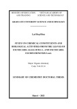
Four copper-containing amine oxidases have yielded to crystallization: Arthrobacter globiformis phenethylamine oxidase (AGAO), pea seedling amine oxidase (PSAO), Hansenula polymorpha amine oxidase (HPAO), and Escherichia coli amine oxidase (ECAO). These amine oxidases are all (cid:1)mushroom(cid:2)-shaped homodimers with two embracing (cid:1)arms(cid:2) (Fig. 1) [13,33]. The b ribbon of Arm I extends to the active site channel, and residues at the end of Arm I form part of the entrance to the channel. These residues vary among the structurally characterized amine oxidases and among amine oxidase primary sequences [13,33]. Overall, the crystal structures differ with respect to the apparent dimensions and electrostatic properties of the active site channel, whether access to the channel is (cid:1)gated(cid:2), and the degree of accessibility to the TPQ cofactor. For example, whereas residues Phe298 in PSAO, Tyr381 in ECAO and Tyr296 in AGAO may act as (cid:1)gates(cid:2) to the active site [13], the corresponding residue in HPAO is Ala317 so the TPQ cofactor is more solvent exposed in this case [33]. Tyr396 has been identified as the corresponding (cid:1)gate(cid:2) residue in a structural model of Pichia pastoris lysyl oxidase (PPLO) [34]. All these differences must contribute to the molecular basis for substrate specificity among these enzymes, and thus observed preferences among mechan- ism-based inhibitors should be attributable to structural
Fig. 1. Two views of AGAO. (A) Ribbon diagram depicting the arms. The Cu atom of each active site is shown as a green sphere. (B) Active site channel represented as a space-filling model. Residues are color-coded [13]: acidic, red; hydrophobic, green; (cid:1)gate(cid:2), white; yellow and blue represent the two monomers. Asp357, Leu358, and Trp359 are located on Arm I (yellow) but are colored red and green, respectively.
Mechanism-based inhibition of amine oxidases (Eur. J. Biochem. 269) 3647
(cid:2) FEBS 2002
dihydrochloride (3), 2-chloro-5-phthalimidopentylamine hydrochloride (4), and 1,4-diamino-2-butyne dihydrochlor- ide (DABY) were synthesized and characterized as described previously [35,36].
Absorption spectra and steady-state kinetic assays
differences in a manner that may eventually become predictable.
Absorption spectra and kinetic assays were recorded at 25 (cid:3)C using a Hewlett Packard 8453 diode array spectro- photometer equipped with a thermostatted cell chamber connected to an Endocal RTE-5 circulating water bath. Protein concentrations were calculated using extinction coefficients at 280 nm as previously reported for ECAO [41], AGAO [42], EPAO [39], PSAO [37], BPAO [43], and PPLO [34]. Stock inhibitor solutions were prepared by dissolving required amount of compound in doubly deionized H2O to yield a final concentration of 1 mM. Experiments were run with freshly thawed protein aliquots from the same enzyme stock. All solutions were stirred during assays, with benzylamine used as a control substrate (Table 1). Enzyme activity is reported as spectroscopic units per mg of protein.
Method A. To analyze general compound effectiveness, enzymes were incubated with varying concentrations of inhibitor for 30 min at 4 (cid:3)C. Amine oxidase activity was determined using benzylamine as substrate and monitoring benzaldehyde production for 3 min at 250 nm using an )1Æcm)1 [23,44]. Excess extinction coefficient of 12800 M inhibitor was not removed before assaying, but was diluted by at least a factor of 30 in the final assay mixture. Assays were run in triplicate for each inhibitor concentration (Table 2). A control was prepared by incubating equivalent concentrations of enzyme and doubly deionized H2O at 4 (cid:3)C for 30 min. All assays were performed at 25 (cid:3)C in 100 mM phosphate buffer, pH 7.2.
To initiate such analysis, we report here the inhibitory effects of four different inactivators with six different copper-containing amine oxidases: two mammalian amine oxidases, equine and bovine plasma amine oxidase (EPAO and BPAO), one plant amine oxidase (PSAO), two bacterial amine oxidases (AGAO and ECAO), and one yeast amine oxidase, Pichia pastoris lysyl oxidase (PPLO). Our choice of the four compounds, 4-(2-naphthyloxy)-2-butyn-1-amine (1), 1,4-diamino-2-chloro-2-butene (2), 1,6-diamino-2,4- hexadiyne (3), and 2-chloro-5-phthalimidopentylamine (4) (Fig. 2), was based on promising preliminary results from screening a larger set of synthetic inhibitors against BPAO [35,36]. In addition, 1,4-diamino-2-butyne (DABY) was tested against selected enzymes. Comparative data have been obtained wherever possible in terms of rates of inactivation, partition ratios, and degree of reversibility. To our knowledge, this is the first systematic comparison among a broad collection of copper amine oxidases to critically explore the extent to which selective inhibition might be possible. These data point to examples of especially potent and selective inhibition which are amen- able for an in depth investigation of the structural and mechanistic basis of inhibition.
Fig. 2. Inhibitor compounds.
M A T E R I A L S A N D M E T H O D S
Enzyme isolation and purification
Method B. Inactivation of amine oxidases over time by selected inhibitors was measured using the assay described above. Buffered enzyme and inhibitor mixtures were prepared at 4 (cid:3)C. Upon addition of inhibitor, time was noted and an aliquot was correspondingly taken from the mixture and diluted 30-fold into the assay cuvette. This was repeated until no activity remained or decreases in activity leveled off.
Method C. Determination of compound turnover was measured by monitoring H2O2 production using a horse- radish peroxidase (HRP)–ABTS (2,2¢-azino-bis(3-ethyl)- benzthiazoline-6-sulfonic acid) coupled assay. HRP reacts with H2O2 to give an activated form which oxidizes ABTS to a product with a kmax of 414 nm [5,45]. Assays were performed at 30 (cid:3)C in 100 mM phosphate buffer, pH 7.0.
)1)
Table 1. Benzylamine kinetics.
PSAO was purified from pea seedling, as described previously [37]. BPAO was purified from bovine plasma using minor modifications of a published procedure [38]. EPAO was purified by a revised protocol (B. O. Elmore & D. M. Dooley, unpublished protocol) based on that initially specified previously [39]. ECAO was a generous gift from M. McPherson, University of Leeds (UK), and was purified as described previously [10]. PPLO was purified as initially described in [40]. AGAO was purified as described previously [2], with slight modifications specified below. Buffer contained no metal chelators, and induction occurred with isopropyl thio-b-D-galactoside and copper supplement to a final concentration of 30 lM. Cell lysate was incubated in 50 mM Hepes, pH 6.8, 0.1 mgÆmL)1 phenylmethanesul- fonyl fluoride, 1 mgÆmL)1 TCPK, and 0.8 mM CuSO4 (final concentration) for 2 h at 30 (cid:3)C.
Inhibitors
Enzyme kcat (min)1) Km (lM) kcat/Km (min)1ÆlM
The five compounds used in this study, 4-(2-naphthyloxy)- 2-butyn-1-amine hydrochloride (1), 1,4-diamino-2-chloro-2- (2), 1,6-diamino-2,4-hexadiyne butene dihydrochloride
EPAO AGAO PSAO BPAO PPLO 13 ± 0.5 32 ± 0.5 131 ± 3 69 ± 5 46 ± 1 87 ± 8 34 ± 1 215 ± 10 1500 ± 200 29 ± 2 0.15 0.94 0.609 0.046 1.6
3648 E. M. Shepard et al. (Eur. J. Biochem. 269)
(cid:2) FEBS 2002
Table 2. Inhibition of six amine oxidases using compounds in Fig. 2.
% Activityb % Activityb Compound Inhibitor: proteina Enzyme Compound Inhibitor: proteina Enzyme
AGAOc PSAOc 14 1 2 1 2 1.3 2.6 1 1.6 9 17 3 3d 4
BPAOc 4 1
ECAOe 1 2
2 3
3 4
4 EPAOg 1
PPLOf 1
2 3
2 3 4
a Reported as excess inhibitor relative to active site concentration. b Percent activity remaining relative to control rates (rounded to nearest integer), following a 30-min incubation (refer to Materials and methods). Numbers shown are averages of three assays. c Active site concentrations ranged between 4 and 7 lM. d Percent activity after a 60-min incubation. e Active site concentrations ranged between 20 and 33 lM. f Active site concentrations were at 5 lM, but compound 4 studies were carried out at 11 lM. g Active site concentrations ranged between 5 and 6 lM, but compound 4 studies were carried out at 47 lM.
a control substrate [46]. A comparative set of assays was run using vanillic acid in place of phenol. Reactions were carried out at 30 (cid:3)C, and the A495 was monitored.
Total assay volume was 1 mL, and substrate concentration was adjusted accordingly to yield a final concentration of 300 lM. Benzylamine was used as a control substrate, and all assays were run in triplicate (Table 3).
Nitroblue tetrazolium assay for TPQ redox competency
Method D. In order to check the results of Shimizu et al. (1997), who reported no benzylamine turnover by AGAO, 4-aminoantipyrine assays were run using phenethylamine as
Purified BPAO or AGAO was incubated with a given molar excess of inhibitor determined to achieve loss of 92–97%
14 27 14 27 1 8 16 5 10 15 1 9 7 17 34 85 2 10 19 2 2 20 38 2 4 11 100 6 0 74 17 100 92 86 38 12 56 32 7 27 8 81 73 53 25 89 77 66 2.5 78 70 53 62 62 50 4 1.5 5 8 17 34 1 2.5 5 1 3 15 2 7 19 4 7 19 3 7 14 1 9 19 37 9 45 0 63 22 8 42 25 18 95 90 100 86 67 95 69 20 63 20 6 100 100 100 70 14 6 5 77 65 13 100 100
Table 3. Inhibitor Turnovera,b. ND, not determined.
Enzyme Benzylamine Ratec 1c 2c 3c 4c
a Reported rates are recorded as lmoles of product per minute per mg of protein. b Numbers in parentheses represent compound turnover rate relative to benzylamine rate. c Substrate concentration was 300 lM. d Active site concentration was 0.78 lM. e Active site concentration was 0.67 lM. f Active site concentrations ranged between 0.53 and 0.6 lM. g Active site concentration was 0.47 lM. h,i Two different grades of enzyme were employed; both purified as described previously [37].
EPAOd AGAOe PSAOf PPLOg 0.066 0.26 0.58h /0.74i 0.48 0.014 (0.21)b 0.0 (0.0) 1.60h (2.8) 0.20 (0.42) 0.034 (0.52) ND 0.57i (0.77) 0.16 (0.33) 0.018 (0.27) 0.008 (0.031) 1.74h (2.4) 0.31 (0.65) 0.0165 (0.25) 0.031 (0.12) 0.11i (0.19) 0.33 (0.69)
Mechanism-based inhibition of amine oxidases (Eur. J. Biochem. 269) 3649
(cid:2) FEBS 2002
dividing Vmax by e250 for benzaldehyde and accounting for protein concentration [48]. Inactivation constants (kinact) and inhibition constants (Ki) were determined by Kitz and Wilson plots of the 4 (cid:3)C incubation [49]. The software program ORIGIN 6.0 (Microcal, MA, USA) was used to analyze data.
Computer modeling
Solvent accessibility models for PSAO (1KSI), AGAO (1AV4), ECAO (1SPU) and PPLO [34]), were determined using a Connolly surface calculation with a probe radius of 1.4 A˚ (Insight98, MSI, CA, USA). AGAO coordinates were appropriately modified to create a dimer. Heteroatoms except for the equatorial copper ligating water molecule, were not included in calculations.
activity in 2 h at 30 (cid:3)C. SDS/PAGE analysis of the inactivated and control enzyme and incubations were performed by standard methods on a polyacrylamide slab gel (6% acrylamide with 0.16% bis-acrylamide). Control samples run were both native and phenylhydrazine- inactivated enzyme. Inhibited and control enzyme samples (40 lL) were mixed with 20 lL of standard denaturing buffer (10% SDS with 0.5 M 2-mercaptoethanol) and the mixture was heated at 100 (cid:3)C for 6 min before application in duplicate to two halves of the gel. After running the gel (at 0.02 amp constant current, voltage near 200 V), the gel was cut in half and the two halves were stained with either Coomassie blue (0.25% Coomassie in 50% methanol, 7% acetic acid) or 0.24 mM Nitroblue tetrazolium in 2 M potassium glycinate pH 10 for 120 min (BPAO) or 180 min (AGAO) in the dark [47]. No alteration of the electrophoretic properties of the enzyme or the intensity of Coomassie staining was observed for any inhibitor. The intensity of the Nitroblue tetrazolium staining relative to the control was judged to the nearest decade percentage by the naked eye by three independent observers, and the value listed (Table 4) was the average of the three estimates.
Phenylhydrazine titration
it
Modeling 1 into the active site of AGAO and PSAO used the same protein coordinates utilized for solvent accessibility calculations. TPQ was rotated about the Ca–Cb bond in both PSAO and AGAO such that C5 was in close proximity to the active site base. Inhibitor 1 was bound to TPQ as shown in Scheme 1, step 2 (substrate Schiff base). The model was based on the crystal structure of the 2-hydrazi- nopyridine (2HP) derivative of ECAO (Fig. 3), which is described by Wilmot et al. [9] to mimic a substrate Schiff base. The O4 position of TPQ is hydrogen bonded (2.46 A˚ ) to Tyr369. In the models, O4 was protonated to account for this hydrogen bond. Although the ECAO-2HP adduct is believed to exist as a hydrazone derivative, is conceivable that it may tautomerize into the azo form. Minimization calculations were run using Steepest Descent and Conjugate algorithms until RMS derivatives were
Reactive TPQ can be quantified by its titration with phenylhydrazine, which forms a stable, intensely yellow- coloured adduct with kmax (cid:5) 450 nm [38]. In order to determine possible modification of the cofactor by inhibi- tors, respective samples were titrated with phenylhydrazine. AGAO (32.1 lM protein) was incubated with 0.47 mM of 1 for 1 h at 4 (cid:3)C in 100 mM phosphate buffer, pH 7.2. Incubation of AGAO (18.3 lM protein) with 5 mM of 3 was carried out for 21 h at 4 (cid:3)C in 100 mM phosphate buffer, pH 7.2. Samples were then assayed according to method A and titrated with substoichiometric additions of freshly prepared phenylhydrazine at 25 (cid:3)C. A control experiment of native enzyme from the same protein stock was also titrated. Adduct formation was monitored by following the change in absorbance at 442 nm.
Data analysis
Km and Vmax values (Table 1) were obtained by graphing rates (DA min)1) vs. benzylamine concentration and non- linear curve fitting to the Michaelis–Menten equation. Catalytic constants (kcat) for benzylamine were found by
Table 4. Nitroblue tetrazolium results.
Enzyme Inhibitor Inhibited enzyme stained with Nitroblue tetrazolium (% of control)
BPAO
AGAO
1 2 3 4 1,4-Diamino-2-butyne 1 2 3 1,4-Diamino-2-butyne 100 6 85 100 10 100 10 100 100 Fig. 3. ECAO-2-hydrazinopyridine adduct.
3650 E. M. Shepard et al. (Eur. J. Biochem. 269)
(cid:2) FEBS 2002
0.041 kcalÆmol)1ÆA˚ )1 for PSAO and 0.0099 kcalÆmol)1ÆA˚ )1 for AGAO (Insight98, MSI, CA, USA).
R E S U L T S
Standard substrate observations
enzyme preparation was titrated against phenylhydrazine, which reacts stoichiometrically with the TPQ quinone C5 carbonyl in the unmodified enzymes, forming a stable hydrazone complex intensely absorbing at 450 nm [38]. Secondly, the redox integrity of the TPQ cofactor was measured by running the inactivated enzyme samples on a denaturing SDS/polyacrylamide gel and then staining the enzyme band according to the redox cycling Nitroblue tetrazolium assay [47], which assesses the ability of the exposed TPQ cofactor to mediate O2-dependent redox cycling oxidation of glycinate. Table 4 summarizes the latter results.
4-(2-Naphthyloxy)-2-butyn-1-amine (1)
In order to examine general trends of inhibition, benzyla- mine was chosen as the substrate for all enzymes. Analysis of benzylamine kinetics (Table 1) enabled us to obtain a basis for turnover rate comparison of different amine oxidase enzymes. Contrary to previous results [46], we report benzylamine turnover by AGAO. The turnover rate of benzylamine is only about 3% that of phenethylamine using method D (data not shown). Due to the low kcat value (Table 1), it is quite likely that benzylamine turnover was not detected by Shimizu et al. [46]. Our measurement of the Km for BPAO, 1.5 mM, agrees quite well with the previously reported value of 1.4 mM determined under identical conditions [50].
Concentration dependencies and time-based inhibition
This compound displays potent and rapid time-dependent inhibition with AGAO; stoichiometric concentrations completely abolish enzymatic activity. Consistent with this observation, no turnover oxidation of 1 by AGAO could be detected. Although AGAO inhibited by 1 displayed no detectable reactivity towards phenylhydrazine, Nitroblue tetrazolium staining of the inactivated enzyme on the SDS/ polyacrylamide gel was indistinguishable from the control enzyme preparation.
inactivation;
To determine the relative effectiveness of the inhibitors, inhibition data were obtained concentration-dependent according to methods A and B. Activities in Table 2 are reported as percentages relative to control assays. Inhibitor excesses reported are relative to monomer protein concen- trations, i.e. the number of active sites. Concentrations of the enzymes are given in the legend. In some cases, the rate of loss of enzyme activity over time slowed and the activity eventually plateaued at a level that was successively lower with increasing starting concentration of inhibitor. This behavior signifies a partitioning of the noncovalent enzyme and reactive product complex between covalent attachment and release of the product into bulk solvent, allowing for maintenance of enzyme activity as long as the inhibitor is completely metabolized before all enzyme becomes inacti- vated. In these cases, a plot of the plateaued activity vs. the stoichiometric ratio of inhibitor concentration to enzyme concentration permitted an estimate of the partition ratio. Inspection of the data reveal the following general selectivity trends: (a) inhibitor 1 significantly inhibits all enzymes with the exception of PSAO; (b) compounds 2 and 3 extensively decrease activity of all enzymes, but PSAO, PPLO and EPAO are significantly more susceptible to inhibition by 2 than other AOs; (c) compound 4 inhibits only BPAO and EPAO.
Inhibitor turnover
The ability of the inhibitors to act as turnover substrates was determined by a coupled assay, based on production of H2O2 (method C). Table 3 displays turnover of inhibitor compounds relative to that of benzylamine. Oxidation rates can be seen to vary quite dramatically among respective enzymes.
Phenylhydrazine titration and determination of TPQ cofactor redox integrity
Compared to AGAO, complete inhibition of PPLO requires greater amounts of 1, but inhibition occurs almost immediately by a 45-fold excess of inhibitor. The inhibitory potency of 1 on the other enzymes diminishes further, with the overall trend being: AGAO > PPLO > BPAO > ECAO > EPAO (cid:6) PSAO. Both EPAO and BPAO displayed unusual behavior in that following the initial drop in activity, there was partial recovery: for a 38-fold excess of 1 the activity of EPAO increased from 48% at 10 min to a plateau at 75% activity over 2 h; for a 10-fold excess of 1 the activity of BPAO increased from 35% at 10 min to a plateau at 45% over 60 min The most likely explanation for this behavior is that there is potent reversible competitive inhibition superimposed on turnover-depen- the enzyme activity is permanently dent diminished over time due to the latter, but eventual complete metabolism of the inhibitor by the active enzyme still present eliminates the competitive inhibition. With increasing amounts of 1, there is an increasing fraction of enzyme that suffers inactivation before 1 is completely metabolized, and a high concentration of 1 was able to completely inactivate BPAO. Nonetheless, the Nitroblue tetrazolium assay run at this point revealed no loss of the redox viability of the TPQ cofactor in this denaturing assay. In contrast to the other enzymes, PSAO activity is unaffected by a 14-fold excess of 1 (Table 2) and even by a 1-h incubation with a 62-fold excess of 1, though a 135-fold excess results in 17% inhibition after 1 h. At the same time, turnover of 1 by PSAO demonstrates that this compound is oxidized much faster than is benzylamine, while oxidation of 1 by PPLO and especially by EPAO was 2.5–5 times lower than oxidation of benzylamine by these enzymes (Table 3). Taken together, these inhibitory and substrate data suggest first that 1 is an excellent substrate for turnover by PSAO, with inactivation occurring only infrequently, whereas the decreased inactivation of EPAO and PPLO by 1 reflects in part weaker recognition.
In selected cases, the nature of irreversible inhibition was investigated in two different ways. First, the inactivated
The interaction of 1 with AGAO and PSAO was further investigated by UV/Vis spectroscopy, with the hope of
Mechanism-based inhibition of amine oxidases (Eur. J. Biochem. 269) 3651
(cid:2) FEBS 2002
inhibition of AGAO requires (cid:5) 3.5 equivalents of 2, 50% inhibition of BPAO and ECAO requires a 6.5- to 7.0-fold excess of 2 over active sites. Time-dependent studies reveal that a twofold excess of 2 effects 50% inhibition of PPLO in 0.5 min. Inhibition of the other enzymes occurs more slowly: PSAO has a t1/2 value of 7.5 min using a threefold excess of 2, AGAO has a t1/2 value of 15 min at a 4.6-fold excess, while EPAO has a t1/2 value of 6 min at a ninefold excess of inhibitor. Whereas a 10-fold excess of 2 over BPAO active sites results in a rapid drop (t1/2 of 3 min) to a 32% activity plateau, suggesting substantial turnover, a 20-fold excess of inhibitor brings the plateau level of activity down to 5% (with a t1/2 of 2.5 min).
More extensive data obtained in the case of AGAO and BPAO permitted both the construction of partition ratio plots and the determination of kinetic parameters by Kitz and Wilson analysis. The partition ratios were 60 ± 9 and 43 ± 5, the kinact values were 0.188 ± 0.003 min)1 and 1.26 ± 0.11 min)1, and the Ki values were 12.0 ± 0.8 lM and 220 ± 40 lM, respectively, for AGAO and BPAO. For both these enzymes, inhibitor 2 caused (cid:5) 90% reduction of the Nitroblue tetrazolium signal, consistent with an inactivation mechanism that results in cofactor modifica- tion. Interestingly, for the related compound, 1,4-diamino- 2-butyne (DABY), loss of cofactor redox competence as indicated by the Nitroblue tetrazolium assay occurred in the case of BPAO but not AGAO.
UV/Vis spectroscopic analysis of the nature of inhibition induced by 2 was pursued with BPAO as a representative enzyme (Fig. 5). Immediately upon the addition of 2 to the protein, a short-lived band is observed at 600 nm in place of the starting cofactor absorption at 480 (Fig. 5, trace b), which is most likely attributed to a highly conjugated derivative of the TPQ cofactor. This complex then decays into another species with a strong absorbance in the 390 nm region (Fig. 5, traces b–f). Assays of the 3 h sample revealed no remaining kinetic activity.
1,6-Diamino-2,4-hexadiyne (3)
Considerable inhibition is seen for all enzymes with 3, though with differing potencies. A stoichiometric concen-
revealing the basis of divergent substrate vs. inhibitor behavior. As the inhibitor alone absorbs at 322 nm (Fig. 4, trace c), with a calculated extinction coefficient of )1Æcm)1 due to the naphthyl group, absorption in 1003 M this region can serve as a crude reporter of the stoichiometry of binding of the inhibitor. Concentrated solutions of PSAO and 1 showed no noticeable color change from native state (pink), whereas those of AGAO and 1 were a faint yellow. Samples of both enzymes inactivated by exposure to a large excess (140 equiv) of 1 were prepared and run over a Sephadex G-25 column, clearly separating excess 1 from AGAO and PSAO. Spectra of the enzymes at this point were not changed by subsequent extensive dialysis against 100 mM phosphate buffer, pH 7.2. It can be seen that the spectrum for AGAO and 1 (Fig. 4, trace a) displays a small shoulder at 328 nm corresponding to a DA of (cid:5) 0.005 (Fig. 4, inset), which, assuming that the bound form of 1 has the same e as that of free 1, suggests that there is (cid:5) 5 lM of 1 bound to the 5 lM of AGAO in this sample. In contrast, the spectrum for PSAO and 1 (Fig. 4, trace b) displays a more prominent shoulder at 328 nm corresponding to a DA of (cid:7)0.05, which, assuming that the bound form of 1 has the same e as that of free 1, suggests that there is (cid:5) 50 lM of 1 bound to the 3.5 lM of PSAO in this sample. Post-dialysis assays were run according to methods A (no incubation) and C, confirming that AGAO is inhibited completely by 1, whereas PSAO still retained 75% of the control activity, despite the substantially higher amount of bound inhibitor.
1,4-Diamino-2-chloro-2-butene (2)
Fig. 4. Spectral analysis of postdialysis samples with 1. a: AGAO (5 lM); b: PSAO (3.4 lM); c: 1 (370 lM). Graph inset depicts a mag- nified spectrum a.
This compound is the most broadly effective inhibitor against the amine oxidases examined in this study. A stoichiometric quantity of 2 results in 95% inhibition of PPLO, and 1.3 and 2 equivalents of 2 inhibit PSAO and EPAO by 94% and 97.5%, respectively (Table 2). However, the finding that all three enzymes exhibit turnover with this compound (Table 3) suggests that there is some partitioning between product release and inactivation but with low partition ratios. Interpolation of the data listed in Table 2 for the other three enzymes reveals that whereas 50%
Fig. 5. BPAO (8.5 lM) inhibition with 2 (150 lM). Feature at 410 nm is likely a trace amount of catalase.
3652 E. M. Shepard et al. (Eur. J. Biochem. 269)
(cid:2) FEBS 2002
tration of 3 inhibits BPAO by 73%, with 1.5 equivalents of inhibitor reducing AGAO by 58% and 2 equivalents reducing ECAO by 37%. For the other three enzymes, significantly higher concentrations were needed to effect inhibition (Table 2), with the overall rank order being BPAO>AGAO>ECAO>EPAO>PSAO 7 PPLO.Par- tition ratio plots for AGAO and PSAO gave values of 36 ± 6 and 85 ± 7, respectively.
Although phenylhydrazine titration of a preparation of AGAO inactivated with 3 by 96% was accompanied by a 92% loss of the ability to form the cofactor phenylhydra- zone, the Nitroblue tetrazolium assay on the completely inactivated preparation indicated no loss of cofactor redox competence. A 15% reduction in the Nitroblue tetrazolium signal was seen for a preparation of BPAO completely inactivated by 3 (Table 4).
enzyme. Time-based studies were not performed with this compound.
UV/Vis spectra of enzymes incubated with 4 displayed a slight shoulder off the 280 nm peak (data not shown), but this may simply be indicative of a generic interaction with 4, which itself has a strong absorbance at 300 nm. EPAO and BPAO spectral features in the visible range, including that of TPQ, did not appear affected, and thus further analysis of spectral changes seemed unwarranted.
1,4-Diamino-2-butyne (DABY)
Time-course studies reveal interesting differences among the enzymes in the rates of inactivation by 3. Inactivation of AGAO and PSAO exhibit marked plateau behavior within the 30 min time course of the data reported in Table 2. Using t1/2 values as the time needed to decrease activity by half that observed at the plateau, Kitz and Wilson plots yield a kinact of 0.244 ± 0.032 min)1 and Ki of 143 ± 22 lM for AGAO and a kinact of 0.46 ± 0.01 min)1 and Ki of 14.8 ± 1.2 lM for PSAO. For PPLO, although increas- ing amounts of 3 resulted in increasing extents of inhibition (Table 2), an even more prominent plateau behavior precluded a Kitz and Wilson analysis: the time taken to reach 50% of the eventual loss of activity did not vary much ((cid:5) 8 min) over an inhibitor concentration range of 10- to 35-fold in excess over active sites. Inhibition of BPAO by 3 displayed very similar behavior, with an average t1/2 value of 4 min for inhibitor concentrations ranging from twofold to 10-fold in excess over active sites, spanning a plateaued activity from 26% down to 13%, respectively.
The relatedness of chloro inhibitor 2 to DABY (they differ by the elements of HCl), led us to examine the interaction of AGAO and BPAO also with DABY. Whereas AGAO inactivated (97%) by DABY was found to retain full TPQ redox competence in the Nitroblue tetrazolium assay, inactivation of BPAO (99%) by DABY was accompanied by (cid:5) 90% loss of TPQ redox competence.
Modeling
than the inactivated enzyme,
rather
Spectral analysis of amine oxidase inhibition with 3 resulted in remarkably similar spectra for all enzymes. Figure 6 displays inhibition of PSAO and AGAO with 3. Upon immediate addition of 3 to PSAO, an adduct with kmax at 440 nm appears (30 s to 12 min, data not shown), accompanied by a change of the solution color from pink to yellow, but by 2 h, a species with a strong absorbance in the 360 nm region develops, resulting in a shoulder off the strong absorption below 300 nm (Fig. 6, trace a). The absorbance in the 360 nm region continues to increase over time (Fig. 6, traces b and c), yielding the characteristic spectra seen for all enzymes inactivated by 3. After overnight incubation of either AGAO or PSAO with 3 at 4 (cid:3)C, the solutions become deep reddish brown. This, however, may well reflect the build-up in solution of a chromophoric product derived from this highly conjugated molecule, since substantial turnover occurs in both cases.
2-Chloro-5-phthalimidopentylamine (4)
The crystal structure obtained for ECAO derivatized by 2-hydrazinopyridine (2HP) [9], provided the basis for modeling the interaction of the inhibitors in the current crystallographically defined amine study with other oxidases. As described by Wilmot et al. [9], the hydrazone derivative of ECAO is analogous to the substrate Schiff base (Scheme 1, step 2), with Asp383 positioned perfectly to carry out deprotonation of the NH rather than Ca of the substrate Schiff base (Fig. 3). Tyr369 is suggested to stabilize the inhibited complex through a hydrogen bond with O4 on the quinone ring. Tyr381, located at the bottom of the channel to the active site, acts as a (cid:1)gate(cid:2) and is in an (cid:1)open(cid:2) position.
To investigate if differences in the active-site structures of PSAO and AGAO contribute to the striking differences in behavior observed with the naphthyloxy compound 1, the compound was modeled into the enzyme active sites as the Schiff base TPQ derivative (Scheme 1, step 2) in analogy to
AGAO, ECAO, PPLO, and PSAO display no significant loss of activity when incubated with varying amounts of this compound. However, both BPAO and EPAO activities are significantly reduced by 4 (Table 2). A sample of BPAO completely inhibited by 4 exhibited no loss of the Nitroblue tetrazolium signal, indicative of retention of cofactor redox competence in the inactivated
Fig. 6. Inhibition with 3. (A) PSAO 2 h (3.5 lM); (B) PSAO 7 h (3.5 lM); (C) PSAO 24 h (3.5 lM); (D) AGAO 24 h (5 lM). Graph inset: PSAO native enzyme (5.9 lM).
Mechanism-based inhibition of amine oxidases (Eur. J. Biochem. 269) 3653
(cid:2) FEBS 2002
is hydrogen bonded (2.0 A˚ )
highly refined structure. The average distance of these residues from the naphthyl ring is (cid:5) 4.0 A˚ . Although clear p-stacking interactions are not resolved in the model, given the proximity of these aromatic residues to the naphthyl ring, such interactions may be present in the enzyme- inhibitor complex in solution [51].
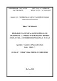
the 2-pyridylhydrazone derivative of ECAO, and the structures were minimized. The C5¼N moiety is seen to be planar, as expected for the substrate Schiff base. In the PSAO–1 structure (Fig. 7), no active site residues come into close proximity with the inhibitor except for Val384 and Gly385. The active site base, Asp300, is in an excellent position to abstract a Ca proton from the compound (2.49 A˚ away). The conserved tyrosine residue (Tyr286 in PSAO) to O4 of TPQ, reminiscent of Tyr369 in the crystal structure of the ECAO-2-pyridylhydrazone (Fig. 3). The active site (cid:1)gate(cid:2), Phe298, is in the (cid:1)open(cid:2) position, much as Tyr381 is in Fig. 3. Figure 8 displays the calculated structure for the AGAO– 1 Schiff base. Asp298, the active site base, is in a suitable position for proton abstraction from Ca (2.68 A˚ ). The in the (cid:1)open(cid:2) position. is active site (cid:1)gate(cid:2), Tyr296, Interestingly, the conserved tyrosine, Tyr284, is hydrogen bonded (2.13 A˚ ) to O2 of TPQ, and Tyr384 is hydrogen bonded (1.54 A˚ ) to Asp298 (Fig. 8A). The most notable difference between Figs 7 and 8 is the depiction of several residues that may directly interact with 1 in the latter case. Tyr296, Tyr302, and Asn381 (residue not shown in figure) all lie in close proximity ((cid:5) 3 A˚ ) to the inhibitor, although no direct interactions were detectable in the model. Residues Trp359 (from b ribbon on Arm I, at entrance to channel), Trp168, Phe105, Tyr302 and Tyr307 (Fig. 8B) appear to outline a hydrophobic pocket, which can accommodate the naphthyl ring. In addition, it appears that Phe105 and Tyr302 are in a good position to form p-stacking interactions with the naphthyl ring of 1. The AGAO–1 model was minimized to a greater degree than that of the PSAO–1 model in order to determine if the apparent p-stacking interactions would be more clearly observed in a
chain amines
(data not
To test the hypothesis that TPQ accessibility may be important in determining compound selectivity, solvent accessibility calculations were performed. Figure 9 displays solvent accessibility calculations for two known enzyme structures (PSAO and AGAO), one homology structural (PPLO), and one enzyme inhibitor complex model (2-hydrazinopyridine-ECAO), performed as described in Materials and methods. Active site accessibility can be seen to vary quite dramatically among these species. Although active site channel sizes for PSAO (Fig. 9A) and AGAO (Fig. 9B) are comparable, TPQ is more clearly visible from the surface in PSAO when respective gate residues are omitted from the model. The TPQ accessi- bility among these four enzymes qualitatively follows the rank order PPLO > PSAO ‡ AGAO > ECAO. The open accessibility of Tyr/TPQ478 in PPLO (Fig. 9C), as predicted by the amino acid sequence based model [34], may help explain why PPLO can oxidize substrates ranging from short to pentapeptides [34,52]. As previously reported for the ECAO 2-pyridyl- hydrazone, the tip of the pyridine ring can be seen protruding into the active site channel (Fig. 9D) [9], and TPQ was determined to be basically inaccessible to solvent without substantial movement of several side chains [13]. In contrast, due to the small size of the active-site (cid:1)gate(cid:2) in HPAO (Ala317), our calculations shown) indicate that both TPQ and the active-site base (Asp319) are solvent accessible in the crystal structure [33].
Fig. 8. Minimized structure of AGAO and 1. Hydrogen bonds are not shown for clarity. TPQ–1/SB: inhibitor 1 bound to TPQ as the sub- strate Schiff base moiety. Panel A: Key interactions with TPQ–1/SB as described in the text. Panel B: Hydrophobic pocket accommodating the naphthyl ring. TPQ–1/SB is rotated counterclockwise to illustrate the planarity of the Schiff base species. Residues in panel A are not displayed for clarity.
D I S C U S S I O N
Amine oxidase inhibition has been extensively studied, but no systematic comparison among different amine oxidases has been conducted to determine if selective inhibition is
Fig. 7. Minimized structure of PSAO and 1. Hydrogen bonds are not shown for clarity. TPQ–1/SB: inhibitor 1 bound to TPQ as the sub- strate Schiff base moiety.
3654 E. M. Shepard et al. (Eur. J. Biochem. 269)
(cid:2) FEBS 2002
substrate. In the latter regard, inactivation of Aspergillus niger amine oxidase [54] and grass pea amine oxidase [55] by DABY is proposed by Frebort et al. to reflect conjugate addition of the aminoynal turnover product to a nucleo- philic residue near the entrance to the channel, resulting in an adduct that cyclizes to a pyrrole. This residue has been implicated as Lys356 in A. niger amine oxidase [54] and Glu in grass pea amine oxidase [55], and homology modeling suggests that Lys113 would be the target in PSAO [55]. Further evidence that covalent modification of residues close to the substrate channel can result in inactivation was obtained in the case of the recombinant phenethylamine oxidase from AGAO, where a lysine chemical modification agent (NBD-F) was found to selectively modify the channel residues Lys184 and Lys354 [56].
feasible. Using six different amine oxidases, four com- pounds that were shown in preliminary studies to inactivate BPAO [35] were found here to inhibit the various enzymes with significantly different potencies and selectivities.
With this background, we can begin to interpret the findings of the current study. In the case of the naphthyloxy compound 1, AGAO is stoichiometrically inactivated (Table 2), and compound turnover does not occur (Ta- ble 3). The findings that the inactivated enzyme complex cannot be derivatized by phenylhydrazine and that its spectrum lacks the 480 nm TPQ absorption band (Fig. 4, trace a), suggests a modification of the TPQ cofactor and occupation of the active site cavity. However, as subjection of the inactivated enzyme to the Nitroblue tetrazolium assay (Table 4) revealed that TPQ is not permanently damaged, it appears that derivatization of TPQ by 1 represents a form that becomes subject to hydrolysis upon denaturation of the protein in the Nitroblue tetrazolium assay. It is possible that the inactive AGAO–1 complex represents merely either (a) a substrate Schiff base that cannot be transformed because of a misorientation between the catalytic base and the aC or (b) a product Schiff base that cannot be hydrolyzed because of inaccessibility of solvent water to the imine linkage. Alternatively, the observed result may reflect rearrangement of the product Schiff base to a more stable, yet ultimately hydrolysable derivative of TPQ that may or may not also involve an active site residue. Structural information on the inactivated enzyme will be needed to resolve these ambiguities.
(Table 1) and the efficiency of
In the case of PSAO, the naphthyloxy compound 1 is an excellent substrate (Table 3) as well as an inactivator. Inhibition thus most likely reflects partitioning of an electrophilic aldehyde turnover product toward alkylation of the active site channel. Assuming the covalently bound form of 1 has the same extinction coefficient as free 1, one calculates from the spectrum (Fig. 4, trace b) that there are (cid:5) 14 molecules of 1 bound to this preparation of PSAO that still has 75% activity. It appears that the reactive turnover product can bind to surface side-chains, such as 32 solvent accessible Lys residues suggested by Frebort et al. [55], in a manner that does not interfere with enzyme function unless there is modification of a side-chain within or at the entrance to the active site.
As a first approximation, one might expect some correlation between the efficiency of substrate activity (kcat/Km) inactivation (kinact/Ki), although little or no substrate turnover will be observed when partition ratios approach zero. Differences in the turnover efficiency will reflect in part the differences noted among the active-site channels that govern accessi- bility to TPQ as a function of substrate/inhibitor size and shape. There are also crucial stereochemical issues govern- ing turnover in terms of the orientation of the catalytic base with respect to that of the substrate/inhibitor when bound in Schiff base linkage to the TPQ cofactor. For any given enzyme-inhibitor Schiff base complex, the catalytic base will likely be positioned for preferential removal of either the pro-R or pro-S Ca proton. Likewise, if there is a Ca methyl group, only one of the two enantiomers will be processed. The finding that different amine oxidases display opposite stereochemical preferences for the same substrates [53] is strong evidence that the respective active-sites engender significant differences in binding modes, consistent with our observations here of rank order reversals in the inhibitory activity of the four compounds studied.
Our modeling results on the PSAO–1 Schiff base (Fig. 7) suggest that 1 is such an excellent substrate for PSAO because the only residues in close proximity to the bound inhibitor are Val384 and Gly385, and there are no unfavorable non–bonded interactions. The (cid:1)gate(cid:2) residue, Phe298, is rotated out of the way leaving room for the aldehyde product to exit after conversion of the substrate to product Schiff base and hydrolysis of the latter. In contrast, modeling of the AGAO–1 Schiff base (Fig. 8) reveals several
Mechanism-based inactivation can result either from covalent derivatization of the TPQ cofactor in manner that precludes reoxidation, or from covalent attachment to residues in or near the active site that block access of
Fig. 9. Solvent accessibility models. (A) PSAO; (B) AGAO; (C) PPLO; (D) ECAO. Blue: monomer 1. Red, b arm of monomer 2. Green, residues of b arm forming entrance to channel. Pink, gate residue. White, TPQ. Yellow, 2-hydrazinopyridine bound to TPQ.
Mechanism-based inhibition of amine oxidases (Eur. J. Biochem. 269) 3655
(cid:2) FEBS 2002
Nitroblue tetrazolium staining results for both AGAO and BPAO (Table 4) indicate, respectively, full or nearly full redox competence of the TPQ cofactor following this denaturing assay. Although one might be inclined to interpret the dramatic spectral changes observed in terms of TPQ modification, especially the initial absorbance at 440 nm seen with PSAO, a unified mechanism for the various enzymes would then require hydrolytic regeneration of redox-active TPQ in the Nitroblue tetrazolium assay, at least in the case of AGAO and BPAO. Thus, a more inactivation reflects conservative interpretation is that covalent modification of active-site or channel residues, with the spectral changes arising from chromophores generated upon rearrangement of the diyne moiety.
intriguing differences. The hydrogen bond between con- served Tyr284 and O2 of TPQ is curious because the hydrogen bond of the conserved tyrosine in ECAO [12] and HPAO [57] is purported to involve O4 of TPQ during normal turnover. We can infer that this hydrogen bond also exists for other amine oxidase species, as Fig. 7 supports. The significance of the hydrogen bond with O2 instead of O4 is not known, nor is the significance of the hydrogen bond between Tyr384 and the active site base, Asp298. The close proximity of nucleophiles Asn381, Tyr296, and Tyr302 may implicate these residues in trapping an intermediate species during turnover. Lastly, the presence of several aromatic residues near the naphthyl ring may potentially be a source for stabilization due to p-stacking, though this was not definitive in the static model depicted in Fig. 8.
The phthalimide inhibitor 4 shows significant turnover though significant tested (Table 3), by all enzymes inhibition was seen only with the two mammalian plasma amine oxidases (Table 2). The mechanism of inactivation by this compound most likely involves alkylation of a protein side-chain by the 2-chloroaldehyde turnover product, based on the result of the Nitroblue tetrazolium assay with BPAO indicating no permanent cofactor modification. The selective vulnerability of the mamma- lian enzymes suggests that they may contain active site features that differ considerably from the other amine oxidases.
C O N C L U S I O N S
The chloro inhibitor 2 is readily processed by all six amine oxidases studied, with good substrate activities relative to benzylamine in the three cases measured (Table 3) and highly efficient inactivation occurring in the cases of PPLO (cid:5) PSAO 7 EPAO > AGAO > BPAO (cid:5) ECAO (Table 2). The Nitroblue tetrazolium assay performed for preparations of AGAO and BPAO inactivated nearly completely by 2, reveals a level of TPQ redox competence consistent only with the small fraction of active cofactor remaining (Table 4). Figure 5 supports modification of the cofactor, as the transient chromophore at 600 nm is attributed to a conjugated complex formed between TPQ and 2. Despite the Nitroblue tetrazolium result, we cannot exclude the possibility that inactivation may also involve an active site residue, in line with Abeles’ proposal of alkylation by an allenic intermediate generated upon expulsion of chloride following Ca-proton abstraction [24].
The importance of this comparative screening study comes to light when considering the development of selective inhibitors of the copper-containing amine oxidases. The study here is one of the first ones to demonstrate such practical selectivity. Distinctions among the active sites must be responsible for differentiating the chemical interactions between the inhibitors and enzymes selected. For example, the unique difference in behavior with the naphthyloxy compound 1 seen between AGAO and PSAO can be understood at least in part from the differences in active–site interactions revealed by the modeling studies. This speaks directly to the importance of subtle differences in the active sites of these enzymes, as channel sizes are quite comparable between AGAO and PSAO. Interestingly, if we compare inhibitor potency for the chloro compound 2, we find that PSAO and PPLO are both substantially more sensitive to this compound than are AGAO and ECAO, whereas the reverse is true for the diyne compound 3. Furthermore, these four enzymes all display similar behavior with the phthalimide 4.
Despite the relatedness of 2 to DABY (by the elements of HCl), our finding that AGAO inactivated by DABY retains full TPQ redox competence in the Nitroblue tetrazolium assay supports a cofactor-sparing side-chain modification mechanism of inhibition in this case, possibly involving cyclocondensation onto a nucleophilic residue at the active site channel demonstrated by Frebort et al. for DABY inactivation of Aspergillus niger AO and grass pea AO [54]. However, inactivation of BPAO by DABY was accompanied by loss of TPQ redox competence, in agreement with what was found for 2 with both BPAO and AGAO. These results are most compatible with two different mechanisms of inactivation by DABY. The mechanism of TPQ modification by DABY in BPAO may be related to how 2 modifies the cofactor for both BPAO and AGAO, as one could envision loss of HCl at a stage subsequent to TPQ Schiff base formation with 2 to give the same intermediate arising from DABY. Even if the mechanisms of inactivation by 2 and DABY do converge in the case of BPAO, this cannot be the case at least for AGAO.
follow a markedly different
(cid:1)gate(cid:2) residues,
It is clear that these trends depart from what would be predicted based strictly on TPQ accessibility (Fig. 9). In addition, in some cases, gate residues must move in order for the inhibitor to bind, as was observed in the ECAO-2HP crystal structure and is further indicated by the AGAO and PSAO model structures with the naphthyloxy compound 1. These observations indicate that factors such as the dynamics of the dimensions/shape of the substrate channel and binding pocket, as well as the orientation of residues that can be alkylated, play at least as important a role in determining inhibitor selectivity as does TPQ accessibility. The clear advantage of mechanism-based inhibitors is that selectivity can arise both from variations in the initial noncovalent binding of the inhibitors with the
The diyne inhibitor 3 is generally less potent than the chloro inhibitor 2, though the selectivities among the six amine oxidases trend: BPAO > AGAO > ECAO > EPAO > PSAO 7 PPLO (Table 2 and kinetics data described). Substrate turnover for 3 is particularly low with AGAO and particularly high with PSAO, consistent with the lower partition ratio measured in the former case (36) and higher in the latter case (85). Despite the inactivated preparation of AGAO being incapable of reaction with phenylhydrazine, the
3656 E. M. Shepard et al. (Eur. J. Biochem. 269)
(cid:2) FEBS 2002
enzymes, as well as from variations in the ensuing chemistry [58].
3. Ruggiero, C.E. & Dooley, D.M. (1999) Stoichiometry of the topa quinone biogenesis reaction in copper amine oxidases. Biochem- istry 38, 2892–2898.
4. Cai, D. & Klinman, J.P. (1994) Copper amine oxidase: hetero- logous expression, purification, and characterization of an active enzyme in Saccharomyces cerevisiae. Biochemistry 33, 7647–7653. 5. Matsuzaki, R., Fukui, T., Sato, H., Ozaki, Y. & Tanizawa, K. (1994) Generation of the topa quinone cofactor in bacterial monoamine oxidase by cupric ion-dependent autooxidation of a specific tyrosyl residue. FEBS Lett. 351, 360–364.
The results presented here for DABY illustrate the point that various amine oxidases may interact with mechanism-based inhibitors by multiple mechanisms: although inhibition of BPAO by DABY is associated with TPQ modification, it appears that AGAO, PSAO, grass pea AO, and Aspergillus niger AO are inhibited by a mechanism distinct from cofactor alteration. This is even more fascinating considering that the closely related chloro inhibitor 2 appears to inactivate both AGAO and BPAO by a similar cofactor modification mechanism. Thus, subtle differences between the inhibitors and between the respective active sites can not only underlie differences in selectivity but also differences in mechanism of inhibition.
6. Cai, D. & Klinman, J.P. (1994) Evidence for a self-catalytic mechanism of 2,4,5- trihydroxyphenylalanine quinone biogenesis in yeast copper amine oxidase. J. Biol. Chem. 269, 32039–32042. 7. Matsuzaki, R., Suzuki, S., Yamaguchi, K., Fukui, T. & Tanizawa, K. (1995) Spectroscopic studies on the mechanism of the topa quinone generation in bacterial monoamine oxidase. Biochemistry 34, 4524–4530.
8. Plastino, J., Green, E.L., Sanders-Loehr, J. & Klinman, J.P. (1999) An unexpected role for the active site base in cofactor orientation and flexibility in the copper amine oxidase from Hansenula poly- morpha. Biochemistry 38, 8204–8216.
9. Wilmot, C.M., Murray, J.M., Alton, G., Parsons, M.R., Convery, M.A., Blakeley, V., Corner, A.S., Palcic, M.M., Knowles, P.F., McPherson, M.J. & Phillips, S.E.V. (1997) Catalytic mechanism of the quinoenzyme amine oxidase from Escherichia coli: exploring the reductive half-reaction. Biochemistry 36, 1608–1620.
10. Parsons, M.R., Convery, M.A., Wilmot, C.M., Yadav, K.D.S., Blakeley, V., Corner, A.S., Phillips, S.E.V., McPherson, M.J. & Knowles, P.F. (1995) Crystal structure of a quinoenzyme: copper amine oxidase of Escherichia coli at 2 A˚ resolution. Structure 3, 1171–1184.
Multiple interesting questions are raised by the results reported here. For example, why do the chloro and diyne compounds 2 and 3 effectively inhibit all enzymes, whereas the phthalimide 4 displays selectivity towards the mamma- lian enzymes? Also, even between two closely related mammalian enzymes, the equine and bovine plasma enzymes, there are notable differences in rank order: BPAO is more sensitive than EPAO to the naphthyloxy and diyne inhibitors 1 and 3, but the reverse is true for the chloro and phthalimide inhibitors 2 and 4. Preliminary results accord- ing to (cid:1)method A(cid:2) indicate that chloro compound 2 is quite selective towards inhibiting human kidney diamine oxidase, which has recently been purified and characterized in our laboratory [59], with 95% inhibition being observed using a fivefold excess of 2 over active sites, whereas even a 40-fold excess of 1, 3, and 4 results in only 32, 66 and 0% inhibition, respectively. These results alone point to the great promise that further work in this area can lead to the development of highly selective human enzyme inhibitors with therapeutic potential.
11. Murray, J.M., Saysell, C.G., Wilmot, C.M., Tambyrajah, W.S., Jaeger, J., Knowles, P.F., Phillips, S.E.V. & McPherson, M.J. (1999) The active site base controls cofactor reactivity in Escherichia coli amine oxidase: X-ray crystallographic studies with mutational variants. Biochemistry 38, 8217–8227.
12. Wilmot, C.M., Hajdu, J., McPherson, M.J., Knowles, P.F. & Phillips, S.E.V. (1999) Visualization of dioxygen bound to copper during enzyme catalysis. Science 286, 1724–1728.
Current work in our laboratories is being directed at identifying other inhibitor examples that display high selectivity for the amine oxidases described here as well as others. At the same time, the current screening study has already revealed particularly striking examples of selective irreversible inhibition, the structural and mechanistic bases of which are being pursued by in depth kinetic and spectroscopic studies, model studies, crystallographic stu- dies, and mass spectrometric analyses.
13. Wilce, M.C.J., Dooley, D.M., Freeman, H.C., Guss, J.M., Matsunami, H., McIntire, W.S., Ruggiero, C.E., Tanizawa, K. & Yamaguchi, H. (1997) Crystal structures of the copper-containing amine oxidase from Arthrobacter globiformis in the holo and apo forms: implications for the biogenesis of topaquinone. Biochem- istry 36, 16116–16133.
A C K N O W L E D G E M E N T S
laboratory)
We thank Dr Gang Sun (L.M.S. lab) for carrying out the Nitroblue tetrazolium staining studies. We also thank Dr Melanie Rogers (D. M. D. for valuable assistance with computer modeling. This work was supported by grants GM 27659 (to D. M. D.) and GM 48812 (to L. M. S.) from the National Institutes of Health.
14. Green, J., Haywood, G.W. & Large, P.J. (1983) Serological differences between the multiple amine oxidases of yeast and comparison of the specificities of the purified enzymes from Candida utilis and Pichia pastoris. Biochem. J. 211, 481–493. 15. Kagan, H.M. (1994) Lysyl oxidase: mechanism, regulation and relationship to liver fibrosis. Pathol. Res. Pract. 190, 910–919. 16. Wang, S.X., Mure, M., Medzihradszky, K.F., Burlingame, A.L., Brown, D.E., Dooley, D.M., Smith, A.J., Kagan, H.M. & Klin- man, J.P. (1996) A crosslinked cofactor in lysyl oxidase: redox function for amino acid side chains. Science 273, 1078–1084. 17. Boomsma, F., Van Veldhuisen, D.J., De Kam, P.J., Man in’t Veld, A.J., Mosterd, A., Lie, K.I. & Schalekamp, M.A. (1997) Plasma semicarbazide-sensitive amine oxidase is elevated in failure. Cardiovasc. Res. 33, patients with congestive heart 387–391.
R E F E R E N C E S
1. Janes, S.M., Mu, D., Wemmer, D., Smith, A.J., Kaur, S., Maltby, D., Burlingame, A.L. & Klinman, J.P. (1990) A new redox co- factor in eukaryotic enzymes: 6-hydroxydopa at the active site of bovine serum amine oxidase. Science 248, 981–987. 18. Salminen, T.A., Smith, D.J., Jalkanen, S. & Johnson, M.S. (1998) Structural model of the catalytic domain of an enzyme with cell adhesion activity: human vascular adhesion protein-1 (HVAP-1) D4 domain is an amine oxidase. Protein Eng. 11, 1195–1204.
19. Bono, P., Salmi, M., Smith, D.J. & Jalkanen, S. (1998) Cloning and characterization of mouse vascular adhesion protein-1 reveals 2. Ruggiero, C.E., Smith, J.A., Tanizawa, K. & Dooley, D.M. (1997) Mechanistic studies of topa quinone biogenesis in phenylethyla- mine oxidase. Biochemistry 36, 1953–1959.
Mechanism-based inhibition of amine oxidases (Eur. J. Biochem. 269) 3657
(cid:2) FEBS 2002
a novel molecule with enzymatic activity. J. Immunol. 160, 5563–5571. 39. Carter, S.R., McGuirl, M.A., Brown, D.E. & Dooley, D.M. (1994) Purification and active-site characterization of equine plasma amine oxidase. J. Inorg. Biochem. 56, 127–141.
40. Dove, J.E., Smith, A.J., Kuchar, J., Brown, D.E., Dooley, D.M. & Klinman, J.P. (1996) Identification of the quinone cofactor in a lysyl oxidase from Pichia pastoris. FEBS Lett. 398, 231–234. 20. Smith, D.J., Salmi, M., Bono, P., Hellman, J., Leu, T. & Jalkanen, S. (1998) Cloning of vascular adhesion protein-1 reveals a novel multifunctional adhesion molecule. J. Exp. Med. 188, 17–27. 21. Salmi, M., Yegutkin, G.G., Lehvonen, R., Koskinen, K., Salminen, T. & Jalkanen, S. (2001) A cell surface amine oxidase directly controls lymphocyte migration. Immunity 14, 265–276. 22. Abeles, R.H. & Maycock, A.L. (1976) Suicide enzyme inactivators. Accounts Chem Res. 8, 313–319.
41. Moenne-Loccoz, P., Nakamura, N., Steinebach, V., Duine, J.A., Mure, M., Klinman, J.P. & Sanders-Loehr, J. (1995) Character- ization of the topa quinone cofactor in amine oxidase from Escherichia coli by resonance Raman spectroscopy. Biochemistry 34, 7020–7026.
23. Neumann, R., Hevey, R. & Abeles, R.H. (1975) Action of plasma amine oxidase on beta-haloamines. Evidence for proton abstrac- tion in the oxidative reaction. J. Biol. Chem. 250, 6362–6367. 24. Hevey, R.C., Babson, J., Maycock, A.L. & Abeles, R.H. (1973) Highly specific enzyme inhibitors. Inhibition of plasma amine oxidase. J. Am. Chem. Soc. 95, 6125–6127. 42. Tanizawa, K., Matsuzaki, R., Shimizu, E., Yorifuji, T. & Fukui, T. (1994) Cloning and sequencing of phenylethylamine oxidase from Arthrobacter globiformis and implication of Tyr-382 as the precursor to its covalently bound quinone cofactor. Biochem. Biophys. Res. Commun. 199, 1096–1102.
25. Tang, S.S., Simpson, D.E. & Kagan, H.M. (1984) Beta-substituted ethylamine derivatives as suicide inhibitors of lysyl oxidase. J. Biol. Chem. 259, 975–979. 43. Suzuki, S., Sakurai, T., Nakahara, A., Manabe, T. & Okuyama, T. (1983) Effect of metal substitution on the chromophore of bovine serum amine oxidase. Biochemistry 22, 1630–1635.
26. Kumagai, H., Uchida, H. & Yamada, H. (1979) Reaction of fungal amine oxidase with beta-bromoethylamine. J. Biol. Chem. 254, 10913–10919.
27. Medda, R., Padiglia, A., Pedersen, J.Z., Agro, A.F., Rotilio, G. & Floris, G. (1997) Inhibition of copper amine oxidase by haloamines: a killer product mechanism. Biochemistry 36, 2595–2602. 44. Tabor, C.W., Tabor, H. & Rosenthal, S.M. (1954) Purification of amine oxidase from beef plasma. J. Biol. Chem. 208, 645–661. 45. Szutowicz, A., Kobes, R.D. & Orsulak, P.J. (1984) Colorimetric assay for monoamine oxidase in tissues using peroxidase and 2,2¢- azinodi (3-ethylbenzthiazoline-6-sulfonic acid) as chromogen. Anal. Biochem. 138, 86–94.
46. Shimizu, E., Ohta, K., Takayama, S., Kitagaki, Y., Tanizawa, K. & Yorifuji, T. (1997) Purification and properties of phenylethyl- amine oxidase of Arthrobacter globiformis. Biosci. Biotechnol. Biochem. 61, 501–505. 28. Pino, R. & Lyles, G.A. (1997) Effect of activity of semicarbazide- sensitive aminooxidases and cellular glutathione on the cytotoxic effect of allylamine, acrolein, and formaldehyde in human cultured endothelial cells. Vopr. Med. Khim. 43, 537–547.
47. Fluckiger, R., Paz, M.A. & Gallop, P.M. (1995) Redox-cycling detection of dialyzable pyrroloquinoline quinone and quinopro- teins. Methods Enzymol. 258, 14–19. 29. Blicharski, J.R. & Lyles, G.A. (1990) Semicarbazide-sensitive amine oxidase activity in rat aortic cultured smooth muscle cells. J. Neural Transm. Suppl. 32, 337–339. 48. Segel, I.H. (1976) Biochemical Calculations, 2nd edn. John Wiley & Sons, New York, NY.
49. Silverman, R.B. (1988) Mechanism-Based Enzyme Inactivation: Chemistry and Enzymology. CRC Press, Inc, Boca Raton, FL. 50. Oi, S., Inamasu, M. & Yasunobu, K.T. (1970) Mechanistic studies 30. Yu, P.H., Davis, B.A. & Boulton, A.A. (1995) Aliphatic pro- pargylamines, a new series of potent selective, irreversible non- amphetamine-like MAO-B inhibitors. Their structures, function and pharmacological implications. Adv. Exp. Med. Biol. 363, 17–23. of beef plasma amine oxidase. Biochemistry 9, 3378–3383.
51. McGaughey, G., Gagne, M. & Rappe, A. (1998) p-Stacking in proteins. J. Biol. Chem 273, interactions. Alive and well 15458–15463. 31. Abeles, R.H. & Tashjian, A.H. Jr (1975) Comparison of the inhibitory effects of propargylamine and pargyline on brain and liver monoamine oxidase activity. Biochem. Pharmacol. 24, 307–308. 52. Tur, S.S. & Lerch, K.
(1988) Unprecedented lysyl oxidase activity of Pichia Pastoris benzylamine oxidase. FEBS Lett. 238, 74–76. 32. Boulton, A.A., Davis, B.A., Durden, D.A., Dyck, L.E., Juorio, A.V., Li, X.M., Paterson, I.A. & Yu, P.H. (1997) Aliphatic propargylamines: new antiapoptotic drugs. Drug Dev. Res. 42, 150–156.
the crystal 53. Battersby, A.R., Staunton, J., Klinman, J. & Summers, M.C. (1979) Stereochemistry of oxidation of benzylamine by the amine oxidase from beef plasma. FEBS Lett. 99, 297–298.
33. Li, R.B., Klinman, J.P. & Mathews, F.S. (1998) Copper amine oxidase from Hansenula polymorpha: structure determined at 2.4 A˚ resolution reveals the active conformation. Structure 6, 293–307.
34. Kuchar, J. & Dooley, D. (2001) Cloning, sequence analysis, and characterization of the (cid:1)lysyl oxidase(cid:2) from Pichia pastoris. J. Inorg. Biochem. 83, 193–204. 54. Frebort, I., Pec, P., Luhova, L., Matsushita, K., Toyama, H. & Adachi, O. (1994) Active-site covalent modifications of quino- protein amine oxidases from Aspergillus niger: evidence for bind- ing of the mechanism-based inhibitor, 1,4-diamino-2-butyne, to residue Lys356 involved in the catalytic cycle. Eur. J. Biochem. 225, 959–965.
35. Sayre, L.M., Wang, F., Lee, Y., Huang, H., Greenaway, F.T., He, Z., Lightning, A. & Kagan, H.M. (1994) Biochemistry of Vitamin B6 and PQQ. Birkhauser-Verlag, Basel, Switzerland.
55. Frebort, I., Sebela, M., Svendsen, I., Hirota, S., Endo, M., Yamauchi, O., Bellelli, A., Lemr, K. & Pec, P. (2000) Molecular mode of interaction of plant amine oxidase with the mechanism- based inhibitor 2-butyne-1,4-diamine. Eur. J. Biochem. 267, 1423–1433. 36. Jeon, H.B. (2000) Synthesis and evaluation of mechanism-based inhibitors of copper-containing amine oxidases. PhD Thesis, Case Western Reserve University, Cleveland, OH.
37. McGuirl, M.A., McCahon, C.D., McKeown, K.A. & Dooley, D.M. (1994) Purification and characterization of pea seedling amine oxidase for crystallization studies. Plant Physiol. 106, 1205–1211. 56. Matsuzaki, R. & Tanizawa, K. (1998) Exploring a channel to the active site of copper/topaquinone-containing phenylethylamine oxidase by chemical modification and site-specific mutagenesis. Biochemistry 37, 13947–13957.
57. Hevel, J.M., Mills, S.A. & Klinman, J.P. (1999) Mutation of a strictly conserved, active-site residue alters substrate specificity and cofactor biogenesis in a copper amine oxidase. Biochemistry 38, 3683–3693. 38. Janes, S.M. & Klinman, J.P. (1991) An investigation of bovine serum amine oxidase active site stoichiometry: evidence for an aminotransferase mechanism involving two carbonyl cofactors per enzyme dimer. Biochemistry 30, 4599–4605.
3658 E. M. Shepard et al. (Eur. J. Biochem. 269)
(cid:2) FEBS 2002
59. Elmore, B., Bollinger, J.A. & Dooley, D.M. (2002) Human kidney diamine oxidase: heterologous expression, purification, and char- acterization. J. Biol. Inorg. Chem 7, 565–579.
58. Lee, Y., Shepard, E.M., Smith, J., Dooley, D.M. & Sayre, L.M. (2001) Catalytic turnover of substrate benzylamines by the qui- none-dependent plasma amine oxidase leads to H2O2-dependent inactivation. Evidence for generation of a cofactor-derived ben- zoxazole. Biochemistry 40, 822–829.