Selective detection of superoxide anion radicals generated from macrophages by using a novel fluorescent probe Jing Jing Gao1, Ke Hua Xu1, Bo Tang1, Ling Ling Yin1, Gui Wen Yang2 and Li Guo An2
1 College of Chemistry, Chemical Engineering and Materials Science, Shandong Normal University, Jinan China 2 College of Life Science, Shandong Normal University, Jinan, China
Keywords 2-chloro-1, 3-dibenzothiazolinecyclohexene; fluorescence image; fluorescent probe; macrophages; superoxide anion radical
Correspondence B. Tang, College of Chemistry, Chemical Engineering and Materials Science, Engineering Research Center of Pesticide and Medicine Intermediate Clean Production, Ministry of Education, Shandong Normal University, Jinan 250014, China Fax: +86 531 861 80017 Tel: +86 531 861 80010 E-mail: tangb@sdnu.edu.cn
–Æ) production at the site of radical Quantitation of superoxide radical (O 2 generation remains challenging. A simple method to detect nanomolar to micromolar levels of superoxide radical in aqueous solution has been devel- –Æ oped and optimized. This method is based on the efficient trapping of O2 using a novel fluorescent probe (2-chloro-1,3-dibenzothiazolinecyclohex- ene), coupled with a spectra character-signaling increase event. A high-spe- cificity and high-sensitivity fluorescent probe was synthesized in-house and –Æ over competing –Æ in living cells. Better selectivity for O2 used to image O2 cellular reactive oxygen species and some biological compounds illustrates the advantages of our method. Under optimal conditions, the linear calib- ration range for superoxide anion radicals was 5.03 · 10)9)3.33 · 10)6 m. The detection limit was 1.68 · 10)9 m. Fluorescence images of probe- stained macrophages stimulated with 4b-phorbol 12-myristate 13-acetate were obtained successfully using a confocal laser scanning microscope.
(Received 24 October 2006, revised 11 January 2007, accepted 29 January 2007)
doi:10.1111/j.1742-4658.2007.05720.x
in situ using confocal
–Æ include ESR [7], superoxide dismutase (SOD)-inhibitable Nitro Blue tetrazolium [8], chemiluminescence [9,10] and fluor- [11]. Among these methods, fluorescence escence
Reactive oxygen species (ROS) such as the superoxide –Æ), hydroxyl radical (HOÆ) and hydro- anion radical (O2 gen peroxide (H2O2) are important mediators in various pathological diseases [1]. Free radicals, in par- –Æ, have been found extensively in myocardial ticular O2 ischemia and reperfusion injury in recent years [2]. –Æ, a toxic reactive oxygen, not only harms Excessive O2 many biological molecules, but can also be converted to other more toxic radicals such as HOÆ, H2O2, 1O2, and so on [3]. Under consenescence or special physical –Æ can lead to coronary arterio- conditions, excessive O2 sclerosis and tumors [4–6]. Therefore, real-time monit- –Æ under physiological conditions is of oring of O2 increasing importance. To date, methods to detect of O2
Abbreviations DBZTC, 2-chloro-1,3-dibenzothiazolinecyclohexene; ROS, reactive oxygen species; SOD, superoxide dismutase.
FEBS Journal 274 (2007) 1725–1733 ª 2007 The Authors Journal compilation ª 2007 FEBS
1725
detection appears to be particularly attractive because it is able make superoxide anion radicals in living cells ‘visible’ laser scanning micro- scopy. Although several types of fluorescent probe for –Æ have been described [11– detecting and imaging O2 13], those with high selectivity, sensitivity and practi- cality are rare. For example, hydroethidine is the most –Æ [14], but its commonly used fluorescent probe for O2 selectivity could be improved [15]. In addition, owing to the short half-life of the superoxide anion radical, there is an exigent need for researchers to develop fast- –Æ in order to investigate its response probes to trap O2 mode of production, metabolism and trafficking. Therefore, considering the design-applicable probes, it is important that the reaction rate and selectivity are improved to avoid potential side reactions from other ROS under conditions that guarantee high sensiti- vity. If novel fluorescence probes that overcome these
J. J. Gao et al.
Selective detection of superoxide anion radicals
Scheme 1. Synthesis of DBZTC.
–Æ in living cells.
problems were available, they would contribute greatly to the elucidation of the roles of O2
increases in fluorescence
–Æ [17].
system provided sustained O2
(Hepes, 0.02 m, pH 7.4). As indicated in Fig. 1, DBZTC showed low blank fluorescence, although the addition of different concentrations of XA ⁄ XO trig- gered prompt (kex ⁄ em ¼ 485 ⁄ 559 nm). The XA ⁄ XO system was used as the –. It has been reported that on the main source of O2 catalysis of XO at pH 7.40, XA is oxidized to uric acid –Æ + 2H+ [16]. XA + 2O2 + H2O ¼ uric acid + 2O2 (single electron transfer). XA can also be oxidized by O2 with double electrons XA + O2 + H2O ¼ uric acid + H2O2 (double electron transfer). During the process, five of the six electron transfers are double electron transfers, and one electron transfer is single. Namely, one unit of XO can catalyze the conversion of 1.00 · 10)6 mol XA into 0.33 · 10)6 mol O2
–Æ.
the fluorescence intensity increased As expected, –Æ concentration (Fig. 1). Moreover, with increasing O2 there was a good linear correlation (R ¼ 0.9941)
between
In this
In this study, a fast-reaction fluorescent probe for –Æ, 2-chloro-1,3-dibenzothiazolinecyclohexene (DBZTC; O2 Scheme 1), was synthesized in-house and characterized using 1H NMR, IR spectra and elemental analysis. In an experiment, a xanthine ⁄ xanthine oxidase (XA ⁄ XO) –Æ production. model –Æ and yield a strong DBZTC was able to react with O2 fluorescence product with an emission maximum at –Æ was accom- 559 nm. Reaction of DBZTC with O2 plished within 10 min, making it more practical than –Æ the probes reported previously [12] for detecting O2 directly in living cells. More promisingly, the proposed –Æ over other ROS method has better selectivity for O2 and biological compounds, especially H2O2. Because –Æ, due to the level of H2O2 is high compared with O2 accumulation in biological systems, H2O2 may be the most competitive ROS in the quantitative detection of study, DBZTC showed a selective In this O2 –Æ that was > 500-fold greater than the response to O2 response to H2O2 (ratio in mol), which provides a unique opportunity to develop a chemical tool to mon- –Æ in a specific manner. Under optimal condi- itor O2 tions, relative relationship linear a –Æ concentration was fluorescence intensity and O2 obtained in the range 5.03 · 10)9)3.33 · 10)6 m. The detection limit was 1.68 · 10)9 m. regard, DBZTC is well suited as a fluorescent reagent that –Æ to be examined at allows the cellular chemistry of O2 the molecular level.
Results and Discussion
Fig. 1. Emission spectra (kex ¼ 485 nm) of DBZTC (10 lM) in the –Æ (0–6.66 lM) at 37 (cid:2)C in presence of various concentrations of O2 Hepes buffer (pH 7.40). Spectra were obtained 10 min after the addition of different concentrations XA ⁄ XO (final concentration: 0 ⁄ 0, 2 ⁄ 2, 3 ⁄ 3, 4 ⁄ 4, 6 ⁄ 6, 10 ⁄ 10, 15 ⁄ 15 and 20 ⁄ 20 lM ⁄ mU) to a solution of DBZTC.
Spectral properties
FEBS Journal 274 (2007) 1725–1733 ª 2007 The Authors Journal compilation ª 2007 FEBS
1726
Initially, we investigated the spectral properties of the probe under simulated physiological conditions
J. J. Gao et al.
Selective detection of superoxide anion radicals
A
–Æ concentration in between fluorescent intensity and O2 the range 5.03 · 10)9)6.66 · 10)6 m. The regression –Æ] (lm) +2579.1 (Fig. 2). equation was F ¼ 3815.8 [O2 The detection limit was 1.68 · 10)9 m. This result –Æ both qualita- showed that DBZTC could detect O2 tively and quantitatively.
Reaction conditions
concentration of To determine the optimum reaction conditions for –Æ, the effect of buffer solution and the analysis of O2 the the fluorescent probe were investigated.
Effect of pH and buffer concentration
B
Fig. 3. (A) Effect of pH. (B) Effect of buffer concentration: DBZTC (10 lM), XA (10 lM), XO (10 mU), Hepes (20 mM).
The pH of the medium has a large effect on fluores- cence intensity. Hepes was used because it has been shown to be a better buffer for the incubation of mammalian cells, and may improve the planting efficiency of cell incubation and breed ability of cell. –Æ detection We showed that the optimal pH for O2 was in the range 7.20–8.20 (Fig. 3A). Buffer concen- tration also affects fluorescence intensity. We showed that the relative fluorescence intensity of the system was high and stable at buffer concentrations of 16–22 (Fig. 3B). Thus 20 (v ⁄ v %) of Hepes (v ⁄ v %) (pH 7.40) was used throughout. In fact, a lower concentration of Hepes has a poor buffer capacity, that and superfluous Hepes leads to a salt effect decreases fluorescence intensity.
Effect of the fluorescent probe concentration
The concentration of DBZTC directly decided whether –Æ was trapped completely, which determined the O2
precision and sensitivity of the analytical method. The relative fluorescence intensity increased as the DBZTC concentration increased (< 9 lm), remained constant at DBZTC concentrations of 9–18 lm, then decreased (Fig. 4). A suitable concentration of DBZTC was advantageous, whereas superfluous DBZTC could quench fluorescence. Therefore, 10 lm of DBZTC was used throughout.
–Æ concentration.
Fig. 2. A linear correlation between the fluorescence intensity and O2
Effects of other ROS and biological compounds
FEBS Journal 274 (2007) 1725–1733 ª 2007 The Authors Journal compilation ª 2007 FEBS
1727
To assess the selectivity of the method, the effect of other ROS and biological compounds on the determin- ation of 3.33 lm O2 –Æ was examined individually. An error of ± 5.0% in the relative fluorescence intensity was considered tolerable. Little or no interference was encountered with (tolerance ratio in mol): NaClO, tert- butyl hydroperoxide, H2O2 (500); 1O2, glutathione, 1,4-hydroquinone (100); VC (50); HOÆ (5); ONOO–, –Æ was created by the enzymatic reaction of NO (1). O2
J. J. Gao et al.
Selective detection of superoxide anion radicals
Fig. 4. Effect of the fluorescent probe concentration: XA (10 lM), XO (10 mU), Hepes (20 mM).
–Æ.
Effect of SOD on fluorescence intensity of the reaction of DBZTC with superoxide
In order to further confirm that the changes in fluores- –Æ, SOD, cence of the probe solution were caused by O2 –Æ, was used in the reactive system. a scavenger of O2 After reaction of SOD (150 U) with XA ⁄ XO (10 lm ⁄ 10 mU) in Hepes buffer (20 mm) had been car- ried out for 30 min at 37 (cid:2)C, DBZTC was added and the reaction was immediately diluted with doubly dis- tilled water. The mixture was equilibrated and kept for 5 min before measurement. As can be seen from Fig. 6, fluorescence intensity was markedly suppressed by addition of SOD. However, when SOD was replaced by heat-inactivated SOD (90 (cid:2)C for 5 min) [18] or catalase, the fluorescence intensity of the reac- tion system barely changed. Overall, the results sub- stantiated that the proposed method was effective for detecting O2
–Æ.
Reaction mechanism
XA ⁄ XO (10 lm ⁄ 10 mU) at 25 (cid:2)C for 5 min. HOÆ and single oxygen (1O2) were generated by reacting H2O2 with Co2+ or NaOCl, respectively. Peroxynitrite (ONOO–) and nitric oxide (NOÆ) were obtained from 3- morpholinosydnonimine hydrochloride (SIN-1), and 3-(aminopropyl)-1-hydroxy-3-isopropyl-2-oxo-1-triazene, respectively. The results are summarized in Fig. 5. DBZTC appears to be a highly selective fluorescent probe for O2
–Æ to simulate bio- We used XA ⁄ XO as the source of O2 logical systems. In order to confirm the reaction mech- anism, IR and 1H NMR spectra of DBZTC and shown in the DBZTC oxide were analyzed. As 1H NMR spectra of DBZTC oxide, peaks correspond- ing to N–H (4.5) and C–H (4.0) disappeared, and in and C–H the (3110 cm)1) also disappeared, while a C ¼ N absorp- tion band at 1618 cm)1 appeared. All spectral data indicated that a larger conjugated structure of the DBZTC oxide was formed. From product analysis and the fluorescence properties, we propose that the mode
–Æ at pH 7.40 (20 mM Hepes). Fig. 5. Selectivity of DBZTC for O2 –Æ, other ROS and biological Fluorescence response of DBZTC to O2 compounds. Bars represent integrated fluorescence the final response (F) over the initial integrated emission (F0). Initial spectra were acquired in a 10 lM solution of DBZTC. Light grey bars repre- sent the addition of an excess of the appropriate other ROS and biological compounds (1.66 mM for H2O2, NaClO, and tert-butyl hydroperoxide, 0.33 mM for 1O2, GSH, and 1,4-hydroquinone, 0.17 mM for VC, 16.7 lM for HOÆ, 3.33 lM for ONOO–, NOÆ and –Æ) to a 10 lM solution of DBZTC. Black bars represent the sub- O2 –Æ to the solution. Excitation was sequent addition of 3.33 lM O2 provided at 485 nm.
Fig. 6. Fluorescence response of 10 lM DBZTC to XA ⁄ XO (10 ⁄ 10 lM ⁄ mU) in the presence of SOD (150 U), heat-inactivataed SOD (150 U), catalase (150 U) or the absence of SOD, respectively (kex ¼ 485 nm).
FEBS Journal 274 (2007) 1725–1733 ª 2007 The Authors Journal compilation ª 2007 FEBS
1728
IR spectrum, N–H (3245 cm)1)
J. J. Gao et al.
Selective detection of superoxide anion radicals
–Æ.
Scheme 2. A mechanism for the reaction of DBZTC with O2
–Æ with DBZTC is as
reaction of O2
–Æ (1 : 1 molar tion mechanism of DBZTC towards O2 ratio), the level of DBZTC was sufficient enough to –Æ completely. As shown in Fig. 8, the fluo- capture O2 rescence intensity of reaction system increased with increasing time up to 10 min and became constant –Æ thereafter. This shows that the probe can capture O2 quickly within 10 min, which attests to DBZTC being a ‘fast response’ probe. The fluorescence of the reagent blank remained unchanged to 40 min, which indicates that the probe has good stability.
–Æ in cells
follows of –Æ, DBZTC, an (Scheme 2). Upon treatment with O2 almost nonfluorescent compound, was oxidized at pH 7.40 by deleting hydrogen [19] to yield DBZTC oxide, which has better rigidity and a larger conjugated system (free electron pairs on S also conjugating with phenyl ring).
–Æ in a 1 : 1 molar ratio.
Confocal fluorescence imaging and detection of O2
In order to validate the molar ratio of the reaction –Æ, we synthesized pure between the probe and O2 DBZTC oxide and tested the fluorescence intensity of the oxide at different concentrations. A linear relation- ship between DBZTC oxide concentration and the fluor- escence intensity was obtained (Fig. 7). The linear equation was as followed: F ¼ 4268 [DBZTC oxide] + 288. Comparing this with the linear correlation between intensity and the concentration of the fluorescent –Æ] + 2579.1), we may deduce that –Æ (F ¼ 3815.8 [O2 O2 DBZTC reacts with O2
Kinetic assay
reaction time. 1, DBZTC (10 lM) + Hepes Fig. 8. Effect of (20 mM) + XA (10 lM) + XO (10 mU); 2, DBZTC (10 lM) + Hepes (20 mM).
Fig. 7. Linear correlation between the fluorescence intensity and the concentration of DBZTC oxide.
FEBS Journal 274 (2007) 1725–1733 ª 2007 The Authors Journal compilation ª 2007 FEBS
1729
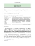
We used RAW264.7 macrophages to investigate the –Æ in living cells. Macr- potential of DBZTC to detect O2 ophages were seeded onto a glass slide and the concen- tration adjusted to 1 · 106 cellsÆmL)1 with Dulbecco’s modified Eagle’s medium containing 10% fetal bovine serum, 1% penicillin and 1% streptomycin. Cells were loaded with DBZTC (10 lm, Hepes, pH 7.40) by incubation at 37 (cid:2)C for 15 min and showed negligible intracellular background fluorescence (Fig. 9A). Macr- ophages, which were loaded with DBZTC (10 lm), were stimulated with 4b-phorbol 12 myristate 13 acet- ate (2 ngÆmL)1) at 37 (cid:2)C for 12 h; an obvious increase Different reaction times were investigated. XA ⁄ XO (10 lm ⁄ 10 mU) was added to buffer solutions of DBZTC (10 lm) maintained in a 37 (cid:2)C water bath. The fluorescence emission of each reaction mixture was measured at 5-min intervals against a reagent blank using a fluorescence spectrometer. Based on the reac-
J. J. Gao et al.
Selective detection of superoxide anion radicals
B
A
D
C
Fig. 9. Confocal fluorescence and brightfield images of live RAW264.7 macrophages. (A) Fluorescence image of RAW264.7 macrophages incubated with DBZTC (10 lM) at 37 (cid:2)C for 15 min. (B) Fluorescence image of probe-stained RAW264.7 macrophages stimulated with 4b-phorbol 12 myristate 13 acetate (2 ngÆmL)1) at 37 (cid:2)C for 12 h. (C) Bright-field image of probe-stained RAW264.7 macrophages stimulated with 4b-phorbol 12 myristate 13 acetate (2 ngÆmL)1) at 37 (cid:2)C for 12 h to confirm viability. (D) Fluorescence image of cells incubated with 100 lM Tiron at 37 (cid:2)C for 1 h after 4b-phorbol 12 myristate 13 acetate stimulation for 12 h, followed by loading with probe at 37 (cid:2)C for 15 min (scale bar ¼ 15 lM).
FEBS Journal 274 (2007) 1725–1733 ª 2007 The Authors Journal compilation ª 2007 FEBS
1730
DBZTC for 15 min, fluorescence intensity decreased markedly (Fig. 9D) Specificity was confirmed by add- ing a nonenzymatic superoxide scavenger, Tiron. The data show that DBZTC is membrane permeable, and –Æ concen- able to respond to micromolar changes in O2 tration in living cells. in fluorescence was seen (Fig. 9B). A bright-field image of macrophages stimulated with 4b-phorbol 12 myri- state 13 acetate for 12 h is shown in Fig. 9C. When 4b-phorbol 12 myristate 13 acetate-stimulated cells were further treated with a Tiron solution (a cell-per- –Æ scavenger) [20] for 1 h and incubated with meable O2
J. J. Gao et al.
Selective detection of superoxide anion radicals
pH meter (Shanghai Lei Ci Device Works, Shanghai, China) with a combined glass–calomel electrode. Fluorimet- ric spectra were obtained with a FLS-920 Edinburgh fluor- escence spectrometer with a xenon lamp and 1.0 cm quartz cells. Confocal fluorescence imaging was captured using a Zeiss LSM 510 META scanning microscope containing an Axiopian 2 MOT upright microscope and a 20· water- immersion objective lens. Excitation at 488 nm was carried out with an argon ion laser. Acquired images were analyzed using image-pro plus 4.5 software.
Materials
by
dissolving
–Æ in the cell extracts were satisfactory. To summarize, the proposed method can be applied –Æ by cell extract –Æ ‘visible’ in living cells using confo-
In order to further investigate the feasibility of the proposed method in biological systems, determination –Æ in cell extracts was performed. The fluorescence of O2 intensity of cell extracts treated with a Tiron solution –Æ mean was used as a blank value. The detected O2 content of 4b-phorbol 12 myristate 13 acetate-stimula- ted cell extracts was 0.92 · 0.02 lm, based on a corre- lation between the fluorescence intensity of the extracts and the regression equation. The average recovery test was carried out using the standard addition method, and the RSD was obtained from a series of six cell extracts (Table 1). The results indicated that the recov- ery and precision of the method applied to determine O2
to the quantitative determination of O2 test, and make O2 cal fluorescence imaging.
Conclusion
o-Aminobenzenothiol was purchased from Fluka (Shang- hai, China). A stock solution (1 mm) of DBZTC (synthes- ized in prepared was in-house) dimethylsulfoxide. This stock solution was diluted to 1.00 · 10)4 m before use. The XA solution (1.00 mm) was prepared by dissolving an appropriate amount of XA in 1.00 · 10)2 m NaOH; XO was from Sigma (St. Louis, MO), a stock solution of XO (1.00 UÆmL)1) was prepared in 2.30 m (NH4)2SO4, 1.00 · 10)2 m sodium salicylate bio- logy buffer, stored at 2–8 (cid:2)C. Hepes was from Sigma. GSH, superoxide dismutase, tert-butyl hydroperoxide (70% aqueous solution), H2O2 (30% aqueous solution), sodium hypochlorite (NaOCl, 5% aqueous solution), dimethyl- sulfoxide, 3-morpholinosydnonimine hydrochloride, 3-(ami- nopropyl)-1-hydroxy-3-isopropyl-2-oxo-1-triazene, phorbol 12-myristate 13-acetate, and RPMI-1640 medium were from Sigma. 1,4-Hydroquinone was from Fluka. 4,5-Dihydroxy- 1,3-benzenedisulfonic acid disodium salt (Tiron) was from Shanghai Reagent Co. Ltd (Shanghai, China). All chemi- cals were of analytical reagent grade, and double-distilled water was used throughout. RAW264.7 cells were from American Type Culture Collection (Manassas, VA).
We have developed a novel fluorescence probe, that can selectively and dose dependently DBZTC, –Æ in cellular systems. The probe has good sta- detect O2 bility, quick reaction, high sensitivity and exhibits –Æ than for other ROS and biolo- better selectivity for O2 gical compounds (no interference was encountered from a 500-fold molar excess of H2O2). Furthermore, DBZTC was confirmed as a cell-permeable probe and –Æ con- was able to respond to micromolar changes in O2 centration within living cells. Overall results establish the potential value of the probe for facilitating investi- gations of the generation, metabolism, and mechanisms of superoxide-mediated cellular homeostasis and injury.
Synthesis and properties of DBZTC
Experimental procedures
Apparatus
1H NMR spectra were recorded on a Bruker Avance 300. Elemental analysis was performed on a Perkin–Elmer Series II CHNS ⁄ O analyzer. IR spectra were recorded on PE-983 IR spectrometer (KBr discs cm)1, Perkin–Elmer, Norwalk, CT). All pH measurements were made using a pH-3c digital
Table 1. Determination of superoxide anion radicals in cell extracts (n ¼ 6). DBZTC (0.01 mM), Tiron solution (1 lM), 4b-phorbol 12 myristate 13 acetate (PMA; 2.0 ngÆL)1), XA (1.00 mM, 30 lL) ⁄ XO (1 U, 30 lL), Hepes (0.10 M, pH 7.40).
–Æ content of PMA-stimulated
Sample
Average recovery (%)
RSD (%)
O2 cells (lM)
Found (lM)
Mean (lM)
Added (lM)
Cell extracts
0.92 ± 0.02
1.00
1.89 ± 0.05
97.1
2.76
1.89, 1.85, 1.95, 1.83, 1.91, 1.96
FEBS Journal 274 (2007) 1725–1733 ª 2007 The Authors Journal compilation ª 2007 FEBS
1731
2-chloro-1-formyl-3-hydroxymethylenecyclohexene [21] A solution of 40.0 mL of dimethylformamide in 40.0 mL of methylene chloride was chilled in an ice bath. A solution of 37.0 mL of phosphorus oxychloride in 35.0 mL of methy- lene chloride was added dropwise with stirring. Then 10.0 g of cyclohexanone was added to the mixture. The solution was refluxed for 3 h, cooled, poured onto 200 g of ice, and
J. J. Gao et al.
Selective detection of superoxide anion radicals
excitation and emission slit were set to 3.5 and 3.5 nm, respectively.
allowed to stand overnight. The yellow solid was crystal- lized from a small volume of acetone cooled with dry ice, to give 14.50 g (82.39% yield) with a melting point of 130– 131 (cid:2)C. Anal. Calcd for C8H9ClO2: C, 55.67; H, 5.25; Cl, 20.54. Found: C, 55.41; H, 5.43; Cl, 20.47.
Preparation and staining of RAW264.7 macrophages cultures
We added 2.50 g (0.02 mmol) of o-aminobenzenothiol, 1.73 g (0.01 mmol) of 2-chloro-1-formyl-3-hydroxymethyl- enecyclohexene and 50.0 mL of a mixture of 1-butanol and benzene (7 : 3 v ⁄ v) to a 100 mL flask. The mixed solution was heated under reflux for 3 h. After the solvent was removed under reduced pressure, a yellow crude solid was obtained. This was recrystallized from benzene and dried under a vacuum (2.087 g, 54% yield). 1H NMR (300 MHz, DMSO-d6, 25 (cid:2)C, TMS): d 8.10–7.90 (m, 2H, benzol-H), 7.75 (d, 2H, benzol-H), 7.47–7.39 (m, 2H, benzol-H), 7,24 (d, 2H, benzol-H), 4.10 (m, 2H, N-H), 3.12 (m, 2H, methine-H), 2.3 (m, 3H, cyclohexene-H), 1.15 (m, 4H, cyclohexene-H). IR (KBr pellet): (cm)1) 3245 (N–H), 3110 (C–H), 1592 (C ¼ C). Melting point: 95–97 (cid:2)C. Elemental analysis (%) calcd for C20H19N2S2Cl (found): C 62.10 (62.02), H 4.92 (4.84), N 7.24 (7.36).
RAW264.7 macrophages were cultured in Dulbecco’s modi- fied Eagle’s medium containing 10% fetal bovine serum, 1% penicillin, and 1% streptomycin at 37 (cid:2)C (w/v) in a 5% CO2 ⁄ 95% air incubator MCO-15AC (SANYO, Tokyo, Japan). The concentration of counted cells was adjusted to 1 · 106 cellsÆmL)1 and cells were passed and plated on glass slide at 37 (cid:2)C, 5% CO2/95% air for 4 h. A set of cells was stimulated with 4b-phorbol 12 myristate 13 acetate (2 ngÆL)1) at 37 (cid:2)C for 12 h. Some of the stimulated cells were washed with serum-free Dulbecco’s modified Eagle’s medium, and a Tiron solution added (0.1 mm in serum-free Dulbecco’s modified Eagle’s medium, 2.5 mL). After 1 h, cells were washed with Dulbecco’s modified Eagle’s med- ium, and a solution of DBZTC (0.1 mm, 0.2 mL) was added to each dish loaded 2.5 mL serum-free Dulbecco’s modified Eagle’s medium, and incubated for 15 min. Before (0.10 m, imaging, cover-slips were washed with Hepes pH 7.40).
DBZTC
The oxidization product, DBZTC oxide, was obtained as follows: 0.050 g DBZTC was dissolved in 10 mL of eth- anol, 0.20 mL of 2.00 · 10)3 m KO2 solution (0.014 g KO2 was dissolved in 100 mL dimethylsulfoxide) and 20 mL of buffer solution (pH 7.40) were mixed together, and stirred for 15 min. The solvent was removed, and the residue was extracted by dichloroethane to give a crude product. The pure brown product was obtained by recrystallization in ethanol: 1H NMR (90 MHz, DMSO-d6, 25 (cid:2)C, TMS): d 8.11–7.93 (m, 2H, benzol-H), 7.66–7.58 (m, 2H, benzol-H), 7.40 (m, 2H, benzol-H), 7,16 (m, 2H, benzol-H), 1.83 (m, 3H, cyclohexene-H), 1.22 (m, 4H, cyclohexene-H). IR (KBr pellet): (cm)1) 1618 (C ¼ N), 1590 (C ¼ C). Elemental ana- lysis (%) calcd for C20H15N2S2Cl (found): C 62.75 (62.71), H 3.92 (3.84), N 7.32 (7.37).
–Æ
Synthesis and characteristics of DBZTC oxide RAW264.7 macrophage extracts
RAW264.7 macrophages were cultured in Dulbecco’s modi- fied Eagle’s medium, with the concentration of counted cells at 1 · 106 cellsÆmL)1. Some cells were added to a –. scavenger, Tiron solution and incubated for 1 h as an O2 others were stimulated with 4b-phorbol 12 myristate 13 acetate at 37 (cid:2)C for 12 h. All cells were then incubated with DBZTC for 30 min at 37 (cid:2)C and harvested by centrifuga- tion in the cold, and washed twice with 0.9% NaCl solu- tion. These cells were suspended again in a volume of Hepes equal to that in which they had been grown, and dis- rupted for 10 min in a VC 130PB ultrasonic disintegrator (Sonics & Materials Inc., Newtown, CT, USA). During sonic disruption, the temperature was maintained below 4 (cid:2)C with circulating ice water. The broken cell suspension was centrifuged at 1435 g for 5 min and the pellet discar- ded. Cell extracts that had been added to a Tiron solution were divided into six parts. The 4b-phorbol 12 myristate 13 acetate-stimulated cell suspension was divided into 12 parts and XA ⁄ XO was added into six parts in turn.
Determination of O2
Acknowledgements
Into a 10 mL color comparison tube were added 1.00 mL of DBZTC (1.00 · 10)4 m), 0.10 mL of XA solution (1.00 mm), 0.10 mL of XO (1.00 U) and 2.00 mL of Hepes buffer (0.10 m) in turn. After diluted to 10.00 mL volume with double-distilled water, the mixture was equilibrated and was laid aside at 37 (cid:2)C for 10 min before determin- ation. The fluorescence intensity was measured at kex ⁄ em ¼ 485 ⁄ 559 nm against a reagent blank at the same time. The
FEBS Journal 274 (2007) 1725–1733 ª 2007 The Authors Journal compilation ª 2007 FEBS
1732
This work was supported by Program for New Cen- tury Excellent Talents in University (NCET-04–0651), the National Natural Science Foundation of China (Nos 20335030 and 20575036).
J. J. Gao et al.
Selective detection of superoxide anion radicals
References
11 Mu¨ nzel T, Afanas’ev IB, Klescchyov AL & Harrison DG (2002) Detection of superoxide in vascular tissue. Arterioscler Thromb Vasc Biol 22, 1761–1768.
1 Chu CT, Levinthal DJ, Kulich SM, Chalovich EM & DeFranco DB (2004) Oxidative neuronal injury. Eur J Biochem 271, 2060–2066.
12 Hatsuo M, Kayko Y, Yoko N, Iho K, Leila H, Narit- sugu U, Shoko Y, Masako F, Yuka F, Yuji Y et al. (2005) A design of fluorescent for superoxide based on a nonredox mechanism. J Am Chem Soc 127, 68–69. 13 Xu KH, Liu X, Tang B, Yang G, Yang Y & An LG
2 Lowe RD & Snook RD (1991) Sensitive spectrophoto- metric determination of aluminium using thermal lens spectrometry. Anal Chim Acta 250, 95–104.
(2006) Design of a phosphinate-based fluorescent probe for superoxide detection in mouse peritoneal macrophages. Eur J Chem 13, 1411–1416.
3 Yi XF & Ben GY (2000) Seasonal variation in antioxi- dants of Polygonum viviparum and its relation to solar radiation in alpine meadow. Acta Bot Boreal Occident Sin 20, 201–205.
14 Tarpey MM & Fridovich I (2001) Methods of detection of vascular reactive species: nitric oxide, superoxide, hydrogen peroxide, and peroxynitrite. Circ Res 89, 224–236.
4 Chaudhury S & Sarkar PK (1983) Stimulation of tubu- lin synthesis by thyroid hormone in the developing rat brain. Biochem Biophys Acta 763, 93–98.
15 Benov L, Sztejnberg L & Fridovich I (1998) Critical
5 Fang RZ (1989) Radicals and Enzymes. Science Press,
Beijing.
evaluation of the use of hydroethidine as a measure of superoxide anion radical. Free Radical Biol Med 25, 826–831.
6 Kozumbo WJ, Trush MA & Kensler TW (1985) Are free radicals involved in tumor promotion? Chem Biol Interact 54, 199–207.
7 Nilsson UA, Haraldsson G, Bratell S, Sorensen V,
16 Fridovich I (1982) Measuing the activity of superoxide dismutase: an embarrassment of riches. In Superoxide Dismutase (Oberley LW, ed.), p. 15. CRC Press, Boca Raton, FL.
17 Fang YZ & Zheng RL (2002) Theory and Application of
Free Radical Biology. Scientific Press, Beijing.
18 Wilson MR, Stone V, Cullen RT, Searl A, Maynard
Akerlund S, Pettersson S, Schersten T & Jonsson O (1993) ESR-measurement of oxygen radical in vivo after renal ischemia in the rabbit: effects of pretreatment with superoxide dismutase and heparin. Acta Physiol Scand 147, 263–270.
8 Armstead WM, Mirro R, Busija DW & Leffler
RL & Donaldson K (2000) In vitro toxicology of respir- able Montserrat volcanic ash. Occup Environ Med 57, 727–733.
CW (1988) Postischemic generation of superoxide anion by newborn pig brain. Am J Physiol 255, H401–H403.
19 Tang B, Zhang L & Zhang LL (2004) Studies and appli- cation of flow injection spectrofluorimetry based on 2-(2-pyridil)-benzothiazoline as fluorescent probe trapping superoxide anion radicals. Anal Biochem 326, 176–182.
9 Dirnagl U, Lindauer U, Them A, Schreiber S, Pfister HW, Koedel U, Reszka R, Freyer D & Villringer A (1995) Global cerebral ischemia in the rat: online monitoring of oxygen free radical production using chemiluminescence in vivo. J Cereb Blood Flow Metab 15, 929–940.
20 Vaquero EC, Edderkaoui M, Pandol SJ, Gukovsky I & Gukovskaya AS (2004) Reactive oxygen species pro- duced by NAD(P)H oxidase inhibit apoptosis in pan- creatic cancer cells. J Biol Chem 279, 34643–34654. 21 Reynolds GA & Drexhage KH (1977) Stable hepta- methine pyrylium dyes that absorb in the infrared. J Org Chem 42, 885–888.
10 Peters O, Back T, Lindauer U, Busch C, Megow D, Dreier J & Dirnagl U (1998) Increased formation of reactive oxygen species after permanent and reversible middle cerebral artery occlusion in the rat. J Cereb Blood Flow Metab 18, 196–205.
FEBS Journal 274 (2007) 1725–1733 ª 2007 The Authors Journal compilation ª 2007 FEBS
1733