
Int.J.Curr.Microbiol.App.Sci (2017) 6(5): 807-815
807
Original Research Article https://doi.org/10.20546/ijcmas.2017.605.091
Study on Bacterial Flora of Burn Wound Infection: A Need for
Microbiological Surveillance in Burn Units
T. Sabetha1, A.V.M. Balaji2, J. Nithyalakshmi3*, K. Mohanakrishnan3 and G. Sumathi3
1Institute of Venerology, Madras Medical College, Chennai, India
2Stanley Medical College, Chennai, India
3Sri Muthukumaran Medical College and Research Institute, Mangadu, India
*Corresponding author:
A B S T R A C T
Introduction
Patients with burn injuries are highly
susceptible for infection as a result of
disruption of the normal skin barrier and
accompanying depression of immune
response. The burn surface contains a large
amount of necrotic tissue and the protein rich
wound exudates provides a rich growth
medium. So, following the initial period of
shock, infection is the major complication and
about 75% of the mortality associated with
burn injuries is related to infection. The
organisms are mainly derived from the
patient’s gastro intestinal and upper
respiratory tracts as well as from the hospital
environment (Al-Aali et al., 2016).
Infection, the risk of which is proportional to
the extent of injury, continues to be the
predominant determinant of outcome in
thermally injured patients. Most of the
International Journal of Current Microbiology and Applied Sciences
ISSN: 2319-7706 Volume 6 Number 5 (2017) pp. 807-815
Journal homepage: http://www.ijcmas.com
75% of the mortality associated with burn injuries is related to infection The aim of the
present study was to identify the bacterial profile of burn wound infection (BWI) in our
setting and determine their susceptibility pattern to commonly used antibiotics.This
prospective study was conducted over a period of one year in a teaching tertiary care
hospital, Chennai. A total of 100 patients with burns of total body surface area (TBSA) of
20% to 40% were included. Three wound swabs on 1st, 4th and 7th day were collected
aseptically and processed. Among the 274 samples collected, 191 swabs revealed
growth while 83 showed no growth. Overall isolation rate was found to be
69.7% and was predominantly monomicrobial with Gram positive cocci in early swabs.
Subsequent swabs showed 100% colonization with a shift to polymicrobial infection with
predominant isolation of Gram negative bacilli. The most common isolate was
Pseudomonas aeruginosa (35.84%), followed by Klebsiella pneumoniae (27.30%)
Acinetobacter spp. (20.13%), Staphylococcus aureus (8.87%), Escherichia coli (2.38%).
Gram negative bacteria were found to be highly susceptible to Imipenem and Piperacillin
/Tazobactum. Staphylococcus aureus was 100% sensitive to Linezolid. Knowledge about
specific pattern of burn wound infection and their resistant profile not only enable us to
plan empirical antibiotics to prevent imminent septic episodes but also reduce infection
related mortality in burns patients.
Keywords
Bacterial Flora,
Burn Wound
Infection,
Microbiological
Surveillance.
Accepted:
04 April 2017
Available Online:
10 May 2017
Article Info

Int.J.Curr.Microbiol.App.Sci (2017) 6(5): 807-815
808
infections are thought to be of nosocomial
origin wherein hand and clothing of
attending staff has been implicated in many
cases. The control of invasive burn wound
infection through the use of effective topical
chemotherapy, prompt surgical excision, and
timely closure of the burn wound has
resulted in unsurpassed survival rates. Even
so, these measures can cause emergence of
antibiotics resistant isolates and treatment
failures (Saaiq et al., 2015).
Several studies about the microbial flora
have revealed that immediately following
burn injury it is predominantly Gram-
positive organisms, within a week it is
replaced by Gram-negative organisms. The
distribution of infective agents varies with
time and is unique to different hospitals
(Mundhada et al., 2015).
The analysis of the isolates and their
sensitivity patterns helps us to track the
emerging trends to formulate an institutional
drug policy for the patients admitted in Burn
Unit. Rational antibiotic therapy according to
the prevalent strains of organisms should
help in reducing the mortality and morbidity
associated with burns (Shahzad et al., 2012).
In view of the above literature, this study
aims to identify the bacterial profile of burn
wound infection (BWI) in our setting and
determine their susceptibility pattern to
commonly used antibiotics.
Materials and Methods
This prospective study was conducted over a
period of one year in a teaching tertiary care
hospital, Chennai. A total of 100 patients with
burns of total body surface area (TBSA) of
20% to 40% (according to rule of nine) were
included. Specimens were three wound swabs
collected aseptically from burn area after
thorough cleaning with sterile saline. First
swab was collected immediately after
admission before start of antibiotics on Day 1`
and thereafter on Day 4 and Day 10.
Sample processing
Samples were processed as per standard
microbiological procedure. The specimens
were subjected to direct gram staining and
culture. Identification of aerobic bacteria and
its antimicrobial susceptibility pattern was
detected as per standard CLSI guidelines.
Antibiotic susceptibility was done by Kirby
Bauer disk diffusion method. Among gram
negative bacteria, Enterobacteriaceae were
tested against Ampicillin 10 µg, Amikacin 30
µg, Tetracycline 30 µg, Levofloxacin 5 µg,
Cefotaxime 30 µg, Ceftazidime 30 µg,
Ciprofloxacin 5 µg Imipenem 10 µg, and
Piperacillin-Tazobactum 100/10 µg. For
Pseudomonas species and Acinetobacter
species, antibiotic discs like Piperacillin-
Tazobactum 100/10 µg, Cefepime 30 µg,
Ceftazidime 30 µg, Imipenem 10 µg,
Gentamicin 10 µg, Amikacin 30 µg and
Ciprofloxacin 5 µg were used. For
Staphylococcus spp.. Cefoxitin 30 µg,
Erythromycin 15 µg,, Gentamicin 10 µg,
Amikacin 30 µg,Levofloxacin 5 µg,
Clindamycin 2 µg, Linezolid 30 µg,
Teicoplanin 30 µg were used.
For Enterobacteriaceae – Isolates were
considered a potential ESBL producer if the
zone of inhibition for ceftazidime was
observed to be <22mm.Potential ESBL
producer was then subjected for ESBL
Phenotypic confirmatory test –Disc Diffusion
method as recommended by CLSI guidelines
for antimicrobial disc susceptibility tests
(NCCLS, 2003b).
Phenotypic confirmatory disc diffusion test
(PCDDT) for ESBL
A Mueller Hinton agar plate was taken and a
lawn culture of potential ESBL producing

Int.J.Curr.Microbiol.App.Sci (2017) 6(5): 807-815
809
isolate was made. Then ceftazidime (30μg)
disc alone and with clavulanic acid (10μg)
were placed at an appropriate distance from
each other on the plate and incubated
aerobically at 37°C overnight. A ≥ 5mm
increase in zone diameter for antimicrobial
Ceftazidime tested in combination with
clavulanic acid in comparison to the zone
diameter when tested alone confirmed the
organisms to be an ESBL producer by
PCDDT.
Detection of MRSA
Methicillin resistant Staphylococcus
aureus (MRSA) detection was done using
cefoxitin 30 μg. Those isolates showed zone
of inhibition <21 mm considered as MRSA.
Results and Discussion
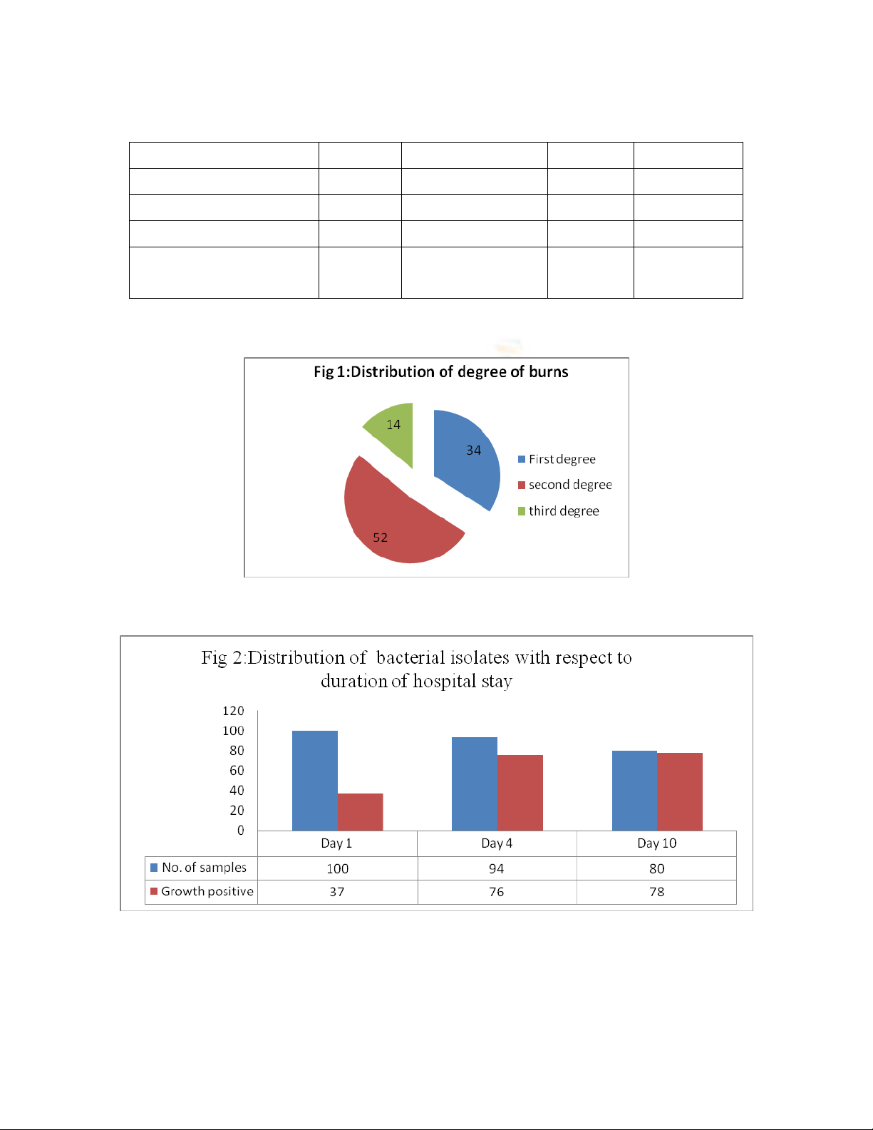
A total of 100 patients (44 were males and 56
were females) with 20% to 40% burns were
included in this study. Majority of the
subjects included in our study had sustained
second degree burns (52%) followed by first
degree (34%).(Fig 1)
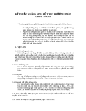
A total of 274 wound swabs were collected
from 100 patients. The reason for less number
of samples collected on day 4 and day10 were
due to the fact that patients were either
discharged or expired. 191 swabs revealed
growth while 83 showed no growth. Isolation
rate was found to be 69.7%.(Fig 2)
On admission Monomicrobial infection was
common and polymicrobial type of infection
was less and it was more with the patients
who stayed in the hospital for more than 2
days (Table 1).
The initial swabs were predominantly
monomicrobial with gram positive isolates
and which is replaced by gram negative
isolates in the later swabs, which were also
polymicrobial. (Table2). Overall, total
number of bacterial isolates obtained was
293.Among them, the most common isolate
was Pseudomonas aeruginosa 105 (35.84%),
followed by Klebsiella pneumoniae 80
(27.30%) Acinetobacter spp. 61(20.13%),
Staphylococcus aureus 22(8.87%),
Escherichia coli 7(2.38%).
To ensure early and appropriate therapy in
burn patients, a frequent evaluation of the
wound is necessary. Therefore, a continuous
surveillance of microorganisms and a regular
update of their antibiotic resistance pattern is
essential to maintain good infection control
program in the burn unit, thus improving the
overall infection-related morbidity and
mortality.
In this study the pattern of burn wound
microbial colonization was evaluated. The
time related changes in the predominant flora
was also evaluated throughout the patients
hospital stay.
Our study revealed slight female
preponderance (56%) compared to male.
This result was in agreement with the finding
reported by Mundhada et al., (2015), who
observed 54% in male and 46% in female.
Also, Rajput et al., (1998) found that burn
infection in females was (60%) while burn
infection in males was (40%). In contrast,
DeMacedo and Santos et al., (2005) found
that BWI in males 59.1% was more than
females 40.9%. In our country this is likely
due to occupational hazards of women
working in the kitchen as the kitchen is the
most common place prone to burn accidents.
In this study, mortality rate was low (8%)
against 19.6% by Lari et al., (2000). This low
rate might be due the fact that we are dealing
with patients having TBSA of burn between
20% and 40%. Majority of the subjects
included in our study had sustained second
degree burns (52%) followed by first degree
(34%) (Fig. 1) This was similar to the results

Int.J.Curr.Microbiol.App.Sci (2017) 6(5): 807-815
810
reported by Al- Akayleh et al., (1999) who
showed highest distribution of burn wound
infection in burn patients who had sustained
second-degree burn (53.9%).
Isolation rate was found to be 69.7% (Fig 2)
which is comparable to the isolation rate
observed by Srinivasan et al., (2009) (86.3%)
and Modi et al., (2013) (85.07). Irrespective
of duration of stay, monomicrobial pattern of
growth was found to be common than
polymicrobial which was in agreement with
other studies by Mundhada et al., (2015)and
Shahzad et al., (2012)(Table 1)
In a recent study on time-related changes in
aerobic bacterial pattern of burn wound
infection by Saha et al., (2011), it was found
that in burn wounds initially it was gram
positive organisms which are gradually
superceded by gram negative opportunists
that have greater propensity to invade.
Table.1 Type of Growth on wound swab
Day 1
n=37
%
Day 4
n=76
%
Day10
n=78
%
Monomicrobial
30
81.08%
42
55.26%
42
53.84%
Polymicrobial
7
18.91%
34
44.73%
36
46.15%
Table.2 Time related changes in bacterial profile of organisms Isolated
Organisms Isolated
Day 1
Day10
Day 4
Day 10
Monomicrobial
30
42
42
Pseudomonas aeruginosa
7
20
26
Klebsiellaspp
4
13
11
Acinetobacter spp
2
1
3
Escherichia coli
--
4
--
CONS
12
---
----
Staphylococcus aureus
5
4
2
Polymicrobial
7
34
36
Pseudomonas + Acinetobacter
---
6
8
Pseudomonas + Escherichia coli
---
3
---
Acinetobacter+ Klebsiella
---
11
10
Klebsiella+ CONS
4
---
----
Pseudomonas + Klebsiella
2
6
Pseudomonas + CONS
2
----
----
Pseudomonas + Acinetobacter+ Klebsiella
-----
6
8
Pseudomonas + Acinetobacter+ S. aureus
-----
2
4
Pseudomonas + S. aureus+ Klebsiella
1
4
-----
Int.J.Curr.Microbiol.App.Sci (2017) 6(5): 807-815
811
Table.3 Resistant Profile of the Organisms
Organisms
Total No
Resistance type
Positive
Percentage
Klebsiella Species
80
ESBL
36
45
Escherichia coli
7
ESBL
4
57
Staphylococcus aureus
22
MRSA
8
36.36
CoNS
18
Methicillin
resistant
5
27.77
Fig.1
Fig.2

![Phương pháp ELISA: [Thêm từ mô tả/định tính để tăng CTR]](https://cdn.tailieu.vn/images/document/thumbnail/2014/20140329/phamtrong91/135x160/8501396068823.jpg)
![Tài liệu Khảo sát trực tiếp [mới nhất]](https://cdn.tailieu.vn/images/document/thumbnail/2014/20140329/phamtrong91/135x160/3301396068801.jpg)


%20--%3e%3cdefs%3e%3cstyle%3e%20.st0%20{%20fill:%20%23fff;%20}%20.st1%20{%20fill:%20%237800fa;%20}%20%3c/style%3e%3c/defs%3e%3cpath%20class='st1'%20d='M117.78,12.18H43.11c2.9,3.47,4.65,7.94,4.65,12.82,0,5.6-2.3,10.66-6.01,14.29h76.02l7.22-13.56-7.22-13.56Z'/%3e%3cg%3e%3cpath%20class='st0'%20d='M53.58,26.17h-.59v-1.46h.59v-4.96h2.83c1.78,0,2.67.94,2.67,2.82v5.76c0,1.87-.89,2.81-2.67,2.81h-2.83v-4.96ZM55.36,21.37v3.34h1.1v1.46h-1.1v3.34h1.01c.61,0,.91-.37.91-1.1v-5.93c0-.74-.3-1.1-.91-1.1h-1.01Z'/%3e%3cpath%20class='st0'%20d='M65.99,31.14h-1.8l-.31-2.07h-2.19l-.31,2.07h-1.64l1.82-11.39h2.62l1.82,11.39ZM65.28,18.04c-.25.46-.51.77-.75.94-.21.15-.47.22-.79.22-.26,0-.57-.07-.92-.22l-.38-.15c-.14-.05-.26-.07-.37-.07-.3,0-.53.18-.71.54l-.91-.68c.25-.46.51-.77.75-.94.21-.14.48-.21.79-.21.26,0,.57.07.92.21l.38.15c.14.05.26.07.37.07.3,0,.53-.18.71-.54l.91.68ZM61.91,27.52h1.73l-.87-5.76-.87,5.76Z'/%3e%3cpath%20class='st0'%20d='M74.53,26.89v1.52c0,1.91-.89,2.86-2.67,2.86s-2.67-.95-2.67-2.86v-5.93c0-1.91.89-2.86,2.67-2.86s2.67.95,2.67,2.86v1.11h-1.69v-1.22c0-.75-.31-1.12-.93-1.12s-.93.37-.93,1.12v6.15c0,.74.31,1.11.93,1.11s.93-.37.93-1.11v-1.63h1.69Z'/%3e%3cpath%20class='st0'%20d='M81.4,31.14h-1.8l-.31-2.07h-2.19l-.31,2.07h-1.64l1.82-11.39h2.62l1.82,11.39ZM75.9,19.2l1.52-1.91h1.71l1.51,1.91h-1.61l-.76-.95-.75.95h-1.61ZM77.32,27.52h1.73l-.87-5.76-.87,5.76ZM83.1,15.99l-1.76,1.91h-1.26l1.17-1.91h1.86Z'/%3e%3cpath%20class='st0'%20d='M84.86,19.75c1.78,0,2.67.94,2.67,2.82v1.48c0,1.87-.89,2.81-2.67,2.81h-.85v4.28h-1.79v-11.39h2.64ZM84.01,21.37v3.86h.85c.58,0,.87-.36.87-1.08v-1.71c0-.71-.29-1.07-.87-1.07h-.85Z'/%3e%3cpath%20class='st0'%20d='M93.51,19.75c1.78,0,2.67.94,2.67,2.82v1.48c0,1.87-.89,2.81-2.67,2.81h-.85v4.28h-1.79v-11.39h2.64ZM92.66,21.37v3.86h.85c.58,0,.87-.36.87-1.08v-1.71c0-.71-.29-1.07-.87-1.07h-.85Z'/%3e%3cpath%20class='st0'%20d='M98.8,31.14h-1.79v-11.39h1.79v4.88h2.03v-4.88h1.83v11.39h-1.83v-4.88h-2.03v4.88Z'/%3e%3cpath%20class='st0'%20d='M105.36,24.55h2.46v1.62h-2.46v3.34h3.09v1.63h-4.88v-11.39h4.88v1.63h-3.09v3.18ZM108.17,17.29l-1.76,1.91h-1.26l1.17-1.91h1.86Z'/%3e%3cpath%20class='st0'%20d='M112.2,19.75c1.78,0,2.67.94,2.67,2.82v1.48c0,1.87-.89,2.81-2.67,2.81h-.85v4.28h-1.79v-11.39h2.64ZM111.35,21.37v3.86h.85c.58,0,.87-.36.87-1.08v-1.71c0-.71-.29-1.07-.87-1.07h-.85Z'/%3e%3c/g%3e%3ccircle%20class='st1'%20cx='25'%20cy='25'%20r='20'/%3e%3cpath%20class='st0'%20d='M32.78,19.27c2.92,0,4.43,2.55,5.28,5.33l.71,2.17c.14.38-.33.75-.71.75h-5.61c.19-.33.24-.71.09-1.08l-.75-2.45c-.43-1.32-.99-2.64-1.79-3.77.75-.57,1.65-.94,2.78-.94h0ZM25,18.38c3.25,0,4.9,2.78,5.89,5.89l.76,2.45c.14.42-.33.8-.8.8h-11.69c-.42,0-.94-.38-.8-.8l.75-2.45c.99-3.11,2.64-5.89,5.89-5.89h0ZM25,11.35c1.74,0,3.11,1.37,3.11,3.11s-1.37,3.11-3.11,3.11-3.11-1.41-3.11-3.11,1.41-3.11,3.11-3.11h0ZM17.27,19.27c1.08,0,1.98.38,2.73.94-.8,1.13-1.37,2.45-1.74,3.77l-.8,2.45c-.14.38-.05.75.09,1.08h-5.56c-.42,0-.9-.38-.75-.75l.71-2.17c.9-2.78,2.41-5.33,5.33-5.33h0ZM17.27,12.91c1.51,0,2.78,1.27,2.78,2.83s-1.27,2.83-2.78,2.83-2.83-1.27-2.83-2.83,1.27-2.83,2.83-2.83h0ZM32.78,12.91c1.56,0,2.78,1.27,2.78,2.83s-1.23,2.83-2.78,2.83-2.83-1.27-2.83-2.83,1.27-2.83,2.83-2.83h0ZM27.07,28.56v.09c0,.57-.24,1.08-.61,1.46h0v.05c-.38.33-.9.57-1.46.57s-1.08-.24-1.46-.61h0c-.38-.38-.61-.9-.61-1.46v-.09h1.41v.09c0,.19.05.38.19.47v.05c.09.09.28.19.47.19s.38-.09.47-.19v-.05c.14-.09.24-.28.24-.47t-.05-.09h1.41ZM30.99,28.56v.09c0,1.65-.66,3.16-1.74,4.24-1.08,1.08-2.59,1.79-4.24,1.79s-3.16-.71-4.24-1.79l-.05-.05c-1.04-1.08-1.7-2.55-1.7-4.2v-.09h1.41v.09c0,1.27.47,2.4,1.27,3.25h.05c.85.85,1.98,1.37,3.25,1.37s2.4-.52,3.25-1.37c.85-.8,1.37-1.98,1.37-3.25v-.09h1.37ZM34.99,28.56v.09c0,2.78-1.13,5.28-2.92,7.07-1.79,1.79-4.29,2.92-7.07,2.92s-5.23-1.13-7.07-2.92c-1.79-1.79-2.92-4.29-2.92-7.07v-.09h1.41v.09c0,2.4.94,4.53,2.5,6.08,1.56,1.56,3.72,2.5,6.08,2.5s4.52-.94,6.08-2.5c1.56-1.56,2.5-3.68,2.5-6.08v-.09h1.41Z'/%3e%3c/svg%3e)