Sulfated polysaccharides inhibit the catabolism and loss of both large and small proteoglycans in explant cultures of tendon Tom Samiric, Mirna Z. Ilic and Christopher J. Handley
School of Human Biosciences, La Trobe University, Melbourne, Australia
Keywords aggrecan; calcium pentosan polysulfate; decorin; heparin; proteoglycan; tendon
Correspondence T. Samiric, School of Human Biosciences, La Trobe University, 3086, Victoria, Australia Fax: +61 394795784 Tel: +61 394793417 E-mail: T.Samiric@latrobe.edu.au
(Received 10 May 2006, accepted 2 June 2006)
doi:10.1111/j.1742-4658.2006.05348.x
This study investigated the effects of two highly sulfated polysaccharides, calcium pentosan polysulfate and heparin, on the loss of newly synthesized proteoglycans from the matrix of explant cultures of bovine tendon. The tensional region of deep flexor tendon was incubated with [35S]sulfate for 6 h and then placed in culture for up to 15 days. The amount of radiolabel associated with proteoglycans lost to the medium and retained in the mat- rix was determined for each day in culture. It was shown that both sulfated polysaccharides at concentrations of 1000 lgÆmL)1 inhibited the loss of 35S-labeled large and small proteoglycans from the matrix and concomitant with this was a retention of chemical levels of proteoglycans in the explant cultures. In other explant cultures that were maintained in culture in the presence of both agents for more than 5 days after incubation with [35S]sul- fate, inhibition of the intracellular catabolic pathway was evident, indica- ting that these highly sulfated polysaccharides also interfered with the intracellular uptake of small proteoglycans by tendon cells.
found in similar proportions in tendon, and together make up almost 20% of the total proteoglycan content of the tissue [5]. These proteoglycans can bind to hya- luronan and form hydrated aggregates within the extracellular matrix or interact with other matrix mac- romolecules and contribute to the organization of the extracellular matrix [7].
The role of tendons is to absorb and transmit force generated by muscles to bones, the basis of joint move- ment. Tendons are fibrous connective tissues that are sparsely populated with cells that are responsible for the synthesis and maintenance of the extracellular mat- rix. Type I collagen is the major macromolecule pre- sent in the extracellular matrix of tendon, comprising over 50% of the dry weight and providing the tissue with its tensile strength. The remainder of the extracel- lular matrix is made up of proteoglycans and noncolla- genous proteins [1,2]. We and others have shown that the small proteoglycans, predominantly decorin, con- stitute (cid:1)80% of the total proteoglycan content in the proximal ⁄ tensional regions of tendon [3–5]. The core proteins of small proteoglycans are characterized by leucine-rich repeat structures that have been implicated in the binding of these proteoglycans to collagen [6]. The large proteoglycans aggrecan and versican are
We have previously shown in both cartilage and fibrous connective tissues such as tendon and ligament that the loss of newly synthesized large proteoglycans from the matrix occurs at a faster rate than that of small proteoglycans [8–10]. In arthritic diseases, the large proteoglycans are one of the first matrix compo- nents to be degraded and lost from the articular carti- lage and this is associated with impairment of the biomechanical function of the tissue [11]. An increased rate of aggrecan degradation can be attributed to pro- teolytic cleavage at specific sites along the core protein.
Abbreviations ADAMTS, a disintegrin and metalloproteinase with thrombospondin motifs; CaPPS, calcium pentosan polysulfate; DMEM, Dulbecco’s modified Eagle’s medium; GnHCl, guanidinium chloride.
FEBS Journal 273 (2006) 3479–3488 ª 2006 The Authors Journal compilation ª 2006 FEBS
3479
T. Samiric et al.
Inhibition of proteoglycan catabolism in tendon
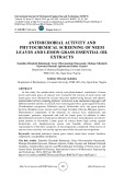
described in the Experimental procedures. The tissue was then maintained in Dulbecco’s modified Eagle’s medium (DMEM) in the presence of 0, 100 and 1000 lgÆmL)1 CaPPS or heparin for 10 days. After predetermined times in culture, the tissue was extracted with 4 m GnHCl for 72 h at 4 (cid:1)C followed by 0.5 m NaOH as described in the Experimental procedures. The percentage of 35S-labeled proteoglycans remaining in the matrix was determined and plotted as a function of time in culture. Figure 1A shows that CaPPS inhib- ited the catabolism of 35S-labeled proteoglycans in these cultures in a dose-dependent manner. On day 10 of the culture period, (cid:1)60% of 35S-labeled proteogly- cans remained in the matrix of cultures that had been
100
We and others have shown that the predominant activ- ity responsible for the degradation of aggrecan in con- nective tissues in vitro is aggrecanase, which has been shown to be attributable to a number of proteinases the ADAM-TS family of proteinases namely of ADAMTS-1, -4 and -5 [12–17]. In contrast, the degra- dation of small proteoglycans in cartilage, ligament and tendon appears to occur at a slower rate, even in the presence of catabolic agents [8,10,18]. This may suggest that the interaction of the small proteoglycans with the surface of the collagen fibrils may protect these pro- teoglycans from proteolysis during the early phases of connective tissue degradation [18]. We have shown that in ligament and tendon, the loss of small proteoglycans from the tissue involves either the loss of intact or par- tially degraded core proteins, or internalization and subsequent degradation by the cells [10,19].
CaPPS
A
90
80
70
60
50
40
30
x i r t a m e h t n
i
20
i
10
i
0
100
Heparin
B
l
90
80
g n n a m e r s n a c y g o e t o r p
70
l
60
d e e b a l -
S 5 3
50
%
40
30
20
10
0
0
1
2
3
4
5
6
7
8
9
10
Days in Culture
A number of highly sulfated polysaccharides have been shown to slow the degradation of articular carti- lage in animal models of osteoarthritis [20]. One such polymer is pentosan polysulfate, a sulfated polyxylan that has been shown to modify both the symptoms and the progression of osteoarthritis in both humans and animals [21]. We and others have shown that the calcium salt of pentosan polysulfate (CaPPS) will directly inhibit aggrecanase activity by interacting with aggrecanase proteinases and will suppress aggrecanase- mediated degradation of newly synthesized aggrecan by cartilage explant cultures [22,23]. Heparin, which is recognized for its anticoagulant properties, is another highly sulfated polysaccharide that has been shown to have inhibitory effects on aggrecanase activity [22–24]. Although these highly sulfated polysaccharides have been shown to suppress the catabolism of proteogly- cans by cartilage [25], the effects of these agents on joint fibrous connective tissues such as tendon where the small proteoglycans are predominant are lacking. In order to elucidate further the mechanisms regulating proteoglycan loss from the tissue, we have investigated the effects of two highly sulfated polysaccharides, CaPPS and heparin, on the degradation of newly syn- thesized proteoglycans from the extracellular matrix of bovine tendon explant cultures.
Results
Measurement of loss of 35S-labeled proteoglycans from bovine tendon explant cultures with varying concentrations of CaPPS or heparin
Fig. 1. Kinetics of loss of 35S-labeled proteoglycans by bovine ten- don explant cultures maintained in DMEM in the presence of vary- ing concentrations of CaPPS or heparin. The proximal region of bovine deep flexor tendon was incubated with [35S]sulfate for 6 h and maintained in DMEM in the presence of 0 (d), 100 (.), or 1000 (s) lgÆmL)1 CaPPS (A) or in the presence of 0 (d), 100 (.), or 1000 (s) lgÆmL)1 heparin (B) for 10 days. The percentage of 35S-labeled proteoglycans remaining in the matrix of tendon cul- tures at each day after incubation with [35S]sulfate was determined as described in the Experimental procedures. The positive error bars represent the range of duplicate samples over the 10 days.
The proximal region of deep flexor tendon was incuba- ted with 200 lCiÆmL)1 [35S]sulfate at 37 (cid:1)C for 6 h as
FEBS Journal 273 (2006) 3479–3488 ª 2006 The Authors Journal compilation ª 2006 FEBS
3480
T. Samiric et al.
Inhibition of proteoglycan catabolism in tendon
Table 2. Hexuronate concentration of bovine tendon explants main- tained in DMEM alone or DMEM containing 1000 lgÆmL)1 CaPPS or heparin. The proximal region of bovine deep flexor tendon was maintained in DMEM alone, or DMEM containing 1000 lgÆmL)1 CaPPS or heparin for up to 10 days. Tissue was extracted on day 5 and 10 of the culture period with 0.5 M NaOH as described in Experimental procedures. The values represent the median ± range of duplicate samples.
Hexuronate concentration (lg ⁄ 100 mg wet weight of tissue)
Day 0
Day 5
Day 10
Treatment
114 ± 8
– –
60.4 ± 18 75.7 ± 5 86.5 ± 8
62.4 ± 2 76.2 ± 2 102.9 ± 22
DMEM alone DMEM +1000 lgÆmL)1 CaPPS DMEM +1000 lgÆmL)1 heparin
in cultures maintained in DMEM alone. However, and 100 maintained in DMEM containing 1000 lgÆmL)1 CaPPS, the percentage of 35S-labeled proteoglycans remaining in the matrix by day 10 of culture was 65 and 72%, respectively. This was also observed in repeated experiments using tissue from dif- ferent animals. Figure 1B shows that in the presence of heparin, the catabolism of 35S-labeled proteoglycans in these cultures was also inhibited. By day 10 of the 35S-labeled proteoglycans culture period, (cid:1)62% of remained in the matrix of cultures that had been main- tained in DMEM alone. However, in cultures main- tained in DMEM containing 100 and 1000 lgÆmL)1 heparin, the percentage of 35S-labeled proteoglycans remaining in the matrix after 10 days in culture was 77 and 79%, respectively. This was also observed in other cultures. In addition, these data indicate that heparin was more potent than CaPPS in inhibiting the loss of 35S-labeled proteoglycans from the matrix of tendon explants.
the loss of
absence or presence of CaPPS or heparin on days 5 and 10, as determined by hexuronate, are shown in Table 2. The levels of endogenous glycosaminoglycans in the matrix on day 10, represented by the chemical pools of both large and small proteoglycans, were greater in cultures maintained in DMEM containing 1000 lgÆmL)1 CaPPS or heparin. Heparin appeared to inhibit tissue glycosaminoglycans to a greater extent than CaPPS in these cultures.
Effects of polysulfated polysaccharides on the loss of 35S-labeled large and small proteoglycans from bovine tendon explants
The rate of incorporation of [3H]serine into proteins of tendon explant cultures was measured on days 5 and 10 of the culture period in the conditions above (Table 1). The addition of CaPPS or heparin, at con- centrations of up to 1000 lgÆmL)1, to the culture medium did not affect the rate of incorporation of [3H]serine into protein on days 5 and 10, indicating that neither sulfated polysaccharide had an adverse affect on cellular metabolism. This lack of effect on the cellular metabolism has also been observed in explant cultures of articular cartilage maintained in both agents at concentrations of up to 1000 lgÆmL)1 [25].
The endogenous glycosaminoglycan levels present in tendon cultures maintained in the
the matrix of
Table 1. Incorporation of [3H]serine into macromolecules of bovine tendon explants maintained in DMEM containing varying concentra- tions of CaPPS or heparin. The proximal region of bovine deep flexor tendon was maintained in DMEM alone or DMEM containing 100 or 1000 lgÆmL)1 CaPPS or heparin. Tissue cultures were incubated with 30 lCiÆmL)1 [3H]serine for 2 h on days 5 and 10, as described in Experimental procedures. The values represent the median ± range of duplicate samples.
[3H]Serine incorporation (dpm ⁄ mg wet weight of tissue per 2 h)
Day 5
Day 10
Treatment
In order to determine whether highly sulfated polysac- charides inhibited the loss of either 35S-labeled large or small proteoglycans from bovine tendon matrix, the hydrodynamic size of 35S-labeled proteoglycans present in tendon matrix at various times during culture maintained in DMEM alone or DMEM containing 1000 lgÆmL)1 CaPPS or heparin was determined. Tis- sue from the experiment described in Fig. 1 was extracted on days 0, 2, 4, 6, 8, and 10 with 4 m GnHCl for 72 h at 4 (cid:1)C as described in the Experimental pro- cedures. These samples were subjected to size-exclusion chromatography on a column of Sepharose CL-4B eluted under dissociative conditions as described in the Experimental procedures (data not shown). From this, the percentage of 35S-labeled large and small proteo- glycans remaining in the matrix at various times after incubation of tendon with [35S]sulfate was determined as previously described [10].
1593 ± 748 1654 ± 499 1822 ± 219 1708 ± 371 1498 ± 456
1356 ± 57 1064 ± 168 1224 ± 129 1452 ± 522 1390 ± 284
DMEM alone DMEM +100 lgÆmL)1 CaPPS DMEM +1000 lgÆmL)1 CaPPS DMEM +100 lgÆmL)1 heparin DMEM +1000 lgÆmL)1 heparin
Figure 2 shows that CaPPS (Fig. 2A) and heparin (Fig. 2B) inhibited the loss of both the 35S-labeled large and small proteoglycans from the extracellular matrix. On day 10 of the culture period, (cid:1)10% of
FEBS Journal 273 (2006) 3479–3488 ª 2006 The Authors Journal compilation ª 2006 FEBS
3481
T. Samiric et al.
Inhibition of proteoglycan catabolism in tendon
small proteoglycans remained in the matrix that had been maintained in DMEM alone, but in cultures main- tained in DMEM containing 1000 lgÆmL)1 of either CaPPS (Fig. 2A) or heparin (Fig. 2B), the percentage of 35S-labeled small proteoglycans remaining in the matrix by day 10 of culture was (cid:1)80%. This indicates that these sulfated polysaccharides inhibited the loss of the radiolabeled small proteoglycans by (cid:1)33%.
Analysis of radiolabeled 35S-proteoglycan core proteins remaining in the matrix
To determine the effects of CaPPS and heparin, at con- centrations of 1000 lgÆmL)1, on the proteolytic pro- cessing of 35S-labeled proteoglycans remaining in the tendon matrix after 9 days in culture, the 35S-labeled proteoglycans core protein fragments were isolated on anion-exchange chromatography prior to being ana- lyzed on SDS ⁄ PAGE followed by fluorography. Figure 3 shows that a high molecular weight band above 200 kDa, representing the large proteoglycans, is retained in the matrix after 9 days in culture in the presence of CaPPS (Fig. 3A) or heparin (Fig. 3B). This further indicates that these highly sulfated agents inhi- bit the loss of 35S-labeled large proteoglycans.
Effects of polysulfated polysaccharides on the intracellular catabolism of 35S-labeled small proteoglycans of tendon explant cultures
and
small
(,)
Fig. 2. The effects of CaPPS and heparin on the percentage of each 35S-labeled proteoglycan species remaining in the matrix of bovine tendon cultures. For the experiment outlined in the legend of Fig. 1, the percentage of 35S-labeled large and small proteogly- cans remaining in the tissue at each time after incubation of bovine tendon with [35S]sulfate was determined in (A) DMEM alone [large (s) or DMEM containing proteoglycans] 1000 lgÆmL)1 CaPPS [large (d) and small (.) proteoglycans], or (B) DMEM alone [large (s) and small (,) proteoglycans] or DMEM containing 1000 lgÆmL)1 heparin [large (d) and small (.) proteogly- cans]. The percentage of 35S-labeled proteoglycans remaining in the matrix of tendon cultures at each day after incubation with [35S]sulfate was determined as described in Experimental proce- dures. The positive error bars represent the mean range of dupli- cate samples over the 10 days.
Previous work has shown that approximately one-half of the 35S-labeled small proteoglycans that are lost from the tendon matrix are taken up by the cells and degra- ded [10]; the following experiment was undertaken to determine the effects of CaPPS and heparin, at concen- trations of 1000 lgÆmL)1, on the intracellular catabol- ism of 35S-labeled small proteoglycans by bovine tendon explant cultures. After incubation with [35S]sulfate as described in the Experimental procedures, tissue was maintained in culture in DMEM alone for 5 days to allow time for loss from the cultures of the large 35S-labeled proteoglycans and any unincorporated [35S]sulfate from the tissue [10]. Cultures were then maintained for a further 10 days in either DMEM alone or DMEM containing 1000 lgÆmL)1 CaPPS or heparin. Culture medium was collected daily and the tissue extracted at the end of the culture period with 4 m GnHCl followed by 0.5 m NaOH as described in the Experimental procedures. Both medium and tissue extract samples were analyzed for the presence of 35S-labeled macromolecules and free [35S]sulfate by size exclusion chromatography using Sephadex G-25 the columns, as we have previously shown that
35S-labeled large proteoglycans remained in the matrix of cultures that had been maintained in DMEM alone but, in cultures maintained in DMEM containing 1000 lgÆmL)1 of either CaPPS (Fig. 2A) or heparin (Fig. 2B), the percentage of 35S-labeled large proteo- glycans remaining in the matrix by day 10 of culture was (cid:1)45%. This indicates that with the inclusion of these sulfated agents there was significant inhibition ((cid:1)40%) of rate of loss of 35S-labeled large proteogly- cans from the matrix by the end of the culture period. Similarly, by day 10 of culture, (cid:1)70% of 35S-labeled
FEBS Journal 273 (2006) 3479–3488 ª 2006 The Authors Journal compilation ª 2006 FEBS
3482
T. Samiric et al.
Inhibition of proteoglycan catabolism in tendon
A
B
C
A
i
Stds (kDa)
l
200
m u d e m e r u t l u c e h t n
i
g n i r a e p p a e t a f l u s ] S 5 3 [ e e r f
f o e g a t n e c r e p e v i t a u m u c c A
100 90 80 70 60 50 40 30 20 10 0
5
6
7
8
9
10 11 12 13 14 15
97
68
B
l
i
43
l
m u d e m e r u
f o e g a t n e c r e p
t l
d e s a e e r s n a c y g o e
t
u c
e v i t
o r p
l
e h
t
o
l
t
n
i
a u m u c c A
d e e b a l -
100 90 80 70 60 50 40 30 20 10 0
S 5 3
5
6
7
8
9
10 11 12 13 14 15
100
C
90
x i r t
l
s n a c y g o e
t
80
a m e h
t
o r p
n
i
l
70
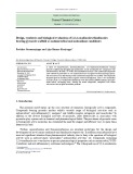
Fig. 3. Analysis of 35S-labeled proteoglycan core proteins present in the matrix or medium of tendon explant cultures in the presence or absence of 1000 lgÆmL)1 CaPPS and heparin. Newly synthesized 35S-labeled proteoglycans remaining in the matrix of tendon explant cultures after 9 days in culture were isolated as described in the Experimental procedures and digested with chondroitinase ABC and keratanase, prior to electrophoresis on a 4–15% polyacryla- mide ⁄ SDS gel. The gel was subjected to fluorography as described in the Experimental procedures. Lanes show peptides present in tissue matrix after 9 days of culture in (A) medium alone, (B) med- ium containing 1000 lgÆmL)1 CaPPS, and (C) medium containing 1000 lgÆmL)1 heparin. The amount of sample applied to each lane corresponds to (cid:1)5000 dpm 35S-radioactivity.
i
i
d e e b a l -
60
g n n a m e r
S 5 3
10
%
0
continued presence of free [35S]sulfate in the medium throughout the culture period results from intracellular degradation of small proteoglycans [10].
5
6
7
8
9
10 11 12 13 14 15
Days in Culture
Fig. 4. Effects of CaPPS and heparin on the rate of formation of [35S]sulfate and release of 35S-labeled proteoglycans from bovine tendon explant cultures. The proximal region of the bovine deep flexor tendon was incubated with [35S]sulfate as described in the Experimental procedures and then maintained in DMEM for 5 days prior to analysis. Tissue was subsequently cultured in DMEM alone (d), DMEM containing 1000 lgÆmL)1 CaPPS (s), or DMEM containing 1000 lgÆmL)1 heparin (.). The culture med- ium was collected daily and analyzed for the presence of [35S]sulfate and 35S-labeled proteoglycans. From this data, (A) the rate of release of [35S]sulfate into the medium, (B) the rate of release of 35S-labeled small proteoglycans into the medium, and (C) the percentage of 35S-labeled small proteoglycans remaining in the matrix were determined as described in the Experimental procedures.
Figure 4A shows that the rate of [35S]sulfate appear- ing in the medium was suppressed by the inclusion of the sulfated polysaccharides. At the end of the culture period, both CaPPS and heparin reduced the appear- ance of [35S]sulfate in the culture medium by (cid:1)5 and (cid:1)11%, respectively. It was also shown that the rate of release of 35S-labeled small proteoglycans into the cul- ture medium was increased by (cid:1)5 and (cid:1)10% in the presence of either CaPPS or heparin, respectively (Fig. 4B). These results indicate that these agents sup- 35S-labeled small press intracellular degradation of proteoglycans, whilst simultaneously increasing the loss of 35S-labeled small proteoglycans into the culture medium. These highly sulfated polysaccharides had lit- tle effect during this time period on the total levels of 35S-labeled small proteoglycans that remained in the extracellular matrix of the tendon cultures (Fig. 4C).
FEBS Journal 273 (2006) 3479–3488 ª 2006 The Authors Journal compilation ª 2006 FEBS
3483
T. Samiric et al.
Inhibition of proteoglycan catabolism in tendon
Discussion
from direct
This study investigated the effects of two highly sulfat- ed polysaccharides, CaPPS and heparin, on the cata- bolism of both newly synthesized large and small proteoglycans in tendon explant cultures. Previous studies have showed that both agents inhibit aggrecan loss from explant cultures of articular cartilage [23]. However, this is the first study to demonstrate that highly sulfated polysaccharides are also able to inhibit proteoglycan catabolism in a connective tissue other than articular cartilage.
effect on soluble aggrecanase activity whereas heparin and heparin sulfate are inhibitory [25]. This relation- ship between the degree of sulfation of polysaccharides and inhibitory activity towards aggrecanase suggests that an ionic interaction may be responsible for the inhibition of aggrecanase by polyanionic molecules [23]. Apart inhibition of aggrecanases, CaPPS or heparin may also be inhibiting aggrecanase activity by altering the nuclear events within tendon fibroblasts that result in decreased expression of aggre- canase activity. Studies have shown that sodium pento- san polysulfate and heparin are internalized by cells and are able to modulate gene expression of matrix proteinases at the transcriptional level [30–32]. It has also been suggested that highly sulfated polysaccha- rides may regulate aggrecanase activity by suppressing the activation of aggrecanase in a similar manner to that suggested for glucosamine [33].
In the case of versican, it is likely that CaPPS and heparin inhibit the catabolism of this proteoglycan by suppressing aggrecanase activity as described above and by inhibiting the activity of other matrix protein- ases since it has been shown that pentosan polysulfate is an inhibitor of serine proteinases as well as a num- ber of metalloproteinases that include MMP-1 (inter- stitial collagenase) and MMP-3 (stromelysin-1) [21].
This study showed that both CaPPS and heparin inhibited the loss of 35S-labeled large proteoglycans in tendon explants by (cid:1)40%. Furthermore, the chemical pool of large proteoglycans was also shown to be inhibited as indicated by hexuronate levels as shown in Table 2. The predominant, large proteoglycans present in the extracellular matrix of tendon have been shown to be aggrecan and versican [3–5]. We have previously analyzed the catabolic products of the large proteogly- cans present in the extracellular matrix of fresh and cultured tendon, as well as medium originating from tendon cultures [10]. It was evident that the catabolism of aggrecan by tendon can be solely attributed to the action of aggrecanase, as all the catabolic products of this proteoglycan that were isolated were derived from the cleavage of the core protein at aggrecanase clea- vage sites [10]. In the case of versican, it appears that the catabolism may involve the action of other matrix proteinases as well as that of aggrecanase [10]. It has been demonstrated that CaPPS and heparin inhibit sol- uble aggrecanase activity as well as recombinant aggre- canase proteinases, and it is therefore likely that the outcome of the inhibition of aggrecanase activity in tendon is the retention of large proteoglycans within the extracellular matrix of the tissue [22–27].
The mechanism by which highly sulfated polysaccha- rides inhibit aggrecanase proteinases remains unclear. The ADAM-TS family of proteinases contain one or more thrombospondin type-1 motifs that are able to bind to sulfated glycosaminoglycans. It has been dem- onstrated in ADAM-TS4 that the interaction between the thrombospondin type-1 motifs and glycosamino- glycans of aggrecan core protein play a critical role in the recognition of specific cleavage sites within the core protein [28,29]. It is possible that when highly sulfated polysaccharides bind to these thrombospondin type-1 motifs this results in the inhibition of proteolytic activ- ity. It should be pointed out that we have demonstra- ted that the inhibition of aggrecanase is dependent on the degree of sulfation of polysaccharides, as chondro- itin or keratan sulfate glycosaminoglycans had no
This study showed that both CaPPS and heparin inhibited the loss of 35S-labeled small proteoglycans in tendon explants by (cid:1)33%. Based on chemical analysis, we have previously shown that (cid:1)85% of the small proteoglycans present in tendon is made up of decorin, and the remainder consists of mainly biglycan [5]. This work also showed that biglyan is lost from the tendon matrix intact, whereas decorin is lost from the tissue either intact or as the result of partial proteolytic clea- vage [5]. Furthermore, using radiolabeled techniques, we have shown that a pool of small proteoglycans are taken up by tendon cells and degraded intracellularly [10]. In the present study, we showed that when tendon explant cultures were incubated with [35S]sulfate for 6 h then maintained in culture for 5 days to allow for the radiolabeled large proteoglycans to be lost, expo- sure of the cultures to either CaPPS or heparin resul- ted in a reduced rate of cellular uptake and subsequent 35S-labeled small proteoglycans as degradation of measured by the appearance of free [35S]sulfate in the medium (Fig. 4A). Inhibition of endocytosis of small proteoglycans by heparin has been reported by previ- ous investigators in cultured skin fibroblasts, however, intracellular degradation of these proteoglycans was not affected by heparin [34–36]. In these cultures, the exposure of tendon to CaPPS or heparin did not affect the levels of radiolabeled small proteoglycans retained
FEBS Journal 273 (2006) 3479–3488 ª 2006 The Authors Journal compilation ª 2006 FEBS
3484
T. Samiric et al.
Inhibition of proteoglycan catabolism in tendon
in the matrix of the tissue but resulted in more radio- labeled small proteoglycans being lost from the tissue presumably from the pool of small proteoglycans that was destined to be internalized by tendon cells (Fig. 4B,C). This is consistent with our previous work using tendon and ligament indicating that if the pool of radiolabeled small proteoglycans that was destined for intracellular degradation is inhibited, then this pool of small proteoglycans is lost from the tissue [10,19].
Preparation of solutions containing sulfated polysaccharides
lear (Boston, MA, USA). Chondroitinase ABC (protease- free from Proteus vulgaris; EC 4.2.2.4) was from ICN Biochemicals (Costa Mesa, CA, USA). Keratanase (from Pseudomonas sp.; EC 3.2.1.103) was obtained from Sigma Chemical Co. (St Louis, MO, USA). Adult bovine meta- carpophalangeal joints were obtained from a local abattoir.
Maintenance of tendon in explant culture
following administration of
tissues
The work presented in this paper supports previous observations that show that highly sulfated polysaccha- rides are able to inhibit the loss of proteoglycans from articular cartilage [25]. However, it is evident in tendon that higher concentrations of these agents are required to observe effects compared with cartilage [25]. The pre- sent study found that both CaPPS and heparin, at a concentration of up to 1000 lgÆmL)1, did not have an adverse impact on cellular metabolism as assessed by the incorporation of [3H]serine into matrix macromole- cules of tendon explant cultures. Although highly sulfat- ed polysaccharides have the potential to cause adverse effects that are related to their anticoagulant and fibrin- olytic activities [37,38], complications involving con- nective these compounds have not been reported [21]. However, it is possible that the inhibition of the catabolism of prote- oglycans in tendon and other fibrous connective tissues such as ligament may result in an increase in the matrix levels of these proteoglycans that would contribute to the increased stiffness of the tissue. Nevertheless, the work described in this paper indicates that highly sulfat- ed polysaccharides have the potential to modulate levels of proteoglycans in joint connective tissues in disease.
The sulfated polysaccharides used for these experiments were dissolved in DMEM at concentrations up to 20 mgÆmL)1. Each solution was then filtered through a ster- ile 0.20 lm Minisart(cid:2) microporous membrane filter before being added to the culture medium to achieve the appropri- ate final concentration.
Experimental procedures
Materials
Measurement of turnover of 35S-labeled proteoglycans in tendon maintained in culture in medium containing varying concentrations of polysulfated polysaccharides
filters were The proximal region of deep flexor tendon was dissected from a single metacarpophalangeal joint obtained from an adult steer. The dissected tendon was chopped into small pieces and incubated in sulfate-free medium (1 g of tissue per 10 mL medium) containing 200 lCiÆmL)1 [35S]sulfate at 37 (cid:1)C for 6 h, as previously described [10]. After washing the tissue exhaustively in culture medium to remove most of the unincorporated radioisotope, duplicate samples con- taining 100 ± 20 mg of tissue were distributed into individ- ual sterile plastic vials containing 4 mL of DMEM with varying concentrations of CaPPS or heparin (0, 100 or 1000 lgÆmL)1) for 10 days. The culture medium used for these experiments was DMEM containing 1 mg glu- coseÆmL)1 and supplemented with organic buffers and amino acids [39]. The medium was collected and replaced daily with fresh media. The collected medium was stored at )20 (cid:1)C in the presence of proteinase inhibitors [40]. After predetermined times in culture, the tissue was rinsed several times with NaCl ⁄ Pi to remove residual exogenous sulfated glycosaminoglycans that had been present in the culture media, before being extracted with 4 m GnHCl at 4 (cid:1)C for 72 h, followed by 0.5 m NaOH at 21 (cid:1)C for 24 h. Individ- ual experiments were repeated at least three times using tis- sue from different animals. In some experiments tissue was extracted with 0.5 m NaOH to determine the amounts of glycosaminoglycans remaining in the matrix.
FEBS Journal 273 (2006) 3479–3488 ª 2006 The Authors Journal compilation ª 2006 FEBS
3485
(carrier-free) [35S]sulfate To determine the proportion of 35S-labeled proteoglycans remaining in the matrix of tendon cultures on each day after incubation with [35S]sulfate, 0.5 mL aliquots of the DMEM, Eagle’s nonessential amino acids, penicillin and streptomycin were from CSL (Melbourne, Victoria, Austra- lia). Sephadex G-25 (as prepacked PD-10 columns) was from Pharmacia (Uppsala, Sweden) and 0.20 lm Minisart(cid:2) from Sartorius microporous membrane (Goettingen, Germany). Amicon(cid:2) Ultra-4 centrifugal filter devices (molecular weight cut-off¼10 000) were purchased from Millipore Corporation, Bedford, MA, USA, and from Amersham Pharmacia (Amersham, Amplify was Buckinghamshire, UK). Heparin (from porcine intestinal mucosa) was purchased from Celsus Laboratories (Cincin- nati, OH, USA). Calcium pentosan polysulfate was a gift from Arthropharm Pty Ltd (Sydney, Australia). Aqueous and [3H]serine solutions of (27.00 CiÆmmol)1) were from DuPont New England Nuc-
T. Samiric et al.
Inhibition of proteoglycan catabolism in tendon
Analysis of 35S-labeled proteoglycan core proteins remaining in the matrix of tendon explant cultures
determined by assaying the extracts for hexuronate as described by Bitter and Muir in 1962 using glucuronic acid as standard [42]. Interference with the hexuronate assay by the polysulfated polysaccharides that were added to the culture media was minimized by thoroughly rinsing the tissue with NaCl ⁄ Pi prior to extraction with NaOH [25].
Separation of 35S-labeled proteoglycans remaining in the matrix of tendon explant cultures by size exclusion chromatography
medium fractions, GnHCl and NaOH extracts were applied to columns of Sephadex G-25 (PD-10 columns) equilibrated and eluted with 4 m GnHCl, 0.1 m Na2SO4, 50 mm sodium acetate, 0.1% (v ⁄ v) Triton X-100, pH 6.1. The rate of loss of 35S-labeled proteoglycans from the matrix of explant cul- tures (which eluted in the excluded volume fractions on Sephadex G-25 columns) was calculated from the amount of 35S-labeled macromolecules that appeared in the medium on each day of culture and that remaining in the matrix at the end of the culture period [41]. From these data, the per- centage of 35S-labeled proteoglycans remaining in the mat- rix on each day was determined and plotted as a function of time in culture [10].
Tissue was dissected from a single metacarpophalangeal joint and incubated with [35S]sulfate for 6 h as described above. The tissue was maintained in DMEM alone or DMEM containing 1000 lgÆmL)1 CaPPS or heparin for 9 days. The culture medium was replaced daily. At the end of the culture period, the tissue was extracted with 4 m GnHCl as described above. Proteoglycans were isolated from tissue extracts by ion-exchange chromatography on Q-Sepharose as described previously [5]. Purified samples were
Measurement of incorporation of [3H]serine into macromolecules by tendon maintained in culture in medium containing varying concentrations of sulfated polysaccharides
Aliquots (1 mL) of the GnHCl extracts obtained from tis- sue after predetermined times in culture were applied to a column of Sepharose CL-4B (1.3 · 87.0 cm) equilibrated and eluted with 4 m GnHCl, 50 mm sodium acetate buffer, 0.1% Triton X-100, pH 5.8. Fractions of 1 mL were collec- ted at a flow rate of 6 mLÆh)1 and assayed for 35S-radioac- tivity. From these data, the percentage of 35S-labeled large and small proteoglycan species remaining in the matrix at different times in culture was determined [9].
Measurement of intracellular degradation of 35S-labeled small proteoglycans in the presence or absence of CaPPS or heparin
concentrated in H2O using Amicon(cid:2) Ultra-4 centrifugal filter devices as described by the manufacturer. Concentrated samples were resuspended and re-concentrated in 0.1 m Tris ⁄ 0.1 m sodium acetate, pH 7.0, and digested with chondroitinase ABC (0.0375 U) and keratanase (0.075 U) at 37 (cid:1)C for 24 h in the pres- ence of proteinase inhibitors [43]. Digested samples were then concentrated, and finally resuspended and then exchanged in H2O using the filter devices as described above. Samples were then lyophilized and subjected to electrophoresis on 4–15% gradient polyacrylamide ⁄ SDS slab gels. The gels were fixed in a solution of 30% (v ⁄ v) methanol and 10% (v ⁄ v) acetic acid for 30 min, soaked in Amplify for a further 30 min, dried and exposed to X-ray film at )20 (cid:1)C for (cid:1)4 weeks.
joint was
Measurement of hexuronate concentration of tendon explants in the presence or absence of 1000 lgÆmL)1 CaPPS or heparin
Bovine tendon was distributed into individual vials ((cid:1)100 mg tissue per vial) containing 4 mL of DMEM and varying concentrations (0, 100 and 1000 lgÆmL)1) of heparin or CaPPS. The tissue was maintained in culture for 10 days and the medium from each vial was replaced daily. The treat- ment groups were maintained as duplicates and explants were incubated with [3H]-serine on days 5 and 10 of the culture period. The tissue was preincubated for 1 h in 2 mL of fresh DMEM, followed by 2 h incubation in DMEM containing [3H]-serine (30 lCiÆmL)1). The rate of incorporation of [3H]serine into macromolecules by the explant cultures was determined for each experimental group as previously des- cribed and expressed as disintegrations per minute (dpm) ⁄ mg wet weight per 2 h [9].
FEBS Journal 273 (2006) 3479–3488 ª 2006 The Authors Journal compilation ª 2006 FEBS
3486
In order to determine the rate of intracellular degradation of 35S-labeled small proteoglycans, bovine tendon from a incubated with single metacarpophalangeal [35S]sulfate for 6 h as described above and then maintained in DMEM alone for 5 days to allow for the loss of the 35S- labeled large proteoglycans from the tissue cultures. After 5 days, the tissue was maintained in culture in the presence or absence of 1000 lgÆmL)1 CaPPS or heparin for a further 10 days. The rate of intracellular proteoglycan catabolism by tendon explants was determined from the amount of ra- diolabeled sulfate appearing in the medium on each day as described previously [10]. glycosaminoglycan concentration of The the NaOH extracts after predetermined times as described above was
T. Samiric et al.
Inhibition of proteoglycan catabolism in tendon
Acknowledgements
This work was supported by a grant from Faculty of Health Sciences, La Trobe University and the Arthritis Foundation of Australia.
13 Ilic MZ, Handley CJ, Robinson HC & Mok MT (1992) Mechanism of catabolism of aggrecan by articular carti- lage. Arch Biochem Biophys 294, 115–122. 14 Loulakis P, Shrikhande A, Davis G & Maniglia CA
References
(1992) N-terminal sequence of proteoglycan fragments isolated from medium of interleukin-1-treated articular cartilage cultures. Putative site(s) of enzymatic cleavage. Biochem J 284, 589–593. 15 Lark MW, Bayne EK & Lohmander LS (1995) Aggre-
1 Vogel KG & Heinegard D (1985) Characterisation of proteoglycans from adult bovine tendon. J Biol Chem 260, 9298–9306. can degradation in osteoarthritis and rheumatoid arthri- tis. Acta Orthop Scand Suppl 266, 92–97. 2 Hess GP, Cappiello WL, Poole RM & Hunter SC 16 Little CB, Flannery CR, Hughes CE, Mort JS,
(1989) Prevention and treatment of overuse tendon inju- ries. Sports Med 8, 371–384.
Roughley PJ, Dent C & Caterson B (1999) Aggrecanase versus matrix metalloproteinases in the catabolism of the interglobular domain of aggrecan in vitro. Biochem J 344, 61–68. 3 Vogel KG & Meyers AB (1999) Proteins in the tensile region of adult bovine deep flexor tendon. Clin Orthop 367, S344–S355. 4 Rees SG, Flannery CR, Little CB, Hughes CE,
17 Kuno K, Okada Y, Kawashima H, Nakamura H, Miyasaka M, Ohno H & Matsushima K (2000) ADAMTS-1 cleaves a cartilage proteoglycan, aggrecan. FEBS Lett 478, 241–245. Caterson B & Dent CM (2000) Catabolism of aggre- can, decorin and biglycan in tendon. Biochem J 350, 181–188.
5 Samiric T, Ilic MZ & Handley CJ (2004) Characteriza- tion of proteoglycans and their catabolic products in tendon and explant cultures of tendon. Matrix Biol 23, 127–140. 18 Sztrolovics R, White RJ, Poole R, Mort JS & Roughley PJ (1999) Resistance of small leucine-rich repeat proteo- glycans to proteolytic degradation during interleukin-1- stimulated cartilage catabolism. Biochem J 339, 571– 577. 6 Carrino DA, Onnerfjord P, Sandy JD, Cs-Szabo G,
19 Winter AD, Campbell MA, Robinson HC & Handley CJ (2000) Catabolism of newly synthesized decorin by explant cultures of bovine ligament. Matrix Biol 19, 129–138.
Scott PG, Sorrell JM, Heinegard D & Caplan AI (2003) Age-related changes in the proteoglycans of human skin. Specific cleavage of decorin to yield a major cata- bolic fragment in adult skin. J Biol Chem 278, 17566– 17572. 20 Burkhardt D & Ghosh P (1987) Laboratory evaluation of antiarthritic drugs as potential chondroprotective agents. Semin Arthritis Rheum 17, 3–34.
7 Wight TN (2002) Versican: a versatile extracellular mat- rix proteoglycan in cell biology. Curr Op Cell Biol 14, 617–623. 21 Ghosh P (1999) The pathobiology of osteoarthritis and the rationale for the use of pentosan polysulfate for its treatment. Semin Arthritis Rheum 28, 211–267. 22 Sugimoto K, Takahashi M, Yamamoto Y, Shimada K
8 Campbell MA & Handley CJ (1987) The effect of retinoic acid on proteoglycan turnover in bovine articular cartilage cultures. Arch Biochem Biophys 258, 143–155. 9 Campbell MA, Winter AD, Ilic MZ & Handley CJ & Tanzawa K (1999) Identification of aggrecanase activ- ity in medium of cartilage culture. Biochem J (Tokyo) 126, 449–455. 23 Munteanu SE, Ilic MZ & Handley CJ (2000) Calcium
(1996) Catabolism and loss of proteoglycans from cul- tures of bovine collateral ligament. Arch Biochem Biophys 328, 64–72. 10 Samiric T, Ilic MZ & Handley CJ (2004) Large aggre- pentosan polysulfate inhibits the catabolism of aggrecan in articular cartilage explant cultures. Arthritis Rheum 43, 2211–2218.
gating and small leucine-rich proteoglycans are degraded by different pathways and at different rates in tendon. Eur J Biochem 271, 3612–3620.
24 Vankemmelbeke MN, Holen I, Wilson AG, Ilic MZ, Handley CJ, Kelner GS, Clark M, Liu C, Maki RA, Burnett D et al. (2001) Expression and activity of ADAMTS-5 in synovium. Eur J Biochem 268, 1259– 1268. 25 Munteanu SE, Ilic MZ & Handley CJ (2002) Highly
FEBS Journal 273 (2006) 3479–3488 ª 2006 The Authors Journal compilation ª 2006 FEBS
3487
11 Arner EC, Pratta MA, Decicco CP, Xue CB, Newton RC, Trzaskos JM, Magolda RL & Tortorella MD (1999) Aggrecanase: a target for the design of inhibitors of cartilage degradation. Ann N Y Acad Sci 878, 92– 107. 12 Sandy JD, Neame PJ, Boyton RE & Flannery CR sulfated glycosaminoglycans inhibit aggrecanase degra- dation of aggrecan by bovine articular cartilage explant cultures. Matrix Biol 21, 429–440. 26 Pratta MA, Yao W, Decicco C, Tortorella MD, Liu RQ, Copeland RA, Magolda R, Newton RC, Trzaskos (1991) Catabolism of aggrecan in cartilage explants: identification of a major cleavage site within the inter- globular domain. J Biol Chem 266, 8683–8685.
T. Samiric et al.
Inhibition of proteoglycan catabolism in tendon
endocytosis receptor proteins leads to different meta- bolic consequences. J Cell Biol 114, 45–52. JM & Arner EC (2003) Aggrecan protects cartilage col- lagen from proteolytic cleavage. J Biol Chem 278, 45539–45545. 35 Hausser H, Schonherr E, Muller M, Liszio C, Bin Z, 27 Mousa SA (2005) Effect of low molecular weight hep-
Fisher LW & Kresse H (1998) Receptor-mediated endo- cytosis of decorin: involvement of leucine-rich repeat structures. Arch Biochem Biophys 349, 363–370.
arin and different heparin molecular weight fractions on the activity of the matrix-degrading enzyme aggreca- nase: structure–function relationship. J Cell Biochem 95, 95–98. 28 Pratta MA, Tortorella MD & Arner EC (2000) Age- 36 Yung S, Hausser H, Thomas G, Schaefer L, Kresse H & Davies M (2004) Catabolism of newly synthesized decorin in vitro by human peritoneal mesothelial cells. Peritoneal Dialysis Int 24, 147–155. related changes in aggrecan glycosylation affect cleavage by aggrecanase. J Biol Chem 275, 39096–39102.
37 Meyer P, Couzi G, Bavle J, Blanc P, Gibelin P, Camous JP & Morand P (1988) Disseminated coronary throm- bosis and thrombopenia induced by pentosan polysul- fate. Arch Mal Coeur Vaiss 81, 913–919.
29 Tortorella MD, Pratta M, Liu RQ, Abbaszade I, Ross H, Burn T & Arner E (2000) The thrombospondin motif of aggrecanase-1 (ADAMTS-4) is critical for aggrecan substrate recognition and cleavage. J Biol Chem 275, 25791–25797.
38 Maffrand JP, Herbert JM, Bernat A, Defreyn G, Delebassee D, Savi P, Pinot JJ & Sampol J (1991) Experimental and clinical pharmacology of pentosan polysulfate. Semin Thromb Hemost 17 (Suppl. 2), 186– 198. 30 Busch SJ, Martin GA, Barnhart RL, Mano M, Cardin AD & Jackson RL (1992) Trans-repressor activity of nuclear glycosaminoglycans on Fos and Jun ⁄ AP-1 onco- protein-mediated transcription. J Cell Biol 116, 31–42. 31 Saito S, Katoh M, Masumoto M, Matsumoto S &
39 Handley CJ & Lowther DA (1977) Extracellular matrix metabolism by chondrocytes. III. Modulation of proteo- glycan synthesis by extracellular levels of proteoglycans in cartilage cells in culture. Biochim Biophys Acta 500, 132–139. Masuho Y (1997) Collagen degradation induced by the combination of IL-1 alpha and plasminogen in rabbit articular cartilage explant culture. Biochem J (Tokyo) 122, 49–54.
40 Oegema TR Jr, Hascall VC & Eisenstein R (1979) Char- acterization of bovine aorta proteoglycan extracted with guanidine hydrochloride in the presence of protease inhibitors. J Biol Chem 254, 1312–1318. 41 Handley CJ & Campbell MA (1987) Catabolism and
turnover of proteoglycans. Methods Enzymol 144, 412– 419. 32 Dudas J, Ramadori G, Knittel T, Neubauer K, Raddatz D, Egedy K & Kovalszky I (2000) Effect of heparin and liver heparan sulphate on interaction of HepG2-derived transcription factors and their cis-acting elements: altered potential of hepatocellular carcinoma heparan sulphate. Biochem J 350, 245–251. 33 Gao G, Plaas A, Thompson VP, Jin S, Zuo F & Sandy 42 Bitter T & Muir HM (1962) A modified uronic acid car- bazole reaction. Anal Biochem 4, 330–334. 43 Oike Y, Kimata K, Shinomura T & Suzuki S (1980)
JD (2004) ADAMTS4 (aggrecanase-1) activation on the cell surface involves C-terminal cleavage by glycosylphos- phatidyl inositol-anchored membrane type 4-matrix metalloproteinase and binding of the activated proteinase to chondroitin sulfate and heparan sulfate on syndecan- 1. J Biol Chem 279, 10042–10051. Proteinase activity in chondroitin lyase (chondroitinase) and endo-beta-d-galactosidase (keratanse) preparations and a method to abolish their proteolytic effect on pro- teoglycan. Biochem J 191, 203–207.
FEBS Journal 273 (2006) 3479–3488 ª 2006 The Authors Journal compilation ª 2006 FEBS
3488
34 Hausser H & Kresse H (1991) Binding of heparin and of the small proteoglycan decorin to the same