Eur. J. Biochem. 269, 1278–1286 (2002) (cid:211) FEBS 2002
Purification and characterization of the thyrotropin-releasing hormone (TRH)-degrading serum enzyme and its identification as a product of liver origin
Stephanie Schmitmeier1,*, Hubert Thole1,†, Augustinus Bader2 and Karl Bauer1 1Max-Planck-Institut fu¨r experimentelle Endokrinologie, Hannover, Germany; 2Gesellschaft fu¨r Biotechnologische Forschung, Abt. Organ und Gewebekulturen, Braunschweig, Germany
analysis indicated that the serum enzyme and the liver enzyme are glycoproteins containing a sugar structure of the complex type, whereas the brain enzyme exhibits an oligo- mannose/hybrid glycostructure. A molecular mass of 97 000 Da could be estimated for all three enzymes after deglycosylation and SDS/PAGE followed by Western blotting. Fragment analysis of the serum TRH-DE revealed that the peptide sequences correspond to the cDNA deduced amino-acid sequences of the membrane-bound brain TRH-DE, whereby two peptides were identified that are encoded by exon 1. These data strongly support the hypo- thesis that the TRH-DEs are all derived from the same gene, whereby the serum enzyme is generated by proteolytic cleavage of the particulate liver enzyme.
Keywords: TRH-degrading enzyme (TRH-DE); serum; liver; brain; characterization.
Previous biochemical studies have indicated that the mem- brane-bound thyrotropin-releasing hormone (TRH)-degra- ding enzyme (TRH-DE) from brain and liver and the serum TRH-DE are derived from the same gene. These studies also suggested that the serum enzyme is of liver origin. The present study was undertaken to verify these hypotheses. In different species, a close relationship between the activities of the serum enzyme and the particulate liver enzyme was noticed. The activity of the serum enzyme decreased when rats were treated with thioacetamide, a known hepatotoxin. With hepatocytes cultured in a sandwich configuration, release of the TRH-DE into the culture medium could also be demonstrated. The trypsin-solubilized particulate liver TRH-DE and the serum TRH-DE were purified to elec- trophoretic homogeneity. Both enzymes and the brain TRH-DE were recognized by a monoclonal antibody gen- erated with the purified brain enzyme as antigen. Lectin blot
signal
substance
kidney and muscle [5–7]. Because the membrane-bound brain TRH-DE and the serum TRH-DE exhibit the same extraordinary high degree of substrate specificity and identical enzyme-chemical characteristics [8–14] it has been suggested that both enzymes are derived from the same gene.
thyrotropin-releasing hormone The (TRH), a hypothalamic hypophysiotropic neuropeptide hormone (reviewed in [1,2]) and a peptidergic neurotrans- mitter/neuromodulator (reviewed in [3,4]), is known to be rapidly inactivated by the brain TRH-degrading enzyme (TRH-DE), an ectoenzyme located on the surface of neuronal cells, as well as by the soluble serum TRH-DE. The highest activity of the membrane-bound TRH-DE is found in brain and significant activities are also detected in retina, lung and liver but not in other tissues such as heart,
Based on the observation that the developmental pattern of the particulate liver TRH-DE and the serum TRH-DE are almost identical it has been proposed that the serum TRH-DE, like most serum enzymes and proteins, might be of liver origin [9,15]. This interpretation was supported by the findings that the activities of the particulate liver enzyme, like the serum enzyme [16–18], is also regulated by thyroid hormones [19]. Moreover, similar enzyme- chemical properties between the particulate liver enzyme and the serum enzyme were noticed [9,15].
To verify the hypothesis that the serum TRH-DE is of liver origin we analyzed the TRH-DE in serum and tissue homogenates of different species and studied the effect of thioacetamide, a hepatotoxin, on the expression of the serum enzyme and the particulate liver enzyme. Furthermore, with hepatocytes in culture we analyzed the release of the TRH-DE into the medium. Finally, we purified the TRH-DE from pig serum and liver to electrophoretic homogeneity and studied the relationship between these enzymes. By sequence analysis we also verified the hypothesis that the membrane-bound brain TRH-DE and the serum TRH-DE are derived from the same gene.
Correspondence to K. Bauer, Max-Planck-Institut fu¨ r experimentelle Endokrinologie, PO Box 610309, D-30603 Hannover, Germany. Fax: + 49 5115359 203, Tel.: + 49 5115359 200, E-mail: karl.bauer@mpihan.mpg.de Abbreviations: TRH, thyrotropin-releasing hormone; TRH-DE, TRH-degrading enzyme; DFP, diisopropyl fluorophosphate; ECL, enhanced chemiluminescence; SNA, Sambucus nigra agglutinin; GNA, Galanthus nivalis agglutinin; MPSP, membrane protein- solubilizing protease; TACE, TNFa protease. *Present address: Department of Biochemistry and Molecular Biology, University of Southern California, Keck School of Medicine and Norris Comprehensive Cancer Center, Cancer Research Laboratory #106, 1303 N. Mission Road, Los Angeles, CA 90033, USA. (cid:160)Present address: Solvay Pharmaceuticals GmbH, PO Box 220, D-30002 Hannover. (Received 18 July 2001, revised 12 December 2001, accepted 8 January 2002)
Characterization of the TRH-DE from serum and liver (Eur. J. Biochem. 269) 1279
(cid:211) FEBS 2002
Hepatocyte isolation and culture
M A T E R I A L S A N D M E T H O D S
Chemicals
(DFP),
fluorophosphate
isolated from young male pigs Hepatocytes were (about 7 weeks old) as described previously [20]. Isolated hepatocytes were adjusted to a density of 2 · 106 viable cells per mL in Williams’ medium E supplemented with fetal bovine serum (10%), insulin (0.17 IUÆmL)1), predni- sone (0.85 lgÆmL)1), glucagon (0.015 lgÆmL)1), penicillin (100 UÆmL)1), streptomycin (100 lgÆmL)1) and glutamine (4.3 mM). The cells were plated onto 60-mm tissue culture dishes coated with collagen and then cultured as described by Bader et al. [21,22]. The rate of albumin secretion into the culture medium was measured by electroimmu- nodiffusion [23] using a polyclonal antibody against porcine albumin. Lactate dehydrogenase activity in the culture medium was determined by a modified method of Bergmeyer & Bernt [24].
Protein and DNA analysis
The DNA content of cultured hepatocytes was determined according to the method described by Downs & Wilfinger [25] using the fluorescent dye bisbenzimidazol (Hoechst dye 33258) and calf thymus DNA as standard. Protein was determined by a modification of the Lowry method as described by Peterson using bovine serum albumin as standard [26].
Determination of TRH-DE activity
Diisopropyl thioacetamide, 2-iodoacetamide, dithioerythritol and calf thymus DNA were obtained from Sigma Aldrich Chemie GmbH (Tauf- kirchen, Germany). Hoechst dye 33258 (bisbenzimidazol) was from Calbiochem-Novabiochem GmbH (Bad Soden, Germany). Glutamine and penicillin/streptomycin were purchased from Life Technologies GmbH (Karlsruhe, Germany). Insulin was from Hoechst AG (Frankfurt, Germany), prednisone from MSD Sharp & Dohme GmbH (Haar, Germany), and glucagon from Novo Nordisk Pharma GmbH (Mainz, Germany). Poly(ethylene glycol) 6000 was obtained from Serva (Heidelberg, Germany). Digoxigenin-labeled lectins, antidigoxigenin antibodies con- jugated either to alkaline phosphatase or to horseradish peroxidase as well as endoglycosidase F/N-glycosidase F enzyme preparation and endoproteinase Lys-C were purchased from Roche Diagnostics GmbH (Mannheim, Germany). Goat anti-(mouse IgG) Ig conjugated to alkaline phosphatase was obtained from Bio-Rad Laboratories GmbH (Munich, Germany). The enhanced chemilumines- cence (ECL)-Western blotting detection kit was from Amersham Pharmacia Biotech (Freiburg, Germany). Nitrocellulose BA-S83 was from Schleicher & Schuell (Dassel, Germany). 5-Bromo-4-chloro-indolylphosphate and Nitro blue tetrazolium were purchased from Biomol Feinchemikalien GmbH (Hamburg, Germany).
Animals
The assay was carried out as described previously using [pyroGlu-3H] TRH as substrate [27]. In brief, samples were incubated at 30 (cid:176)C in a final reaction mixture of 50 lL containing 27 nM [pyroGlu-3H] TRH and the inhibitors of the cytosolic TRH-DEs (1 mM DFP and 1 mM 2-iodoacet- amide for post proline cleaving enzyme and pyroglutamate aminopeptidase, respectively). As a measure for the enzy- matic activity, the initial rate of TRH-degradation was determined by a four-point kinetic test.
Cows (Schwarz-bunte Rasse) and pigs (Deutsche Land- rasse) were raised and maintained at the Institut fu¨ r Tierzucht und-verhalten, Mariensee, Germany. Sprague– Dawley rats were maintained at our institute according to the animal welfare committee of the Medizinische Hochsch- ule Hannover, Germany. All animals had access to standard chow and water ad libitum.
Purification of the TRH-DE from porcine serum
Preparation of tissue homogenates and serum
After the animals were killed, blood and tissues were immediately collected. Serum was obtained after clotting overnight at 4 (cid:176)C and centrifugation. Livers were thor- oughly perfused with cold NaCl/Pi (2.8 mM KH2PO4, 9.4 mM K2HPO4, 150 mM NaCl, pH 7.3). Brains and perfused livers were minced and then homogenized in 3 vol. of 10 mM sodium phosphate buffer, pH 7.3 containing 0.04% NaN3 (buffer A) by use of an Ultra Turrax homogenizer (Jahnke and Kunkel, Staufen, Ger- many).
Porcine serum (1 L) was diluted with 1 L of buffer A. Under constant stirring, 2 L of a poly(ethylene glycol) 6000 solution (dissolved 50% w/v in buffer A) were added through a dropping funnel over a period of 3 h. After an additional hour without stirring, the precipitated protein was pelleted by centrifugation at 17 000 g for 3 h. The supernatant was discarded and the protein pellet was dissolved by stirring overnight with 3 L of buffer A. The clear supernatant obtained after centrifugation at 17 000 g for 1 h was subjected to the purification procedure as described for the trypsin-solubilized membrane-bound TRH-DE from pig brain [28].
Induction of liver cirrhosis in rats
Purification of the membrane-bound TRH-DE from porcine liver
Adult male Sprague–Dawley rats weighing 380–400 g were used. Over the experimental period 10 rats were given tap water containing 0.03% thioacetamide and 10 rats were kept as control. At given time intervals, approximately 1 mL of blood was collected by retrobul- bar puncture and after clotting serum was obtained by centrifugation.
After homogenization of thoroughly perfused pig livers and washing of the membranes, the membrane-bound TRH-DE was solubilized by trypsin treatment and purified to homogeneity by following the protocol described for the isolation of TRH-DE from pig brain [28].
1280 S. Schmitmeier et al. (Eur. J. Biochem. 269)
(cid:211) FEBS 2002
SDS/PAGE analysis
SDS/PAGE analysis was carried out according to Laemmli [29]. The proteins were denatured under reducing conditions by boiling for 3 min in sample buffer containing 200 mM dithioerythritol.
Table 1. Specific activities of the TRH-DEs in serum, liver and brain from rat, pig and cow. Serum and tissue homogenates were prepared and analyzed as described in Materials and methods (n (cid:136) 3 for pig and cow, n (cid:136) 8 for rats, values are means (cid:139) SD).
Western blot analysis
Specific activity of the TRH-degrading enzyme (% TRH-degradedÆmin)1Æmg protein)1)
Species Serum Liver Brain
described for the particulate TRH-DE from rat and pig brain [32]. Enzyme fragments were isolated by reverse-phase HPLC on C4 or C8 Vydac columns using acetonitrile in 0.1% trifluoroacetic acid as eluant. Isolated fragments were analyzed by gas-phase sequencing using the Applied Biosystem 477A sequenator.
Rat Pig Cow 3.92 (cid:139) 0.45 11.92 (cid:139) 1.67 0.19 (cid:139) 0.03 0.95 (cid:139) 0.06 1.78 (cid:139) 0.26 0.04 (cid:139) 0.004 8.56 (cid:139) 1.05 10.27 (cid:139) 0.52 6.60 (cid:139) 0.51
R E S U L T S
After electrophoresis proteins were blotted onto a nitrocel- lulose membrane as described by Towbin et al. [30]. After blocking with NaCl/Tris (50 mM Tris/HCl, 150 mM NaCl, pH 7.5) containing 0.1% Tween-20 (NaCl/Tris/Tween), the membrane was incubated overnight at 4 (cid:176)C with a mono- clonal antibody (41H2; 4 lgÆmL)1) generated against the particulate TRH-DE from pig brain. The membrane was then washed with NaCl/Tris/Tween and subsequently incubated for 1 h at room temperature with goat anti- (mouse IgG) Ig conjugated to alkaline phosphatase (1 : 3000 in NaCl/Tris). After washing, the membrane was incubated with a 5-bromo-4-chloro-indolylphosphate/Nitro blue tetrazolium solution (335 lM 5-bromo-4-chloro-indo- lylphosphate, 400 lM Nitro blue tetrazolium in 200 mM Tris/HCl, 100 mM NaCl, 10 mM MgCl2, pH 9.5) for visualization.
Degradation of TRH by serum and tissue homogenates from different species
Lectin blot analysis
For comparative studies the specific activities of the TRH- DEs were determined in serum as well as in brain and liver homogenates from cow, pig and rat (Table 1). For all three species, high enzymatic activities were found in brain. In rat and pig, high enzymatic activities were also detected in serum and significant activities in liver. In contrast, very low activities were measured in liver homogenates and serum from cows.
Lectin blot analysis was performed according to the method described by Haselbeck et al. [31]. After Western blotting and blocking, the membrane was cut and individual strips were incubated for 1 h with digoxigenin-conjugated lectins (1 : 1000 in NaCl/Tris containing 1 mM MgCl2, 1 mM MnCl2, 1 mM CaCl2 and 1 mM ZnCl2, pH 7.5). The strips were then washed with NaCl/Tris/Tween and subsequently incubated for 1 h with sheep anti-digoxigenin Ig conjugated either to alkaline phosphatase (0.75 UÆmL)1) or to horse- radish peroxidase (0.1 UÆmL)1). After washing of the strips with NaCl/Tris/Tween, the reaction products of alkaline phosphatase or peroxidase were visualized by incubation with the 5-bromo-4-chloro-indolylphosphate/Nitro blue tetrazolium solution or by using the ECL-Western blotting detection kit, respectively.
Deglycosylation
Deglycosylation of the purified TRH-DE from liver, serum and brain was performed as described previously [28] using the endoglycosidase F/n-glycosidase F enzyme preparation. Briefly, the enzymes (30 lL containing 0.4–0.5 lg protein) were denatured by boiling for 3 min in the presence of 0.1% SDS. After adding n-octylglycoside in a threefold excess to SDS, the glycosidase mixture (0.2 U in 50 lL) was added and the reaction mixture was incubated for 24 h at 25 (cid:176)C. Following SDS/PAGE and Western blotting, the enzymes were visualized by using the monoclonal antibody 41H2 as described above.
Enzyme fragmentation and peptide sequencing
After isolation, the serum TRH-DE (100 lg, approximately 860 pmol) was either exposed to cyanogen bromide in 70% formic acid or digested by endoproteinase Lys-C as
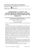
Fig. 1. Effect of thioacetamide, a hepatotoxin, on the activity of the serum TRH-DE. Thioacetamide (0.03%) was either added or not to the drinking water of adult male rats. At the indicated time points 1 mL of blood was collected from control (d) and thioacetamide- treated (s) animals. Serum was prepared and tested for the TRH- degrading activity as described in Materials and methods (values are means(cid:139)SD).
Characterization of the TRH-DE from serum and liver (Eur. J. Biochem. 269) 1281
(cid:211) FEBS 2002
Influence of thioacetamide, a hepatotoxin, on the activity of the serum TRH-DE
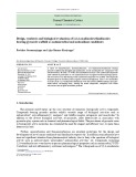
Fig. 2. Activity of the TRH-DE in the medium of cultured hepatocytes. As described in Materials and methods pig hepatocytes were isolated and seeded onto a collagen layer. After cultivation for 24 h, the cells were covered by a second layer of collagen (arrow). After gelatinization of the second layer for 4 h, culture medium was added. The medium was changed every 24 h and used to determine the concentration of albumin (r), the activity of lactate dehydrogenase (d) and the activity of the TRH-DE (s) as described in Materials and methods (n (cid:136) 10; values are mean(cid:139)SD).
When rats were treated with thioacetamide, an agent which is known to induce liver cirrhosis [33,34], we observed a rapid decrease in the activity of the serum TRH-DE within 18 days (Fig. 1). Histological examinations revealed dis- tinctive evidence of damage in liver sections of animals treated for 46 days. In contrast to control animals, in liver of thioacetamide-treated rats loss of the liver cell structure and intercellular granula, increase of connective tissue and appearance of noduli and fibrotic septa were noticed (data not shown).
Synthesis of the TRH-DE by hepatocytes in culture
At a poly(ethylene glycol) concentration of 25% the serum enzyme completely precipitated. The enzyme was recovered almost completely (97%) from the protein pellet and could be applied directly to the Q-Sepharose column. Elution from this column and further purifica- tion followed again the procedure described previously [28].
Characterization of the TRH-DEs from serum and liver
In contrast to hepatocytes kept as monolayers in primary cultures, hepatocytes cultured in a sandwich configuration continue to synthesize and secrete serum enzymes and proteins after an initial lag phase required for cell recovery [35,36]. This lag phase is characterized by a decline of the lactate dehydrogenase activity released into the culture leakage (Fig. 2). After 2 days of medium due to cell cultivation and restoration of the cell membrane integrity, the concentration of albumin and the activity of the TRH-DE in the culture medium increased in a correlative manner (Fig. 2).
Purification of the TRH-DEs from serum and liver
Molecular mass estimation. An approximate molecular mass of 260 000 Da has been determined before for the serum TRH-DE by gel filtration of porcine serum [9]. By the same method, a molecular mass of (cid:25) 250 000 Da could be estimated for the trypsin-solubilized and purified membrane-bound liver TRH-DE (Fig. 3). When the purified enzymes were subjected to SDS/PAGE under reducing conditions a molecular mass of (cid:25) 125 000 Da could be estimated for both enzymes, the serum TRH- DE and the trypsin-solubilized membrane-bound liver TRH-DE (Fig. 4A), indicating that both enzymes consist of two identical subunits. In contrast, but in agreement with our previous data [28] a molecular mass of 116 000 Da could be determined for the trypsin-solubi- lized membrane-bound TRH-DE from pig brain (Fig. 4A).
The membrane-bound liver TRH-DE was solubilized and purified by following exactly the procedure elaborated for the purification of the particulate TRH-DE from rat and pig brain [28]. For the purification of the serum TRH- DE fractionation by poly(ethylene glycol) precipitation was used as the first step not only to partially purify the enzyme but also to reduce the ionic strength due to salt.
Fig. 3. Estimation of the trypsin-solubilized TRH-DE from pig liver by gel filtration on a TSK-G 3000 SW-column. After partial purification, the trypsin-solubilized membrane-bound liver TRH-DE was subjected to gel filtration on a calibrated TSK-G 3000 SW-column. The protein elution profile was monitored by following the absorbance at 280 nm (Æ Æ Æ). The enzyme activity (s) was determined as described in Materials and methods. For calibration, a mixture of standard proteins (d) of known molecular mass, namely: thyroglobulin (1; Mr 669 000), ferritin (2; Mr 450 000), catalase (3; Mr 245 000) and ovalbumin (4; Mr 45 000) was applied to the same column.
1282 S. Schmitmeier et al. (Eur. J. Biochem. 269)
(cid:211) FEBS 2002
are immunologically very similar. At this point, it is worth noting that this antibody is specific to the enzymes of porcine origin and does not react with the enzymes from rat or mouse.
Identification as glycoproteins. As in the case of the brain TRH-DE [28], the TRH-DEs from liver and serum also bind strongly to the Lentil-lectin Sepharose columns which were used for the purification of these enzymes. Thus, both enzymes could be identified as glycoproteins. to the carbohydrate To gain more information as structure, the three enzymes were subjected to lectin blot analysis. As shown in Table 2, the serum enzyme and the liver enzyme exhibit identical properties, distinctly differ- ent from the brain enzyme. For example, the liver enzyme and the serum enzyme are readily recognized by the lectin SNA (Sambucus nigra agglutinin) but not by the lectin GNA (Galanthus nivalis agglutinin), whereas the opposite is true for the brain enzyme. The collected data shown in Table 2 indicate that the brain enzyme con- tains an oligomannose/hybrid glycostructure, whereas the serum enzyme and the liver enzyme belong to the groups of glycoproteins with a glycostructure of the complex type. To substantiate the notion that the TRH-DEs from liver, serum and brain differ only in the carbohydrate moiety, the three enzymes were incubated with the endoglycosidase F/N-glycosidase F enzyme preparation. After Western blotting, a molecular mass of 97 000 Da could be determined for all three enzymes (Fig. 4C) and thus a carbohydrate content of 22% could be estimated for the liver enzyme and the serum enzyme vs. 16% for the brain enzyme.
Peptide sequences of the serum TRH-DE
No sequence information could be obtained when the purified serum enzyme was subjected directly to sequencing, indicating that the aminoterminus is blocked. Therefore, serum TRH-DE was either subjected to cyanogen bromide cleavage or to enzymatic digestion with endoproteinase Lys-C. Overall 25 peptides could be isolated and sequenced. Ten peptides are listed in Table 3. Interestingly, four peptides (3, 4, 8 and 10) were identical with the sequences determined before when fragments of the membrane-bound
Western blot analysis. To verify the hypothesis that the brain TRH-DE, the serum TRH-DE and the liver TRH-DE are derived from the same gene, the enzyme preparations were subjected to Western blotting. All three enzymes were recognized by the monoclonal antibody 41H2 which was generated by using purified TRH-DE from pig brain as antigen (Fig. 4B). This finding indicates that these enzymes
Fig. 4. SDS/PAGE and Western blot analysis of the purified porcine TRH-DE from brain, liver and serum. As described in Materials and methods the solubilized and purified membrane-bound TRH-DE from brain (lane 1) and liver (lane 3) as well as the purified serum TRH-DE (lane 2) were subjected to SDS/PAGE and visualized either by silver staining (A) or immunologically after Western blotting onto a nitro- cellulose membrane by use of the monoclonal antibody 41H2 (B). For the identification as glycoprotein (C), the purified enzymes were either treated (T) or not (NT) with the endoglycosidase F/N-glycosidase F enzyme preparation as described in Materials and methods and then subjected to SDS/PAGE followed by Western blotting. The proteins were again identified by use of the monoclonal antibody 41H2.
Table 2. Lectin blot analysis of the TRH-DEs from pig brain, serum and liver. The enzyme preparations were subjected to SDS/PAGE followed by Western blotting. The nitrocellulose membrane was then cut and individual strips were incubated with digoxigenin-conjugated lectins. Anti- digoxigenin antibodies conjugated either to alkaline phosphatase or to horseradish peroxidase were used for visualization as described in Materials and methods.
TRH-degrading
Brain enzyme Serum enzyme Liver enzyme Lectin
– + – – + + + + + + – – – + + – – – + – – – + + – – – SNA (Sambucus nigra A.) GNA (Galanthus nivalis A.) MAA (Maackia amurensis A.) DSA (Datura stramonium A.) ConA (Concanavalin A) WGA (Wheat germ A.) PHA-L (Phytohaemagglutinin-L) PHA-E (Phytohaemagglutinin-E) RCA120 (Ricinus communis A. I)
Characterization of the TRH-DE from serum and liver (Eur. J. Biochem. 269) 1283
(cid:211) FEBS 2002
Table 3. Sequence of peptide fragments. The serum TRH-DE was either subjected to cyanogen bromide cleavage (+) or digested with endopro- teinase Lys-C ((cid:224)). The peptides were isolated by reverse-phase HPLC and sequenced. The peptides which had been identified before from digests of TRH-DE from pig brain [32] are marked with an asterisk. The numbers refer to the position of the amino acid as deduced from the cDNA of rat [32] or human [37] brain TRH-DE. Differences are found at position 604 (F in pig; L in human and rat), 607 (T in pig and human; M in rat), 614 (I in pig and human; L in rat), 680 (L in pig and human; I in rat), 760 (K in pig; R in rat and human), 991 (A in pig and human; S in rat), 1009 (M in pig and human; R in rat) and 1023 (L in pig and human; M in rat).
bound TRH-DE, whereby the latter peptidase exhibits identical enzyme-chemical characteristics as the serum TRH-DE. Using enzyme-specific conditions to determine the activity of the TRH-specific TRH-DEs indeed we found high enzymatic activities in brain homogenates of all three species examined. In contrast, considerable differences in the TRH-degrading activities were noticed in the serum of these animals, whereby the enzyme activity is almost absent in the serum from cow. Interestingly, we also observed a correlation between the activity of the serum TRH-DE and the activity of the TRH-DE in liver homogenates, suggest- ing that the serum enzyme may be of liver origin.
TRH-DE from pig brain were analyzed [32]. This result clearly demonstrates that both enzymes are derived from the same gene. Comparison of the peptide sequences of the serum TRH-DE with the cDNA deduced amino-acid sequences of the TRH-DE from rat [32] and human [37] brain reveals that only eight amino acids (2.8%) were different out of the 288 amino acids identified, whereby at six positions the amino acids of the enzyme from pig and human were identical and different from the rat enzyme and only at two positions were the amino acids of the porcine peptide sequence different from that of rat and human, which in turn were identical.
Peptide 1+ Peptide 2(cid:224) Peptide 3+ Peptide 4+ Peptide 5+ Peptide 6(cid:224) Peptide 7(cid:224) Peptide 8+ Peptide 9(cid:224) Peptide 10(cid:224) 160-E XFTFSGEVNV EIA 192-VQLAEDRAF GAVPVAGFFL YPQTQVLVVV L *485-EKQRFL TDVLHEV *543-GHSVFQRQ LQDYLTIHKY GNAARNDLWNT LSEA 598-GYP VITIFGNTTA ENRII 677-GSWL LGNI 751-DFLPWHAASK *958-NSK LISGVTEFLN TEGELKELKN 985-SYDGVA AASFSRAVET VEANVRW *1009-M LYQDELFQWL GKALRH
D I S C U S S I O N
importance of
At present we do not have an explanation for the late development of the serum TRH-DE or for the extremely low activity of this enzyme in some species. Nevertheless, these results support the notion that the serum TRH-DE, like most serum enzymes and proteins, is derived from the liver. The decrease of the activity of the TRH-DE in rats treated with thioacetamide, a known hepatotoxin which induces liver cirrhosis [33,34] seemed to be in line with this interpretation. However, a rapid decrease in the enzymatic activity was already observed within a few days after thioacetamide treatment. As liver cirrhosis is generally a long-term process and is observed histolog- ically only after treatment with thioacetamide for several weeks, this decrease in the enzymatic activity seems to be related to other effects of thioacetamide on hepatocytes such as the reported inhibition of respiratory metabolism, binding to metal-containing enzymes, blockade of mRNA transport and loss of the cell’s ability to store glycogen [41].
Even before TRH was finally isolated and structurally elucidated, rapid inactivation of the biologically active material by serum enzyme(s) had been demonstrated [38]. Subsequently, the serum enzyme catalyzing the hydrolysis of TRH at the pyroGlu-His bond has been characterized [8,9]. The findings that the activity of this enzyme is regulated by thyroid hormones [16–18] strongly suggest a physiological role of this peptidase for the inactivation of TRH released into the peripheral circulation. This inter- pretation is also supported by the high substrate specificity of the enzyme [10] which therefore has also been named (cid:212)thyroliberinase(cid:213). The physiological this peptidase was also supported by the observation that the TRH-degrading enzyme (TRH-DE) is absent in the plasma of neonatal rats, whereas TRH is rapidly inactivated by plasma of adult rats [39]. The endocrinological importance of this enzyme was subsequently questioned by the findings that the activity of this peptidase varies considerably among species and is almost absent in the plasma of beagle dogs [5]. In this study, the half-life of TRH after incubation with various homogenates from different species was also examined but a correlation between the half-life of TRH and TRH-degrading activities of the tissue homogenates between species was not observed. This result is not surprising as in tissue homogenates TRH is not only inactivated by one enzyme as in serum or plasma but is degraded by three peptidases (reviewed in [13,40]) namely pyroglutamate aminopeptidase and post proline cleaving enzyme (both are cytosolic enzymes), and the membrane-
Our experiments with hepatocytes in primary culture provided more direct evidence for the notion that the serum TRH-DE is of liver origin. While hepatocytes in monolayer cultures appear to dedifferentiate and rapidly stop secreting liver-derived proteins, these cells maintain their function (e.g. secretion of albumin, transferrin, a1-antitrypsin) when cultured in a collageneous matrix [20–22,35,36]. After seeding and establishing the cultures in a sandwich confi- guration, we observed a decrease in the activity of lactate dehydrogenase (a marker for the restoration of the integrity of the cell membrane) released into the culture medium. Correspondingly, we found an increase in the amount of albumin (a liver specific marker protein) and an increase in
1284 S. Schmitmeier et al. (Eur. J. Biochem. 269)
(cid:211) FEBS 2002
the activity of the TRH-DE released into the culture medium, suggesting that the increase in the enzymatic activity is due to the increased synthetic activity of hepatocytes and not due to cell leakage.
alternative mRNA splicing but must be generated by proteolysis. Whether the serum enzyme is released from the plasma membrane of hepatocytes by proteases acting as sheddases or secretases (also designated as membrane protein-solubilizing proteases, MPSPs) [53,55–57] remains to be elucidated. Preliminary experiments indicate that the release of the serum enzyme is not affected by inhibitors directed against well characterized sheddases [namely b-secretase, c-secretase and TNFa protease (TACE)]. The present results indicate furthermore that the serum enyzme might be generated intracellularly because after homogeni- zation of isolated hepatocytes and high speed centrifuga- tion, 40% of the TRH-degrading activity could be found in the cytosolic fraction and 60% of the enzyme activity was recovered from the particulate fraction (Schmitmeier, S. & Bauer, K., unpublished observation). This aspect warrants further investigation.
A C K N O W L E D G E M E N T S
For direct analysis we purified the membrane-bound liver TRH-DE after solubilization by trypsin and the serum TRH-DE to electrophoretic homogeneity by following the procedure described for the isolation of the membrane- bound TRH-DE from pig brain [28]. By gel filtration a molecular mass of 250 000 Da could be estimated for the truncated liver enzyme, a value which corresponds well with the molecular mass of the papain-solubilized liver enzyme [15] and the molecular mass of the serum enzyme [9] reported before (260 000 Da) but differs from the molecular mass of 230 000 Da determined for the trypsin-solubilized brain enzyme [28]. After SDS/PAGE under reducing conditions a molecular mass of 125 000 Da was estimated for the liver enzyme and the serum enzyme and a molecular mass of 116 000 Da for the brain enzyme, indicating that all these enzymes exist as homodimers, like many surface peptidases [42]. Interestingly, TRH-DEs from brain, liver and serum were all recognized by the monoclonal antibody 41H2 which was generated after immunizing mice with the TRH-DE from pig brain.
We would like to thank Prof Dr P. W. Jungblut for his interest and encouragement and for providing the antibodies against serum albumin. We also thank H. O. Bader, S. Thiele for animal care, P. Affeldt for advice and help, and V. Ashe for typing and for linguistic help in preparing the manuscript. Supported by the Deutsche Forschungsgemeinschaft.
R E F E R E N C E S
1. Guillemin, R. (1978) Peptides in the brain: the new endocrinology of the neuron. Science 202, 390–402. 2. Schally, A.V. (1978) Aspects of hypothalamic regulation of the pituitary gland. Science 202, 18–28. 3. Jackson, I.M. (1982) Thyrotropin-releasing hormone. N. Engl. J. Med. 306, 145–155. 4. Gri(cid:129)ths, E.C. (1985) Thyrotrophin releasing hormone: endocrine and central effects. Psychoneuroendocrinology 10, 225–235.
stable analogues.
5. Brewster, D. (1983) Species variations in TRH inactivation: In Thyrotropin-Releasing Advantages of Hormone (Gri(cid:129)ths, E.C. & Bennett, G.W., eds), pp. 109–118. Raven Press, New York. 6. Bauer, K.
thyrotropin (1989) Multihormonal regulation of releasing hormone-degrading ectoenzyme from rat anterior pituitary. In Recent Advances in Basic and Clinical Neuroendocri- nology (Casanueva, F.F. & Dieguez, C., eds), pp. 135–140. Elsevier Science Publishers, Amsterdam, the Netherlands.
7. Vargas, M.A., Cisneros, M., Herrera, J., Joseph-Bravo, P. & Charli, J.L. (1992) Regional distribution of pyroglutamyl pepti- dase II in rabbit brain, spinal cord, and organs. Peptides 13, 255–260. 8. Taylor, W.L. & Dixon, J.E.
(1978) Characterization of a pyroglutamate aminopeptidase from rat serum that degrades thyrotropin-releasing hormone. J. Biol. Chem. 253, 6934–6940. 9. Bauer, K. & Nowak, P. (1979) Characterization of a thy- roliberin-degrading serum enzyme catalyzing the hydrolysis of thyroliberin at the pyroglutamyl-histidine bond. Eur. J. Biochem. 99, 239–246.
10. Bauer, K., Nowak, P. & Kleinkauf, H. (1981) Specificity of a serum peptidase hydrolyzing thyroliberin at pyroglutamyl-histi- dine bone. Eur. J. Biochem. 118, 173–176.
11. O’Connor, B. & O’Cuinn, G. (1984) Localization of a narrow- specificity thyroliberin hydrolyzing pyroglutamate aminopepti- dase in synaptosomal membranes of guinea-pig brain. Eur. J. Biochem. 144, 271–278.
As the brain TRH-DE has been identified as a glycopro- tein [28], the immunological identity of this enzyme, the serum enzyme and the liver enzyme strongly suggested that these proteins differ only in their carbohydrate moiety. Analysis of the carbohydrate structures revealed that the brain enzyme contains a glycostructure of the oligoman- nose/hybrid type. The occurrence of terminal nonsubstitu- ted mannose and galactose residues is a general feature of most brain glycoproteins [43] and thus the brain TRH-DE is a (cid:212)brain(cid:213) type glycoprotein [44]. In contrast, TRH-DE from both liver and serum contained terminal nonsubstituted a(2–6)-sialic acid units linked to galactose and were thus characterized as glycoproteins of the (cid:212)serum(cid:213) type [45]. The presence of sialic acid units in serum proteins is of biological importance, as on hepatocytes desialyated proteins are recognized by asialoglycoprotein-specific receptors and are thus removed from the circulation by the liver [46]. As glycosylation is a species- and tissue-specific process [47–50] the three enzyme preparations were enzymatically degly- cosylated and subsequently subjected to SDS/PAGE. For all three enzymes a band with a molecular mass of 97 000 Da could be visualized immunologically, indicating the polypeptide chain of these enzymes is very similar or identical. These results strongly support the hypothesis that the serum TRH-DE and the membrane-bound TRH-DE from brain and liver are derived from the same gene, whereby the soluble enzyme might be either generated by alternative splicing of the mRNA (e.g. as reported for immunoglobulin l [51,52]) or by proteolytic cleavage of the membrane-bound liver enzyme as demonstrated for various membrane-bound proteins with soluble counterparts (reviewed in [53]). By fragmentation analysis of the purified serum TRH-DE, two peptide sequences (peptide 1 and 2) (Table 1) could be identified which correspond to the sequences 160–173 and 192–221 of the cDNA deduced amino-acid sequence of the membrane-bound brain TRH-DE. As both peptides are encoded by exon 1 which ends at the position of amino-acid 260 [37,54], we can conclude that the serum enzyme is not a product of
12. Wilk, S. & Wilk, E.K. (1987) Pyroglutamyl peptidase II, a thy- rotropin releasing hormone degrading enzyme: purification and
Characterization of the TRH-DE from serum and liver (Eur. J. Biochem. 269) 1285
(cid:211) FEBS 2002
specificity studies of the rabbit brain enzyme. Neurochem. Int. 15, 81–89. thyrotropin-releasing hormone. Proc. Natl Acad. Sci. USA 91, 9534–9538.
33. Muller, A., Machnik, F., Zimmermann, T. & Schubert, H. (1988) Thioacetamide-induced cirrhosis-like liver lesions in rats – use- fulness and reliability of this animal model. Exp. Pathol. 34, 229–236.
13. O’Cuinn, G., O’Connor, B. & Elmore, M. (1990) Degradation of thyrotropin-releasing hormone and luteinising hormone-releasing hormone by enzymes of brain tissue. J. Neurochem. 54, 1–13. 14. O’Leary, R.M. & O’Connor, B. (1995) A study of a synaptosomal thyrotropin releasing hormone-inactivating pyroglutamate ami- nopeptidase from bovine brain. Int. J. Biochem. Cell Biol. 27, 881–890. 34. Dashti, H., Jeppsson, B., Hagerstrand, I., Hultberg, B., Srinivas, U., Abdulla, M. & Bengmark, S. (1989) Thioacetamide- and carbon tetrachloride-induced liver cirrhosis. Eur. Surg. Res. 21, 83–91.
15. Scharfmann, R., Morgat, J.L. & Aratan-Spire, S. (1989) Presence of a particulate thyrotropin-releasing hormone-degrading pyro- glutamate aminopeptidase activity in rat liver. Neuroendocrinology 49, 442–448. 35. Dunn, J.C., Yarmush, M.L., Koebe, H.G. & Tompkins, R.G. (1989) Hepatocyte function and extracellular matrix geometry: long-term culture in a sandwich configuration. FASEB J. 3, 174–177.
36. Dunn, J.C., Tompkins, R.G. & Yarmush, M.L. (1992) Hepato- cytes in collagen sandwich: evidence for transcriptional and translational regulation. J. Cell Biol. 116, 1043–1053.
16. Bauer, K. (1976) Regulation of degradation of thyrotropin releasing hormone by thyroid hormones. Nature 259, 591–593. 17. Dupont, A., Labrie, F., Levasseur, L., Dussault, J.H. & Schally, A.V. (1976) Effect of thyroxine on the inactivation of thyro- trophin-releasing hormone by rat and human plasma. Clin. Endocrinol. 5, 323–330.
18. White, N., Jeffcoate, S.L., Gri(cid:129)ths, E.C. & Hooper, K.C. (1976) Effect of thyroid status on the thyrotrophin-releasing hormone- degrading activity of rat serum. J. Endocrinol. 71, 13–19. 37. Schomburg, L., Turwitt, S., Prescher, G., Lohmann, D., Horsthemke, B. & Bauer, K. (1999) Human TRH-degrading ectoenzyme cDNA cloning, functional expression, genomic structure and chromosomal assignment. Eur. J. Biochem. 265, 415–422.
19. Scharfmann, R., Ebiou, J.C., Morgat, J.L. & Aratan-Spire, S. (1990) Thyroid status regulates particulate but not soluble TRH- degrading pyroglutamate aminopeptidase activity in the rat liver. Acta Endocrinol. 123, 84–89. 38. Burgus, R., Ward, D.N., Sakiz, E. & Guillemin, R. (1966) Action des enzymes proteolytiques sur des preparations purifiees de l’hormone hypothalamique TSH-hypophysiotrope, TRF. CR Acad. Sci. Paris. 262, 2643–2645.
20. Bader, A., DeBartolo, L. & Haverich, A. (2000) High level ben- zodiazepine and ammonia clearance by flat membrane bioreactors with porcine liver cells. J. Biotechnol. 25, 95–105. 39. Neary, J.T., Kieffer, J.D., Federico, P., Mover, H., Maloof, F. & Soodak, M. (1976) Thyrotropin releasing hormone: development of inactivation system during maturation of the rat. Science 193, 403–405.
40. Bauer, K., Schomburg, L., Heuer, H. & Scha¨ fer, M.K.-H. (1999) Thryotropin-Releasing Hormone (TRH), the TRH-receptor and the TRH-degrading ectoenzyme; three elements of a peptidergic signalling system. Results Probl. Cell Differ. 26, 13–42.
21. Bader, A., Knop, E., Kern, A., Boker, K., Fru¨ hauf, N., Crome, O., Esselmann, H., Pape, C., Kempka, G. & Sewing, K.-F. (1996) 3-D coculture of hepatic sinusoidal cells with primary hepatocytes- design of an organotypical model. Exp. Cell Res. 226, 223.–233. 22. Bader, A., Hansen, T., Kirchner, G., Allmeling, C., Averich, A. & Borlak, J.T. (2000) Primary porcine enterocyte spheroidal cultures to study drug oxidation. Br. J. Pharmacol. 129, 331–342. 23. Laurell, C.B. & McKay, E.J. (1981) Electroimmunoassay. Meth- 41. Nuber, R., Teutsch, H.F. & Sasse, D. (1980) Metabolic zonation in thioacetamide-induced liver cirrhosis. Histochemistry 69, 277–288. ods Enzymol. 73, 339–369.
42. Maraux, S. (1987) Structural and topological aspects. In Mam- malian Ectoenzymes (Kenny, A.J. & Turner, A.J., eds), pp. 15–45. Elsevier, Amsterdam, the Netherlands.
24. Bergmeyer, H.U. & Bernt, E. (1974) Lactate-dehydrogenase: UV-assay with pyruvat and NADH. In Methods of Enzymatic Analysis (Bergmeyer, H.U., ed.), pp. 574–579. Academic Press, New York.
43. Krusius, T. & Finne, J. (1977) Structural features of tissue glycoproteins. Fractionation and methylation analysis of glyco- peptides derived from rat brain, kidney and liver. Eur. J. Biochem. 78, 369–379.
25. Downs, T.R. & Wilfinger, W.W. (1983) Fluorometric quantifica- tion of DNA in cells and tissue. Anal. Biochem. 131, 538–547. 26. Peterson, G.L. (1977) A simplification of the protein assay method of Lowry et al. which is more generally applicable. Anal. Biochem. 83, 346–356. 44. Hoffmann, A., Nimtz, M., Wurster, U. & Conradt, H.S. (1994) Carbohydrate structures of beta-trace protein from human cerebrospinal fluid: evidence for (cid:212)brain-type(cid:213) N-glycosylation. J. Neurochem. 63, 2185–2196. 45. Finne, J. & Krusius, T. (1979) Structural features of
the carbohydrate units of plasma glycoproteins. Eur. J. Biochem. 102, 583–588. 27. Bauer, K., Carmeliet, P., Schulz, M., Baes, M. & Denef, C. (1990) Regulation and cellular localization of the membrane-bound thyrotropin-releasing hormone-degrading enzyme in primary cultures of neuronal, glial and adenohypophyseal cells. Endocrinology 127, 1224–1233. 46. Ashwell, G. & Harford, J. (1982) Carbohydrate-specific receptors of the liver. Ann. Rev. Biochem. 51, 531–554. 47. Rademacher, T.W., Parekh, R.B. & Dwek, R.A. (1988) 28. Bauer, K. (1994) Purification and characterization of the thyro- tropin-releasing-hormone-degrading ectoenzyme. Eur. J. Biochem. 224, 387–396. Glycobiology. Ann. Rev. Biochem. 57, 785–838. 48. Paulson, J.C. (1989) Glycoproteins: what are the sugar chains for? Trends Biochem. Sci. 14, 272–276. 49. Lis, H. & Sharon, N. (1993) Protein glycosylation. Structural and functional aspects. Eur. J. Biochem. 218, 1–27. 50. Parekh, R.B. (1994) Site-specific protein glycosylation. Adv. Drug 29. Laemmli, U.K. (1970) Cleavage of structural proteins during the assembly of the head of bacteriophage T4. Nature 227, 680–685. 30. Towbin, H., Staehelin, T. & Gordon, J. (1979) Electrophoretic transfer of proteins from polyacrylamide gels to nitrocellulose sheets: procedure and some applications. Proc. Natl Acad. Sci. USA 76, 4350–4354. Del. Rev. 13, 251–266.
31. Haselbeck, A., Schickaneder, E., von der Eltz, H. & Hosel, W. (1990) Structural characterization of glycoprotein carbohydrate chains by using digoxigenin-labeled lectins on blots. Anal. Biochem. 191, 25–30. 51. Early, P., Rogers, J., Davis, M., Calame, K., Bond, M., Wall, R. & Hood, L. (1980) Two mRNAs can be produced from a single immunoglobulin l gene by alternative RNA processing pathways. Cell 20, 313–319.
32. Schauder, B., Schomburg, L., Ko¨ hrle, J. & Bauer, K. (1994) Cloning of a cDNA encoding an ectoenzyme that degrades 52. Rogers, J., Early, P., Carter, C., Calame, K., Bond, M., Hood, L. & Wall, R. (1980) Two mRNAs with different 3¢ ends encode
1286 S. Schmitmeier et al. (Eur. J. Biochem. 269)
(cid:211) FEBS 2002
membrane-bound and secreted forms of immunoglobulin l chain. Cell 20, 303–312. 55. Corvol, P., Michaud, A., Soubrier, F. & Williams, T.A. (1995) Recent advances in knowledge of the structure and function of the angiotensin I converting enzyme. J. Hypertens. 13, S3–S10. 56. Corvol, P., Williams, T.A. & Soubrier, F. 53. Ehlers, M.R. & Riordan, J.F. (1991) Membrane proteins with soluble counterparts: role of proteolysis in the release of trans- membrane proteins. Biochemistry 30, 10065–10074. (1995) Peptidyl dipeptidase A: angiotensin I-converting enzyme. Methods Enzymol. 248, 283–305. 57. Hooper, N.M., Karran, E.H. & Turner, A.J. (1997) Membrane protein secretases. Biochem. J. 321, 265–279. 54. Turwitt, S. (1999) Genomische Characterisierung and Promo- toranalyse des Thyrotropin-Releasing Hormon-abbauenden Ektoenzyms (in German). PhD Thesis, University of Hannover, Germany.