The contribution of tryptophan residues to conformational changes in clostridial glutamate dehydrogenase ) W64 and W449 as mediators of the cooperative response to glutamate Muaawia A. Hamza1, Stephen R. Martin2 and Paul C. Engel1
1 School of Biomolecular and Biomedical Science, Conway Institute of Biomolecular and Biomedical Research, University College Dublin,
Ireland
2 Division of Physical Biochemistry, National Institute for Medical Research, Mill Hill, London, UK
Keywords allosteric mechanism; circular dichroism; cooperativity; glutamate dehydrogenase; tryptophan residues
Correspondence P. C. Engel, School of Biomolecular and Biomedical Science, Conway Institute, University College Dublin, Belfield, Dublin 4, Ireland Fax: +353 1283 7211 Tel: +353 1716 6764 E-mail: paul.engel@ucd.ie
(Received 13 April 2007, revised 30 May 2007, accepted 14 June 2007)
doi:10.1111/j.1742-4658.2007.05940.x
Abbreviation GDH, glutamate dehydrogenase.
FEBS Journal 274 (2007) 4126–4134 ª 2007 The Authors Journal compilation ª 2007 FEBS
4126
The hexameric glutamate dehydrogenase of Clostridium symbiosum has pre- viously been shown to undergo a pH-dependent inactivating conformational change that perturbs the environment of one or more Trp residues and is reversed by glutamate in a highly cooperative fashion with a Hill coefficient of almost 6. Five single mutants have now been made in which each of the Trp residues in turn has been replaced by Phe. All five were successfully over-produced as soluble proteins and purified. Far-UV CD showed that none of the mutations significantly affected secondary structure. All five proteins were active, ranging from 13 UÆmg)1 (W64F) to 20.8 UÆmg)1 (W393F), compared to 20 UÆmg)1 for wild-type, and the kinetic parameters at pH 7 were little changed, except for a five- to six-fold increase in Km for glutamate in W243F. Thermostability was also relatively little changed, although W310F and W393F were somewhat more stable and W64F less stable than the unmutated enzyme. All still showed the characteristic reversi- ble, time-dependent high-pH inactivation. Near-UV CD spectra, reflecting the environment of aromatic residues, were recorded at both pH 7 and 8.8, and four of the mutants showed essentially the same perturbation in the 280 nm region as the wild-type enzyme. W64F, however, showed essentially no change. W64 is thus clearly a passive reporter of the pH-dependent con- formational change, and not actually required for the transition to occur. The CD comparisons also suggest that the aromatic CD spectrum is contri- buted almost entirely by W64 and W449. Consistent with the pH-dependent change, all five mutant proteins also showed a positively cooperative response to glutamate at pH 9, reversing the inactivation. However, the Hill coefficient decreased from > 5 for wild-type to approximately 3 for the act- ive site cleft mutation W243F and to approximately 2 for the interfacial mutants W64F and W449F in which the trimer–trimer interaction may be directly interrupted. W64 of each subunit is in contact with W449 in its dimer partner at the trimer–trimer interface. It seems that, although neither of these two residues is required for the pH-dependent change, together, they are essential in mediating the total cooperativity of the hexameric enzyme’s response to glutamate and are presumably directly involved in transmitting conformational information between the two trimers.
M. A. Hamza et al.
Two tryptophans mediating cooperativity
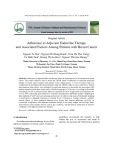
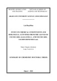
Fig. 1. Distribution of tryptophan residues in the 3D structure of the clostridial GDH subunit. The figure shows a ribbon representa- tion of a single GDH subunit with both glutamate (orange) and NAD+ (violet) bound in the active-site cleft. This is taken from the solved structure of the enzyme–glutamate complex with NAD+ added in the position observed in the enzyme–NAD+ complex [7]. The catalytic residues K125 and D165 on either side of the active site are also highlighted. The side chains of the five Trp residues [14] are shown in grey, with their numbering indicated (white labels). Two (W243 and W310) are in the coenzyme binding domain, with the other three (W64, W393 and W449) located in the glutamate binding domain. Of these last three, two (W64 and W449) are in the intersubunit interface that links the two trimers.
Glutamate dehydrogenases (GDH) which catalyse oxi- dative deamination of l-glutamate using NADP+ as a coenzyme [1], are widely distributed among living organisms [2]. They have attracted considerable atten- their tion from enzymologists not only because of metabolic importance, but also because many GDHs display interesting allosteric properties, still only parti- ally understood [3–5]. GDHs of higher organisms pos- sess auxiliary allosteric sites for regulatory nucleotide ligands [6], and these can be clearly associated with additional structural elements that are missing in bac- terial and some fungal GDHs. Thus, for example, the first GDH to have its 3D structure solved at high reso- lution, that of the anaerobe Clostridium symbiosum [7] (an NAD+-dependent GDH; EC 1.4.1.2), lacks the 48-residue C-terminal extension now shown to form a nucleotide-binding ‘antenna’ in bovine GDH [8]. This does not mean however, that, as recently stated [8], allosteric interaction is confined to the GDHs of higher organisms. It has been known for some time that clostridial GDH is indeed still subject to homotropic allosteric regulation [9–11]. At neutral pH, the rate dependence on NAD+ concentration shows a pattern of negative cooperativity [9] that is strikingly similar to that seen with the bovine enzyme [12]. Lacking the fur- ther layer of structural and kinetic complexity intro- duced by the C-terminal antenna, the bacterial enzyme may offer a better hope of unravelling the basis of intersubunit communication in GDHs. The hexameric clostridial GDH,
like some from other sources [13], also undergoes pH-dependent loss of activity [10] attributed to conformational change [14]. A later study [11], however, showed that the inactivation could be reversed merely by apparent increasing the glutamate concentration. At low fixed coenzyme concentrations, the Hill coefficient for the resulting highly cooperative dependence of rate on glu- tamate concentration approached the theoretical limit- ing value of 6 for a hexamer. This implies, in keeping with the simplest and most extreme version of the Monod–Wyman–Changeux allosteric model [15], that the activating conformational change in any one sub- is compulsorily and symmetrically propagated unit throughout the hexamer, with little or no contribution of hybrid conformational states to the equilibrium state. allosteric transition. The availability of both a high- resolution crystal structure [7] and a cloned over- expressed gene [16] for Clostridium symbiosum offers the opportunity to investigate the role of individual amino acids. Of the five tryptophan residues (Fig. 1), only W310 and W393 are conserved across the GDH family. To better understand the role of individual tryptophan residues in the conformational change and ⁄ or as reporters, a set of five mutants has been constructed by replacing one tryptophan residue at a time with phenylalanine. We report the kinetic, stabil- ity and allosteric properties of these mutant enzymes, including a CD study of the pH-dependent conforma- tional change.
Results
Specific activities
FEBS Journal 274 (2007) 4126–4134 ª 2007 The Authors Journal compilation ª 2007 FEBS
4127
The five mutant enzymes behaved similarly to wild- type GDH during chromatographic purification on a A CD study to confirm and investigate the cooper- ative response of clostridial GDH to glutamate at high pH suggested that at least one of the five tryptophan residues per subunit might be involved [14], and the present investigation attempts to refine our knowledge of that involvement, in the hope that these chromo- for studying the phoric residues may offer a tool
M. A. Hamza et al.
Two tryptophans mediating cooperativity
dye-ligand column. All five were also active, and the specific activities measured in the standard glutamate oxidation assay at pH 7.0 were WT 20 UÆmg)1, W64F 13 UÆmg)1, W243F 15.8 UÆmg)1, W310F 15.8 UÆmg)1, W393F 20.8 UÆmg)1, and W449F 14.2 UÆmg)1. The activities of the mutants are thus remarkably close to that of the unmutated enzyme, with 65% of wild-type activity for W64F being the most significant perturba- tion. Even the conserved Trp residues 310 and 393 therefore are not essential for maintaining an active enzyme structure.
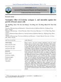
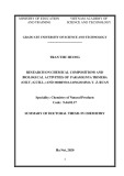
Fig. 2. Changes in the CD signal at 222 nm with increase of tem- perature for wild-type clostridial GDH and the five tryptophan mutants. The six spectra and the identifier for each protein are colour coded for ease of identification.
Thermal stability
Table 1. Kinetic parameters for wild-type clostridial GDH and the five tryptophan mutants. The Km values for wild-type GDH and the single Trp mutants were determined at pH 7 with fixed concentra- tions of either 40 mM glutamate or 1 mM NAD+. The last two col- umns show the Hill coefficient (h) and S0.5 obtained from an analysis of the variation of reaction rate with glutamate concentra- tion at pH 9 with a fixed NAD+ concentration of 1 mM.
Enzyme
Km [NAD+] (mM)
Km [Glut] (mM)
h
S0.5 (mM)
showed greater
Wild-type W64F W243F W310F W393F W449F
0.125 ± 0.01 0.230 ± 0.03 0.40 ± 0.03 0.35 ± 0.05 0.138 ± 0.02 0.22 ± 0.01
3.25 ± 0.033 3.41 ± 0.01 18.2 ± 0.04 3.75 ± 0.02 3.25 ± 0.03 3.30 ± 0.01
5.25 1.95 2.85 4.57 3.29 2.14
180 98 305 200 180 110
After incubation at 50 (cid:2)C for 30 min in KPi (pH 7.0), the wild-type GDH retained only approximately 48% of starting activity. W243F and W449F showed slightly enhanced stability, retaining approximately 5% more activity over the 30 min period. Two other mutants, W310F and W393F, stabilization, retaining 78% and 82% of their activities, respectively. Conservation of Trp at these two positions throughout the GDH family thus cannot be merely related to main- taining protein stability. Of the five mutants, only W64F was less stable than wild-type GDH, retaining only 37% activity after 30 min. This correlates with the problems in recombinant over-expression of this mutant at 37 (cid:2)C. Overall, the stability changes are only moderate, and again, like the specific activities, imply minimal structural perturbation. Thermal stability of the six proteins
above:
stored in Tris ⁄ HCl (pH 7.0) was further examined by following the change in the CD signals at a fixed wavelength temperature. The results (222 nm) with increase of shown in Fig. 2 match the conclusions from activity measurements two mutants, W310F and W393F, showed significantly increased thermal stabil- ity, whereas one, W64F, was considerably less stable compared to all the other proteins. The remaining two, W243F and W449F, showed similar behaviour to the wild-type enzyme, consistent with very similar rates of decline in catalytic activity.
Steady-state kinetics at pH 7.0 site. The Km values of W64F, W310F, W393F and W449F for glutamate were similar to that of wild-type GDH whereas that for W243F showed a 5.6-fold increase. The latter relatively substantial change is not similarly reflected in the specific activity measurement because the standard glutamate concentration, 40 mm, is considerably in excess of all the Km values measured here.
pH-dependent inactivation and conformational change
FEBS Journal 274 (2007) 4126–4134 ª 2007 The Authors Journal compilation ª 2007 FEBS
4128
The primary motivation of this study was to analyse the involvement of Trp residues in the allosteric con- formational changes brought about by alteration in pH or glutamate concentration. The mutant enzymes Table 1 shows the kinetic parameters for the wild-type and the mutated enzymes at 25 (cid:2)C. The Km values of W64F, W393F and W449F for NAD+ are very similar to that of the wild-type enzyme, whereas W243F and W310F showed a two-fold increase. The slight alter- ation in the Km for the coenzyme in the case of these two mutants could perhaps be attributed to the loca- tion of W243 and W310 in the vicinity of the active
M. A. Hamza et al.
Two tryptophans mediating cooperativity
A
show that, at all five positions examined, phenylalanine replacing the bulkier tryptophan has little or no effect on secondary structure. The similarity of the spectra at the two pH values likewise indicates that the conform- ational change involves no major change in secondary structure.
this earlier showing
B
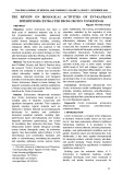
CD spectra in the near-UV region are sensitive to changes in tertiary structure that affect the environ- ment of aromatic amino acid residues. Syed et al. [14] showed that the pH-dependent, inactivating conforma- tional change in this enzyme is accompanied by a large CD perturbation, consistent with an alteration in the environment of one or more Trp residues. Figure 4A confirms a marked result, pH-dependent change in ellipticity in the 280 nm region for the unmutated enzyme. The W64F mutant, by contrast, shows no change in the CD spectrum with change of pH (Fig. 4B). All other mutants show sub- stantial changes that are qualitatively similar to that seen with the wild-type enzyme. This is clearly illustra- ted in Fig. 5, which shows the difference spectra for the wild-type protein and the five mutants. It is poss- ible that the small variations in the difference spectra for the other four mutants reflect minor errors in con- centration determination.
The other surprising feature of these spectra is that three of the mutants (W243F, W310F, and W393F) have pH 8.8 spectra that are very similar in intensity to the wild-type spectrum, suggesting that these three tryptophans make only very small contributions to the total aromatic CD intensity. Inspection of Fig. 4B,F shows that W64, and particularly W449, are the major contributors to the aromatic CD intensity.
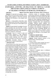
Fig. 3. Far-UV CD spectra of the wild-type clostridial GDH enzyme and the five mutants. These spectra, indicative of secondary struc- ture, were recorded at 20 (cid:2)C both at pH 7.0 (A) and pH 8.8 (B).
Cooperativity at pH 9.0
to mutant
were therefore initially tested to discover whether, like wild-type GDH [10], they undergo apparent inactiva- tion at elevated pH. Under the conditions of this experiment, the unmutated enzyme lost 91% of its activity over a few minutes on transfer from pH 7.0 to pH 8.8. The initial rate and the constant rate reached after 5 min were taken as the basis of this calculation. All five mutated enzymes also showed a marked activ- ity loss and, although the extent of inactivation in the case of W393F (89%) and W449F (90%) was remark- ably close to that in the wild-type enzyme, the other three showed even more severe decreases: 97% for W310F, 98% for W64F, and 99.7% for W243F.
FEBS Journal 274 (2007) 4126–4134 ª 2007 The Authors Journal compilation ª 2007 FEBS
4129
CD spectra (Fig. 3) in the far-UV (195–260 nm) at 20 (cid:2)C and at the two different pH values (7.0 and 8.8) To ascertain whether the mutations interfere with the cooperative reactivation of the enzyme by glutamate, initial rates were measured at pH 9.0 with glutamate concentration varied over a wide range from 4 mm up to 600 mm. All the mutants displayed sigmoidicity in the dependence of initial rates on glutamate concentra- tion over this range, as also shown by the wild-type [11]. However, even though cooperativity is not abol- ished, in every case, it is decreased to an extent that varies greatly from mutant (Table 1). Replacement of the two conserved tryptophans, per- haps surprisingly, gave the smallest change (Hill coeffi- cient, h ¼ 4.53 for W310F and 3.29 for W393F). W243F, with its mutation in the active site region, gave a lower Hill coefficient (h ¼ 2.85 versus 5.25 for the wild-type enzyme), and the saturation curve was shifted to the right (S0.5 increased from 180 mm to
M. A. Hamza et al.
Two tryptophans mediating cooperativity
A
B
C
D
E
F
Fig. 4. Near-UV CD spectra at 20 (cid:2)C for wild-type clostridial GDH and each of the five tryptophan mutants recorded at pH 7 and 8.8. (A) Wild-type CsGDH, (B) W64F, (C) W243F, (D) W310F, (E) W393F and (F) W449F. In each case, the spectrum at pH 7 is shown in grey and the corresponding spectrum at pH 8.8 in black.
305 mm), reflecting overall weaker binding of glutam- ate. W64F and W449F showed larger decreases in their Hill coefficients (to 1.95 and 2.14, respectively), but with the curve now shifted to the left, indicating tigh- ter binding for glutamate (S0.5 ¼ 98 and 110 mm, respectively).
Discussion
FEBS Journal 274 (2007) 4126–4134 ª 2007 The Authors Journal compilation ª 2007 FEBS
4130
The present study was directed towards establishing the role of the five Trp residues in Clostridium symbiosum GDH in various aspects of the protein’s structure and function and, in particular, in the allosteric conforma- tional transition, where it was assumed [14] that one or more Trp residues might be either active partici- pants or at least reporter groups. As already noted, only two of the five Trp residues are conserved. Never- theless, it was essential first of all to establish in each case that the replacement of Trp by a smaller residue did not lead to a wider disruption of the protein struc- ture. All five mutant proteins were successfully purified in good yield. Our standard procedure relies on a highly selective dye-ligand chromatography step [9], which is liable to fail in the case of any disruption of tertiary or quaternary structure [17,18]. Therefore chromatography for all five mutants trouble-free implies that these are well-folded GDH proteins. Simi- larly, native PAGE showed uniformly normal mobility.
M. A. Hamza et al.
Two tryptophans mediating cooperativity
equilibrium. The high-pH state is evidently entirely inactive.
to that of
Fig. 5. Differences between the near-UV CD spectra at pH 7.0 and 8.8. The spectrum for each protein is colour coded. The mutants the wild-type gave difference spectra very similar enzyme, with the exception of W64F for which there was almost no change in the CD spectrum in this region on changing the pH.
from the wild-type (h ¼ 5.25) but,
Initial-rate measurements at pH 9 showed that all the mutant proteins also retained an allosteric response in their dependence on glutamate concentration, but with substantial differences in the Hill coefficients observed. Thus, W310F showed relatively little change (h ¼ 4.57) in W243F and W393F, the cooperativity was less marked with h-values of approximately 3. The remaining two mutants, W64F and W449F, showed a larger decrease in cooperativity with h-values of approximately 2. Both these residues are in the interface region between the two trimers that make up a hexamer (Fig. 1), and it is likely that these two substitutions interfere in the intimate subunit interaction required to propagate the conformational change throughout the hexamer. If the hexamer is in effect converted into two linked but independent trimers, then the maximum Hill coefficient would be decreased to 3. The observed figures for these two mutants were 1.95 and 2.14.
Finally the far-UV CD spectra for the six proteins were indistinguishable, indicating that the mutations do not significantly alter the secondary structure, and that none of the tryptophans make substantial contri- butions to the far-UV CD intensity.
In addition to these structural indicators, the strong- est evidence against any wider disturbance is the fact that all the proteins were catalytically active, with remarkably little variation in the specific activity in the standard assay at pH 7. The kinetic parameters (Table 1) also show a remarkable constancy in the Km for the substrate glutamate, with the sole exception of W243F, for which there is a six-fold increase. This residue is in the active-site cleft and might therefore be expected to have some influence on binding parame- ters.
The effects of the mutations on thermal stability were varied. The direct measurements of activity loss on heating showed that W243F and W449F were slightly more stable than the wild-type (approximately 5% more retention of activity over the 30 min incuba- tion at 50 (cid:2)C). On the other hand, the mutations at 310 and 393 both resulted in substantial stabilization so that approximately 80% activity was retained com- pared with only 48% for the wild-type. Just one of the mutations (at interfacial position 64) resulted in signifi- cant destabilization, with only 37% activity after 30 min. These results correlated very closely with those of the CD study (Fig. 2), showing the change in ellip- ticity at 222 nm with the onset of thermal unfolding. The profiles for wild-type, W243F and W449F are vir- tually indistinguishable, those for W310F and W393F are also very close, and that for W64F shows a very early change as the temperature is raised.
The near-UV CD spectrum was measured at both pH 7 and 8.8, as in the earlier study of the wild-type enzyme [12], in order to assess the contributions of the various residues to the overall CD spectrum and, in particular, to the change in signal accompanying the conformational transition. The pair of spectra shown in Fig. 4A for the wild-type enzyme closely match the result reported earlier, indicating that the pH-depend- ent conformational change perturbs the environment of one or more Trp residues.
FEBS Journal 274 (2007) 4126–4134 ª 2007 The Authors Journal compilation ª 2007 FEBS
4131
In the absence of any gross structural disturbance caused by any of the five mutations, the main thrust of this study was to explore the involvement of the Trp residues in two closely linked phenomena, the inacti- vating conformational change between pH 7 and 9 and its cooperative reversal by increasing concentrations of glutamate. All five mutants, like the wild-type enzyme, show a marked inactivation upon transfer from pH 7 to pH 8.8. For two mutants, W393F and W449F, this change was quite similar to that seen in the unmutated enzyme, with approximately 10% residual activity measurable. All the other mutants showed even lower levels of activity, 2–3% in the case of W64F and in the case of W310F, and even less, only 0.3%, W243F. These differences presumably reflect small changes in the pH dependence of the conformational The difference spectra (Fig. 5) show that W64F alone fails to show pH dependence in its aromatic CD spectrum, even though it undergoes the pH-dependent inactivation. It would therefore appear that, although
M. A. Hamza et al.
Two tryptophans mediating cooperativity
Experimental procedures
Materials
Grade II NAD+ (free acid) was obtained from Roche (Mannheim, Germany) and l-glutamate (monosodium salt), Trizma-base, glycine and EDTA, all of analytical grade, were from Sigma Chemical Co. (St Louis, MO, USA) or Sigma-Aldrich (Tallaght, Ireland). Sepharose CL-6B was from Pharmacia Biotech (Uppsala, Sweden) and IPTG from Melford (Suffolk, UK). Other routine chemicals and reagents were also of analytical grade. Remazol Brilliant Red GG was obtained from DyStar Textilfarben GmbH (Frankfurt, Germany).
W64 is solely responsible for the pH-dependent change in the CD spectrum, it is not required as an active par- ticipant in the conformational transition that brings about that change; it is simply a passive reporter, responding to a changed environment at the trimer– trimer interface. However, as seen earlier, the cooper- ative response to glutamate requires the integrity of W64 in order to be fully expressed. Thus, W64 appears to be a key residue in mediating communication between the two trimers and achieving total coopera- tivity within the hexamer.
coli TG-1[F ¢traD36,
Bacterial strains and vectors
)mk
lacI q, D(lacZ)M15, Escherichia +McrB)), thi1, proA+B+, supE, D(hsdM-mcrB)5, (rk D(lac-proAB)], used as a host strain for all plasmids, was grown routinely at 37 (cid:2)C in Luria–Bertani medium supple- mented with ampicillin 100 lgÆmL)1, unless otherwise sta- ted. Wild-type and mutant GDHs were all expressed using the plasmid expression vector ptac 85 [19,20].
W243F (Fig. 4C), W310F (Fig. 4D), and W393F (Fig. 4E), on the other hand, show pairs of spectra very similar to the wild-type and this indicates not only that none of these Trp residues is required for the observed change, but also that none of them can be a major contributor to the enzyme’s CD signal at either pH.
Construction of mutants
Oligonucleotides, designed to replace the various Trp codons with Phe codons (Table 2), were synthesized by Operon (Cologne, Germany). The lyophilized oligonucleo- tides as supplied were dissolved in 10 mm Tris ⁄ HCl buffer, pH 7.5 containing 1 mm EDTA and diluted for use in the PCR reaction. Site-directed mutations were introduced into the Clostridium symbiosum gdh gene by using the Quick- Change kit by Invitrogen (Paisley, UK). Mutated plasmids, extracted from overnight cell cultures, were sequenced by Agowa or MWG (Ebersberg, Germany), confirming both
Finally, Fig. 4F shows the spectra for W449F; these spectra are the most qualitatively distinct of the set, because clearly W449 is a major contributor to the overall ellipticity. On the other hand, there is still a difference between the pH 7 and 8.8 spectra that is very similar in magnitude to that for the wild-type enzyme (Fig. 5). Thus, it would appear that, even though W449 is a major contributor to the chromo- phoric environment, it is not a major contributor to the pH-dependent change. As with W64, however, the integrity of this residue appears to be essential for full cooperativity throughout the hexamer.
Table 2. Sequences of mutagenic oligonucleotides. The sequences of the sense (forward, Fw) and antisense (reverse, Re) primers used to alter the five tryptophan codons in the clostridial gdh gene to phenylalanine codons are shown. In each case, the mutagenic triplet is underlined.
Oligonucleotide name
Sequence (5¢ to 3¢)
the conformational transition. Inspection of
W64F (Fw) W64F (Re) W243F (Fw) W243F (Re) W310F (Fw) W310F (Re) W393F (Fw) W393F (Re) W449F (Fw) W449F (Re)
GTTCCGTTCGAGGATGACAACG CGTTGTCATCCTCGAACGGAAC GTAACGTAGCATTCGGCGCTGC GCAGCGCCGAATGCTACGTTAC GAGAAACCATTCGGCCAGAAGG CCTTCTGGCCGAATGGTTTCTC GAGACTTTCCTTCACAGCAGAG CTCTGCTGTGAAGGAAAGTCTC GCATTGCTTTCTAATCTGAAGAC GTCTTCAGATTAGAAAGCAATGC
these
FEBS Journal 274 (2007) 4126–4134 ª 2007 The Authors Journal compilation ª 2007 FEBS
4132
In the absence, to date, of a solved structure for the high-pH conformation of clostridial GDH, the precise nature and mechanism of the conformational change remains unclear. However, these studies have now fo- cussed attention on two key interfacial Trp residues, W64 and W449: both are required for full cooperativi- ty in the hexamer and W64 is also a sensitive reporter the of whole hexamer structure reveals that the pairing of these two residues is no coincidence: at the interface of each of the three dimers making up the complete enzyme molecule, they provide paired contacts, where W64 of one residue interacts with W449 of the other. Either of the W fi F mutations would therefore abol- ish six such interactions between the two trimers. The identification of this linkage should provide both a useful tool and a lead in further studies of this alloster- ic enzyme. It is intriguing to note that, although Trp is not uniformly conserved at two positions throughout the GDH family, the NADP+ dependent GDH of Neurospora crassa, which shows somewhat similar allosteric behaviour [4], does indeed have Trp at both positions.
M. A. Hamza et al.
Two tryptophans mediating cooperativity
the presence of the planned mutations and the absence of any other, undesired changes.
contained 1 mm NAD+ and 40 mm glutamate in 0.1 m potassium phosphate buffer (pH 7). Reaction mixtures were incubated at 25 (cid:2)C for 10 min prior to addition of enzyme. At pH 9.0, glutamate concentration was varied over a range up to 400 mm while maintaining the NAD+ concen- tration at 1 mm in glycine ⁄ NaOH buffer. The enzyme was kept in the same buffer. Both the enzymes and assay solu- tions were incubated at 25 (cid:2)C for at least 10 min prior to initiation of the reaction.
In all cases, reaction rates were measured spectrophoto- metrically by following formation of NADH at 340 nm and using an extinction coefficient of 6220 m)1Æcm)1 [22].
Overproduction and purification of the wild-type and mutant enzymes
Steady-state kinetic assays in the oxidative deamination direction were run using standard reactant concentrations as mentioned above in 0.1 m Tris ⁄ HCl at pH 8.8 (Sys- tem II) [10]. Wild-type or mutant enzyme solutions stored at pH 7.0 were used to initiate the reaction. The reaction progress was followed over 7 min. Reaction rates were measured over the first minute and again after 5 min.
pH-dependent inactivation
With the exception of the mutant W64F, the mutant pro- teins, like wild-type GDH, were all over-produced at 37 (cid:2)C in good yield (approximately 80 mg pure soluble protein per litre growth). W64F gave rise to a substantial amount of insoluble protein under these growth conditions but gave a good yield of soluble protein at the lower growth tem- perature of 25 (cid:2)C. Cell pastes were collected by centrifuga- tion, resuspended in 0.1 m potassium phosphate buffer, pH 7 ⁄ 1 mm EDTA, and sonicated to break the cell walls. The broken cells were centrifuged again at 38 700 g for 25 min using a Sorvall RC5 and GSA rotor and the sup- ernatants were loaded onto freshly prepared Remazol-Red chromatographic columns [9] pre-equilibrated with 0.1 m potassium phosphate buffer, pH 7. These columns were washed with ten-fold the column volume of equilibration buffer before eluting the pure proteins with 1 m NaCl in the same buffer. Pure fractions, as judged by SDS ⁄ PAGE, were pooled, precipitated in 60% ammonium sulphate and stored at 4 (cid:2)C till needed. Native PAGE gels (data not shown) showed no significant alteration in mobility for any of the mutants in comparison to the wild-type enzyme.
CD measurements
Protein purity and precise determination of concentration are crucial for CD studies. Separate samples of wild-type GDH and the five mutants were prepared from the stocks in ammonium sulphate by extensive dialysis against Tris ⁄ HCl buffer, either at pH 7.0 or 8.8, with at least four buffer chan- ges in each case. The concentrations were determined from A280 as described above. Samples for far-UV measurement were all approximately 0.15 mgÆmL)1 whereas those for near-UV were 1.2–2 mgÆmL)1. Enzyme solutions were centri- fuged prior to CD measurement to remove any particles.
CD spectra were recorded at 20 (cid:2)C using a JASCO J-715 CD spectropolarimeter (Jasco, Tokyo, Japan) with a thermal Peltier temperature controller. Tertiary structure of the proteins was examined at different pH values (7.0–8.8) by following the changes in the near-UV CD spectra (255– 340 nm). Secondary structure was explored by following changes in the far-UV region (195–250 nm). All reported spectra are averages of multiple scans with appropriate baseline subtraction.
The concentration of the wild-type clostridial GDH was determined spectrophotometrically at 280 nm by using an absorption coefficient of 1.05 LÆg)1Æcm)1 [9]. However, for the tryptophan mutants, in view of the changed content of chromophores, concentrations were determined either by the standard Bradford method [20] with BSA for calibra- tion or else from A280 by the protparam programme based on the method of Gill and von Hippel [21]. Direct compar- ison of these two methods revealed varying levels of agree- ment. The protparam estimation gave concentrations that, respectively, were 11%, 1%, 4% and 10% lower than the Bradford values for W64F, W243F, W310F and W449F and, in the case of W393F, gave a value 20% higher. The protparam method was deemed to be more reliable and therefore calculations based on the Bradford estimates were corrected accordingly.
Concentration measurements
Thermal stability
the wild-type enzyme and the The thermal stability of mutants was studied both enzymatically, by measuring the residual activities after incubating the enzyme at 50 (cid:2)C for 30 min and then cooling in ice for 10 min prior to initiation of the assay reaction, and also by following the change in CD at 222 nm with increasing temperature. For the latter experiments, the temperature was increased by 2 (cid:2)CÆmin)1.
At pH 7.0, the steady-state kinetic parameters for the oxi- dative deamination reaction were determined by measuring initial reaction rates with the concentration of each sub- strate systematically varied in turn. Fixed substrate concen- trations were those of the standard assay solution, which
FEBS Journal 274 (2007) 4126–4134 ª 2007 The Authors Journal compilation ª 2007 FEBS
4133
Enzyme assays
M. A. Hamza et al.
Two tryptophans mediating cooperativity
Acknowledgements
concentration dependence of steady-state reaction rates measured with clostridial glutamate dehydrogenase and mutant A163G at high pH. Biochemistry 36, 11417– 11422.
This work was supported in part by grants from Enterprise Ireland. We also wish to acknowledge the enthusiastic help of Mr Kalyana Venneti with the molecular graphics.
References
12 Engel PC & Dalziel K (1969) Kinetic studies of glutam- ate dehydrogenase with glutamate and norvaline as sub- strates: coenzyme activation and negative homotropic interactions in allosteric enzymes. Biochem J 115, 621– 631.
1 Smith EL, Austen BM, Blumenthal KM & Nyc JF (1975) Glutamate dehydrogenases. In The Enzymes (Boyer PD, ed.), 3rd edn, Vol. 11, pp. 294–367. Academic Press, New York, NY.
13 Ashby B, Wootton JC & Fincham JRS (1974) Slow conformational changes of a Neurospora glutamate dehydrogenase studied by protein fluorescence. Biochem J 143, 317–329.
14 Syed SE-H, Engel PC & Martin SR (1990) A circular
2 Hudson RC & Daniel RM (1993) L-Glutamate dehy- drogenase: distribution, properties and mechanism. Comp Biochem Physiol 106B, 767–792.
3 Tomkins GM, Yielding KL, Talal N & Curran JF
dichroism study of the pH-dependent activation ⁄ inacti- vation equilibrium in the glutamate dehydrogenase of Clostridium symbiosum. FEBS Lett 262, 176–178.
(1963) Protein structure and biological regulation. Cold Spring Harbor Symp Quant Biol 28, 461–470.
4 West DJ, Tuveson RW, Barratt RW & Fincham JRS
15 Monod J, Wyman J & Changeux J-P (1965) On the nat- ure of allosteric transitions: a plausible model. J Mol Biol 12, 88–118.
16 Teller JK, Smith RJ, McPherson MJ, Engel PC &
Guest JR (1992) The glutamate dehydrogenase gene of Clostridium symbiosum. Cloning by polymerase chain reaction, sequence analysis and overexpression in Escherichia coli. Eur J Biochem 206, 151–159.
(1967) Allosteric effects in nicotinamide adenine dinucleotide phosphate-specific glutamate dehydroge- nase from Neurospora. J Biol Chem 242, 2134–2138. 5 Le´ John HB, Stevenson RM & Meuser R (1970) Multi- valent regulation of glutamate dehydrogenases from fungi: effects of adenylates, guanylates and coenzyme A derivatives. J Biol Chem 245, 5569–5576.
6 Goldin BR & Frieden C (1972) L-glutamate dehydro-
genases. Curr Top Cell Reg 4, 77–117.
17 Pasquo A, Britton KL, Stillman TJ, Rice DW, Co¨ lfen H, Harding SE, Scandurra R & Engel PC (1996) Con- struction of a dimeric form of glutamate dehydrogenase from Clostridium symbiosum by site-directed mutagen- esis. Biochim Biophys Acta 1297, 149–158.
18 Dunlevy G (2006) Subunit interaction and communication
7 Baker PJ, Britton KL, Engel PC, Farrants GW, Lilley KS, Rice DW & Stillman TJ (1992) Subunit assembly and active site location in the structure of glutamate dehydrogenase. Proteins Struct Funct Genet 12, 75–86. 8 Peterson PE & Smith TJ (1999) The structure of bovine
in clostridial glutamate dehydrogenase: a study of mutations at the trimer-trimer interface. PhD Thesis, University College Dublin, Ireland.
19 Marsh P (1986) Ptac-85, an E. coli expression vector for
non-fusion proteins. Nucleic Acids Res 14, 3603.
glutamate dehydrogenase provides insights into the mechanism of allostery. Structure Fold Des 7, 769–782. 9 Syed SE-H, Engel PC & Parker DM (1991) Functional
20 Bradford MM (1976) A rapid and sensitive method for
quantitation of microgram quantities of protein utilizing the principle of protein-dye binding. Anal Biochem 72, 248–254.
studies of a glutamate dehydrogenase with known three- dimensional structure: steady-state kinetics of forward and reverse reactions catalysed by the NAD+-depend- ent glutamate dehydrogenase of Clostridium aymbiosum. Biochim Biophys Acta 1115, 123–130.
21 Gill SC & von Hippel PH (1989) Calculation of protein extinction coefficient from amino acid sequence data. Anal Biochem 182, 319–326.
10 Syed SE-H & Engel PC (1991) A pH-dependent activation–inactivation equilibrium in glutamate dehydrogenase of Clostridium symbiosum. Biochem J 271, 351–355.
22 Horecker BL & Kornberg A (1948) The extinction coef- ficients of the reduced band of pyridine nucleotides. J Biol Chem 175, 385–390.
11 Wang X-G & Engel PC (1995) Positive cooperativity with Hill coefficient of up to 6 in the glutamate
FEBS Journal 274 (2007) 4126–4134 ª 2007 The Authors Journal compilation ª 2007 FEBS
4134