HUE JOURNAL OF MEDICINE AND PHARMACY ISSN 3030-4318; eISSN: 3030-4326HUE JOURNAL OF MEDICINE AND PHARMACY ISSN 3030-4318; eISSN: 3030-4326
92 93
Hue Journal of Medicine and Pharmacy, Volume 15, No.2/2025 Hue Journal of Medicine and Pharmacy, Volume 15, No.2/2025
Comparative analysis of antibiotic resistance patterns and virulence
genes in Staphylococcus aureus strains isolated from clinical samples
at Hue University of Medicine and Pharmacy Hospital
Ung Thi Thuy, Nguyen Thi Khanh Linh, Dinh Thi Hai, Hoang Thi Minh Ngoc, Nguyen Hoang Bach*
Department of Microbiology - University of Medicine and Pharmacy, Hue University
Abstract
Introduction: Staphylococcus aureus (S. aureus) is a common pathogen associated with severe infections,
and its antibiotic resistance is potentially associated with various virulence factors. This study explored the
relationship between antibiotic resistance and virulence genes in S. aureus isolates from the clinical samples
of Hue University of Medicine and Pharmacy Hospital. Materials and Methods: Between 2021 and 2022, 122
S. aureus strains were isolated from clinical samples, with 114 non-duplicate strains undergoing antibiogram
and virulence gene analysis. The antimicrobial susceptibility was tested using the Kirby-Bauer method.
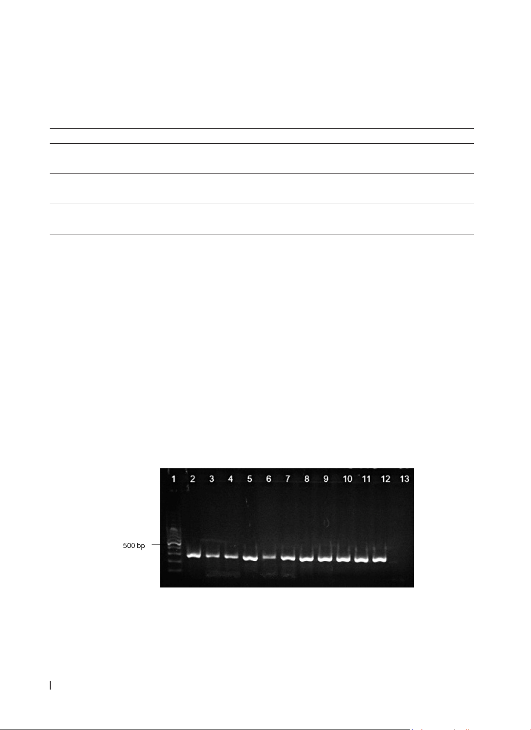
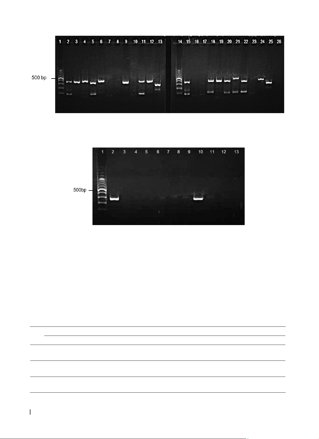
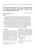
Molecular genotyping of Nuc, mecA, spa, pvl, and tsst-1 was performed using singleplex and multiplex
PCR techniques. Results: Out of 122 isolated S. aureus strains, 114 non-duplicate strains were analyzed,
with MRSA (Methicillin-resistant Staphylococcus aureus) comprising 49.1% and MSSA(Methicillin-sensitive
Staphylococcus aureus) 50.9%. Carriage of the mecA and spa genes was significantly associated with MRSA
infection (p<0.05). The mecA gene was associated with resistance to penicillin, erythromycin, clindamycin,
and tetracycline (p<0.05), whereas the spa gene was associated with oxacillin resistance (p=0.002). tsst-1 was
linked to resistance to penicillin and clindamycin (p<0.001). No correlation was found between the presence
of pvl gene and antibiotic resistance. Conclusion: The presence of methicillin-resistant genes in MSSA poses
significant challenges for its diagnosis and treatment. Investigating the virulence and antimicrobial resistance
of MRSA and MSSA is crucial to improving patient treatment outcomes in the future.
Keywords: Staphylococcus aureus, MRSA, MSSA, nuc, mecA, spa, pvl, tsst-1.
*Corresponding Author: Nguyen Hoang Bach, Email: nhbach@huemed-univ.edu.vn
Received: 8/11/2024; Accepted: 10/3/2025; Published: 28/4/2025
DOI: 10.34071/jmp.2025.2.14
1. INTRODUCTION
Staphylococcus aureus (S. aureus) has emerged
as one of the most common bacteria associated
with hospital- and community-acquired infections. It
is associated with various conditions, including skin
and soft tissue infections, osteomyelitis, invasive
infections, and septicemia [1]. Methicillin-resistant
Staphylococcus aureus (MRSA) strains are known
to have evolved through a combination of virulence
and methicillin-resistant genes, enabling them
to invade healthy individuals and spread quickly
within populations. Therefore, understanding
the molecular characteristics of MRSA isolates is
crucial for effective infection control. While most
studies have focused on MRSA, infections caused by
methicillin-sensitive Staphylococcus aureus (MSSA)
may be even more clinically significant, as MSSA
isolates often possess diverse virulence factors [2].
A previous study indicated that MSSA strains exhibit
similar patterns of enterotoxin distribution, the
methicillin-resistant gene mecA, and the virulence
gene tsst-1 (toxic shock syndrome toxin-1 gene) as
MRSA strains. Consequently, MSSA isolates should
be regarded as similar to MRSA strains because they
can serve as potential sources of infection [3].
S. aureus produces several virulence factors,
including toxins, enzymes, and adhesion factors,
contributing to its pathogenicity. The Panton-
Valentine leukocidin (PVL) toxin, encoded by the pvl
gene, is linked to infections ranging from skin and
soft tissue infections to severe conditions, such as
necrotizing pneumonia. Additionally, Toxic Shock
Syndrome Toxin-1 (TSST-1), encoded by tsst-1, can
cause life-threatening toxic shock syndrome (TSS),
such as rash, fever, and organ failure. Understanding
the prevalence of the tsst-1 gene and its link to
antibiotic resistance is crucial for improving patient
outcomes. Toxins from both the pvl and tsst-1 genes
can lead to severe infections, which are even more
challenging to treat when associated with the mecA
gene-mediated resistance to beta-lactam antibiotics
[4]. The advancement of molecular biological
techniques has been crucial for detecting genetic
patterns in MRSA and MSSA. For example, the nuc
gene is highly specific for identifying S. aureus, along
with the presence of virulence genes such as spa
HUE JOURNAL OF MEDICINE AND PHARMACY ISSN 3030-4318; eISSN: 3030-4326HUE JOURNAL OF MEDICINE AND PHARMACY ISSN 3030-4318; eISSN: 3030-4326
92 93
Hue Journal of Medicine and Pharmacy, Volume 15, No.2/2025 Hue Journal of Medicine and Pharmacy, Volume 15, No.2/2025
(Staphylococcal protein A), pvl (Panton-Valentine
leukocidin), mecA, and tsst-1. Furthermore, these
studies demonstrated a significant correlation
between antibiotic resistance and virulence gene
expression in S. aureus strains [5-7].
While MRSA is often recognized as the primary
cause of moderate to severe staphylococcal
infections, MSSA isolates are increasingly frequently
isolated. The discussion on the role of pathogenesis
in MRSA infections is complicated by the increasing
prevalence of MSSA among patients with similar
infections. Thus, investigating the virulence and
antimicrobial resistance of both MRSA and MSSA
populations is essential, as understanding the
differences between them could enhance patient
treatment outcomes [8]. Therefore, this study aimed
to explore the relationship between the molecular
characterization of virulence genes and antibiotic
resistance phenotypes in MRSA and MSSA isolates
collected from Hue University of Medicine and
Pharmacy Hospital.
2. MATERIALS AND METHODS
2.1.
Study design
Cross-sectional, descriptive, and laboratory
experimental study.
2.2.
Collection and species confirmation of S.
aureus isolates
From April 2021 to November 2022, 122 strains
were collected and confirmed to be S. aureus at the
Hue University of Medicine and Pharmacy Hospital.
The culture and isolation of S. aureus strains were
performed according to routine microbiological
procedures for phenotypic identification, which
included Gram staining, catalase, and coagulase
tests. Then, these strains were confirmed by
conventional PCR with the nuc gene. 114 non-
duplicate S. aureus strains will be analyzed for their
antibiotic resistance patterns and virulence genes [6]
[9]. These strains were stored at -20oC, a retention
medium in a cryovial (to reach an end concentration
of 15% - 20%) at -80°C for long-term storage.
2.3.
Antimicrobial susceptibility testing
Antibiograms were performed using the
Kirby-Bauer disc diffusion method, following the
Clinical and Laboratory Standards Institute (CLSI)
guidelines - Performance standards for antimicrobial
susceptibility testing- M100 30th edition, 2020,
on 114 non-duplicate S. aureus strains. A variety
of eight antibiotics were tested: penicillin (10U),
cefoxitin (30 µg), erythromycin (15 µg), clindamycin
(2 µg), chloramphenicol (30 µg), trimethoprim/
sulfamethoxazole (1.25/23.75 µg), tetracycline
(30 µg), and doxycycline (30 µg). The isolates were
phenotypically classified as MRSA or MSSA based
on the antibiogram results from the cefoxitin (FOX)
disk (30 µg). Oxacillin sensitivity and resistance
were also interpreted according to the results of this
antibiotic [10]
.
2.4.
Preparation of DNA template
Staphylococcus aureus isolates were recovered
on Brain Heart infusion agar (Merck KgaA, Germany)
at 37°C for 24 h. Single colonies were picked and
suspended in a 1.5 ml Eppendorf tube containing
200 µL of nuclease-free water. The cell suspension
was boiled at 100°C for 10 min, and then the
bacterial tubes were placed in an ice bath for 15
min. The samples were centrifuged at 14,000 rpm
for 5 min at room temperature. A volume of 100 µL
of the supernatant containing DNA was transferred
to a new sterile tube and stored at -20°C until PCR
amplification [11, 12].
2.5.
Nuc gene PCR amplification
Nuc gene amplification was
performed using a forward primer
(5’-GCGATTGATGGTGATACGGTT-3’) and reverse
primer (5’-AGCCAAGCCTTGACGAACTAAAGC-3’).
A total 25µL PCR reaction mixture consists of 12.5
µl Master Mix Dream Taq™ (Phu Sa Biochem,
Vietnam), 0.5 µl of each primer (10 µM), 11 µl
nuclease-free water and 0.5 µl of DNA template.
The cycling parameters consisted of 30 cycles of
denaturation at 940C for 30 s, primer annealing at
500C for 1 min, and extension at 720C for 1 min 30
s in a Veriti® Thermal Cycler (Applied Biosystems,
CA, USA) [6]. S. aureus ATCC 29213 strain was used
as a positive control for the PCR reaction. The PCR
products were separated by electrophoresis on a
2% agarose gel pre-colored with SafeView™ Classic
in a VE100 vertical electrophoresis machine (Phu Sa
Biochem, Can Tho, Vietnam). The PCR product was
279 bp [6].
2.6.
Multiplex PCR for detecting resistant and
virulent genes in S. aureus
2.6.1. Multiplex PCR for detecting mecA, spa,
and pvl genes
A total of 114 S. aureus isolates were subjected
to multiplex PCR targeting mecA, spa, and pvl. Briefly,
PCR reactions were performed in a 25 µL volume
containing 12.5 µl Master Mix Dream Taq™ (Phu
Sa Biochem, Can Tho, Vietnam) and 1 µL of each
primer pair (Table 1).
7.5 µl nuclease-free water, and 2 µL DNA
template. PCR was performed on a Veriti® Thermal
HUE JOURNAL OF MEDICINE AND PHARMACY ISSN 3030-4318; eISSN: 3030-4326HUE JOURNAL OF MEDICINE AND PHARMACY ISSN 3030-4318; eISSN: 3030-4326
94 95
Hue Journal of Medicine and Pharmacy, Volume 15, No.2/2025 Hue Journal of Medicine and Pharmacy, Volume 15, No.2/2025
Cycler (Applied Biosystems, CA, USA). The reaction
conditions were as follows: initial denaturation at 94
°C for 5 min, followed by 30 cycles of 94 °C for 30 s,
59 °C for 1 min, and 72 for 1 minute [13]. Following
PCR, 5 µl aliquots of each sample were subjected
to electrophoresis on 2% agarose gel pre-colored
with SafeView™ Classic to validate their identities.
Bands were visualized using a VE100 vertical
electrophoresis machine (Phu Sa Biochem, Can Tho,
Vietnam).
Tabel 1. The primers for amplification of mecA, spa, and pvl genes in S. aureus [13]
Primers Sequences PCR product size (bp)
mecA_F 5’- TCCAGATTACAACTTCACCAGG - 3’ 162
mecA_R 5’- CCACTTCATATCTTGTAACG - 3’
spa_F 5’- TAAAGACGATCCTTCGGTGAGC - 3’ 180-600
spa_R 5’- CAGCAGTAGTGCCGTTTGCTT - 3’
pvl_F 5’- GCTGGACAAAACTTCTTGGAATAT-3’ 83
pvl_R 5’- GATAGGACACCAATAAATTCTGGATTG-3’
2.6.2. PCR amplification of tsst-1 gene
To amplify tsst-1, oligonucleotide primers tsst-
1 forward: 5’-CTGGTATAGTAGTGGGTCTG-3’ and
reverse: 5’-AGGTAGTTCTATTGGAGTAGG-3’ were
used. A volume of 0.5 µL of genomic DNA was
amplified in 25 μL of a reaction mixture consisting
of 12.5 µl Master Mix 2X Dream Taq™ (Phu Sa
Biochem, Can Tho, Vietnam), 0.5 µl of each primer
(10 µM), and 11 µL of nuclease-free water. PCR
was conducted in a Veriti® Thermal Cycler (Applied
Biosystems, CA, USA) with an initial denaturation
step of 5 min at 94°C, 35 cycles of 1 min at 94°C,
2 min at 54°C, and 1 min at 72°C, followed by a 5
minutes final extension at 72°C [5]. To confirm the
presence of the desired amplicon, electrophoresis
was performed on a 2% agarose gel stained
with SafeView™ Classic, and the products were
visualized using a VE100 vertical electrophoresis
machine (Phu Sa Biochem, Can Tho, Vietnam). The
PCR product size for amplification of tsst-1 was 271
bp [5].
2.7.
Research methods
Data were entered and stored in Microsoft Excel.
Data processing and analysis were performed using
SPSS 25.0 software. The Chi-square test was applied
to evaluate the relationship which was considered
significant with p<0.05.
3. RESULTS
3.1.
Species identification
All 122 S. aureus isolates were collected and
identified from various clinical specimens, and >
86.9% were collected from pus samples. All S. aureus
strains in our research identified by conventional
biochemical tests were confirmed by singleplex PCR
with the nuc gene (Figure 1). 114 non-duplicate
strains were selected for antibiogram analysis and
gene amplification.
Figure 1. Electrophoresis of PCR products of nuc gene of S. aureus.
Lane 1:100 bp DNA ladder; lane 2: positive control (S. aureus ATCC 29213); lane 3, 4, 5, 6, 7, 8, 9, 10, 11,
12: amplified product of nuc gene (270 bp); lane 13: negative control.
3.2.
Antimicrobial susceptibility testing and
MRSA/MSSA classification
In total, 114 non-duplicate S. aureus strains were
evaluated for antibiotic resistance. The antibiogram
using a cefoxitin disk (30 µg) revealed that 56 of
114 strains (49.1%) were classified as MRSA, while
HUE JOURNAL OF MEDICINE AND PHARMACY ISSN 3030-4318; eISSN: 3030-4326HUE JOURNAL OF MEDICINE AND PHARMACY ISSN 3030-4318; eISSN: 3030-4326
94 95
Hue Journal of Medicine and Pharmacy, Volume 15, No.2/2025 Hue Journal of Medicine and Pharmacy, Volume 15, No.2/2025
58 strains (50.9%) were identified as MSSA. Table 3
compares the activities of the eight antistaphylococcal
antibiotics against MRSA and MSSA. Resistance
frequencies were higher in MRSA than in MSSA for
all antibiotics tested. Among the 56 MRSA isolates,
the majority were resistant to penicillin (100.0%),
clindamycin (92.9%), and erythromycin (89.3%).
The resistance rates for other antibiotics were
48.2% for tetracycline, 39.3% for trimethoprim/
sulfamethoxazole, 8.9% for chloramphenicol, and
1.8% for doxycycline. MSSA strains showed resistance
to these agents at the following rates: 50%, 32.8%,
32.8%, 22.4%, 17.2%, 6.9%, and 0%, respectively. Our
study showed a significant association between MRSA
strains and resistance to penicillin, erythromycin,
clindamycin, and tetracycline (p<0.05) (Table 2).
Table 2: Antibiotic susceptibility of S. aureus in MRSA and MSSA
Antibiotic
Resistant phenotype (%)
MRSA
(n = 56)
MSSA
(n = 58)
p
n % n %
Penicillin 56 100 29 50 0.000
Oxacillin 56 100 0 0 0.000
Erythromycin 50 89.3 19 32.8 0.000
Clindamycin 52 92.9 19 32.8 0.000
Tetracycline 27 48.2 13 22.4 0.006
Doxycycline 11.8 0 0 0.249
Trimethoprim-sulfamethoxazole 19 33.9 10 17.2 0.121
Chloramphenicol 58.9 4 6.9 0.74
n = total isolates belonging to the same species with the same resistance to antibiotic
3.3.
Gene profile of MRSA and MSSA
The mecA, spa, pvl, and tsst-1 genes were
analyzed in all 114 S. aureus strains, encompassing
both MRSA and MSSA (Chart 2). Multiplex PCR
detected the mecA gene in 48 out of 56 (85.7%) MRSA
strains, whereas only 6.9% (4 out of 58) of MSSA
strains possessed this gene. The spa gene was the
most prevalent among the genes examined and was
present in 55 (98.2%) MRSA strains and 44 (75.9%)
MSSA strains. The pvl virulence gene was detected
more frequently in the MRSA group (35.7%) than in
the MSSA group (22.4%) (Figure 2). Only four tsst-1
genes were identified across all 114 S. aureus strains
in our study (Figure 3), with a notable predominance
in the MSSA group (5.2%) compared to the MRSA
group (1.8%) (Table 3).
Table 3. Frequency and distribution of S. aureus genotyping between MRSA and MSSA isolates
Gene Genotype profile (%)
Total (n = 114) MRSA (n = 56) MSSA (n = 58) p
MecA 52 (45.6%) 48 (85.7%) 4 (6.9%) 0.000
Spa 99 (86.8%) 55 (98.2%) 44 (75.9%) 0.002
Pvl 33 (28.9%) 20 (35.7%) 13 (22.4%) 0.149
Tsst-1 4 (3.5%) 1 (1.8%) 3 (5.2%) 0.619
Panton-Valentine leukocidin (pvl); Staphylococcus protein A (spa); Methicillin-resistant gene (mecA); toxic
shock syndrome toxin (tsst-1).
The M-PCR products of mecA, spa, and pvl were analyzed by electrophoresis, revealing the anticipated
bands: mecA at 162 bp, pvl at 83 bp, and the spa gene varying from 180 to 600 bp (Figure 2).
HUE JOURNAL OF MEDICINE AND PHARMACY ISSN 3030-4318; eISSN: 3030-4326HUE JOURNAL OF MEDICINE AND PHARMACY ISSN 3030-4318; eISSN: 3030-4326
96 97
Hue Journal of Medicine and Pharmacy, Volume 15, No.2/2025 Hue Journal of Medicine and Pharmacy, Volume 15, No.2/2025
Figure 2. Agarose gel electrophoresis of the PCR-amplified products spa, mecA, and pvl
Lanes 1, 14: 100-bp ladder; Lanes 2, 15: positive control; lanes 3, 4, 6, 7, 24, 25: spa gene positive;
lanes 5, 11, 18, 20, 21, 22: spa and pvl positive; lane 13: spa and mecA positive, lanes 7, 8, 10, 16, 17, 23:
negative; lane 26: negative control.
Figure 3. PCR for detecting tsst-1. Lane 1:100-bp ladder; lane 2: positive control; lanes 3, 4, 5, 6, 7, 8, 11, 12:
negative results of samples; lane 9: tsst-1 positive; Lane 13: negative control.
3.4.
The correlation of resistance phenotype and
genotype for S. aureus isolates
Results regarding the prevalence of mecA, spa, pvl,
and tsst-1 genes, along with the antibiotic resistance
patterns of the isolates, are presented in Table 4.
Resistance to penicillin, oxacillin, erythromycin, and
clindamycin significantly increased in the presence
of mecA (p<0.05). Notably, except for oxacillin
(p=0.002), the distribution of the spa gene was not
associated with resistance to the other antibiotics
tested. pvl was only associated with resistance to
tetracycline and doxycycline, with p-values of 0.001
and 0.015, respectively. Additionally, the tsst-1 gene
showed a significant correlation with resistance to
penicillin and clindamycin (p<0.05), but no significant
correlation was found with the other antibiotics in
this study (Table 4).
Table 4: The Comparison of antibiotic resistance Pattern with the presence
or absence of mecA, spa, pvl, and tsst-1
AB mecA spa pvl tsst-1
pos neg ppos neg ppos neg ppos neg p
P51
(98.1%)
34
(54.8%) 0.000 76
(76.8%) 9 (64.3%) 0.354 26
(78.8%)
59
(72.8%) 0.694 2 (50%) 83
(75.5%) 0.000
OX 48
(92.3%) 8 (12.9%) 0.000 55
(55.6%) 1 (7.1%) 0.002 20
(60.6%)
36
(44.4%) 0.149 1
(25.5%) 55 (50%) 0.619
E 45
(86.5%)
24
(38.7%) 0.000 64
(64.6%) 5 (35.7%) 0.139 19
(57.6%)
50
(61.7%) 0.089 1 (25%) 68
(61.8%) 0.177