
JOURNAL OF 108 - CLINICAL MEDICINE AND PHARMACY Vol. 19 - Dec./2024 DOI: https://doi.org/10.52389/ydls.v19ita.2515
78
Case report: Successful treatment of Candidal granuloma
caused by Candida parapsilosis with oral fluconazole and
terbinafine
Le Minh Chau*
, Nguyen Thi Quynh Trang, Luu Ngoc Vi,
Hoang Quoc Tuan, Nguyen Thi Thuy Quynh,
Nguyen Ha Anh, Phung Thi Lan Huong and Tran Phi Hung
108 Military Central
Hospital
Summary
Candidal granuloma is a rare disease and treatment is still difficult in clinical practice. This disease is
common in immunocompromised patients. Herein, we report a case of a 63-year-old man who
presented with Candidal granuloma caused by Candida parapsilosis. The diagnosis was confirmed
according to fungal culture. The patient was successfully treated with oral fluconazole and terbinafine.
After 2 months, the skin lesions were fully resolved and after 6 months of follow-up, there was no
recurrence.
Keywords: Candida parapsilosis, Candidal granuloma, fluconazole, terbinafine.
I. BACKGROUND
In recent years, Candida parapsilosis has emerged
as a leading non-albicans species, with a significant
increase in its isolation9. In reality, there are not many
case reports of Candidal granuloma - a rare type of
cutaneous candidiasis, manifesting as erythematous,
inflammatory papules, nodules, blisters, pustules,
abscesses, and scaly plaques involving the skin,
mucous membranes, nails, and commonly affecting
the face, scalp, hands, and trunk5. Diagnosis and
treatment of this rare disease remain challenging.
Herein, we report a case of primary cutaneous fungal
infections caused by C. parapsilosis successfully treated
with a multimodality approach, including topical and
systemic antifungal therapy (fluconazole + terbinafine)
and antihistamines.
II. CASE PRESENTATION
Received: 03 October 2024, Accepted: 15 November 2024
*Corresponding author: leminhchau@bk.ru -
108 Military Central Hospital
A 63-year-old male with no past medical history,
presented with a 1-centimeter, firm, erythematous
papule with a raised, smooth surface and a well-
defined border on the right cheek, accompanied by
severe pruritus. Dermoscopic manifestation of the
papule revealed telangiectasia and scales on an
erythematous base. The patient sought medical
attention at hospital X and was initially diagnosed
with allergic contact dermatitis based on clinical
symptoms and examination of dermoscopy. He was
treated with oral antihistamine, topical
corticosteroids (Fucicort) and topical tacrolimus,
with no improvement.
After 2 months, the patient presented to our
department with an enlarged skin lesion of 4cm
plaque with numerous papules of 2-3mm in the
diameter at the periphery of this lesion on the right
side of the cheek (figure 2). Microscopic examination
revealed no evidence of fungal infection. A skin
biopsy showed mild hyperkeratosis, the presence of
mononuclear cell infiltrates in the dermis, and no
evidence of malignancy. A diagnosis of Granuloma
annulare was made based on clinical picture,
consequently, oral antihistamine and topical
JOURNAL OF 108 - CLINICAL MEDICINE AND PHARMACY Vol. 19 - Dec./2024 DOI: https://doi.org/10.52389/ydls.v19ita.2515
79
Fucicort twice daily were continued. However, in 2
days pustules rapidly appeared on the surface of the
lesion and fungal culture of the pustules was
negative. The patient was treated with oral cefixime
200mg twice daily as a prophylactic measure against
bacterial infections. After 7 days, a few papules
remained at the periphery of the lesion, and the
central lesion remained unchanged (figure 1). Once
again, a fungal culture of the pustules was taken
from the lesion site and was examined at another
facility, which revealed Candida parapsilosis. The
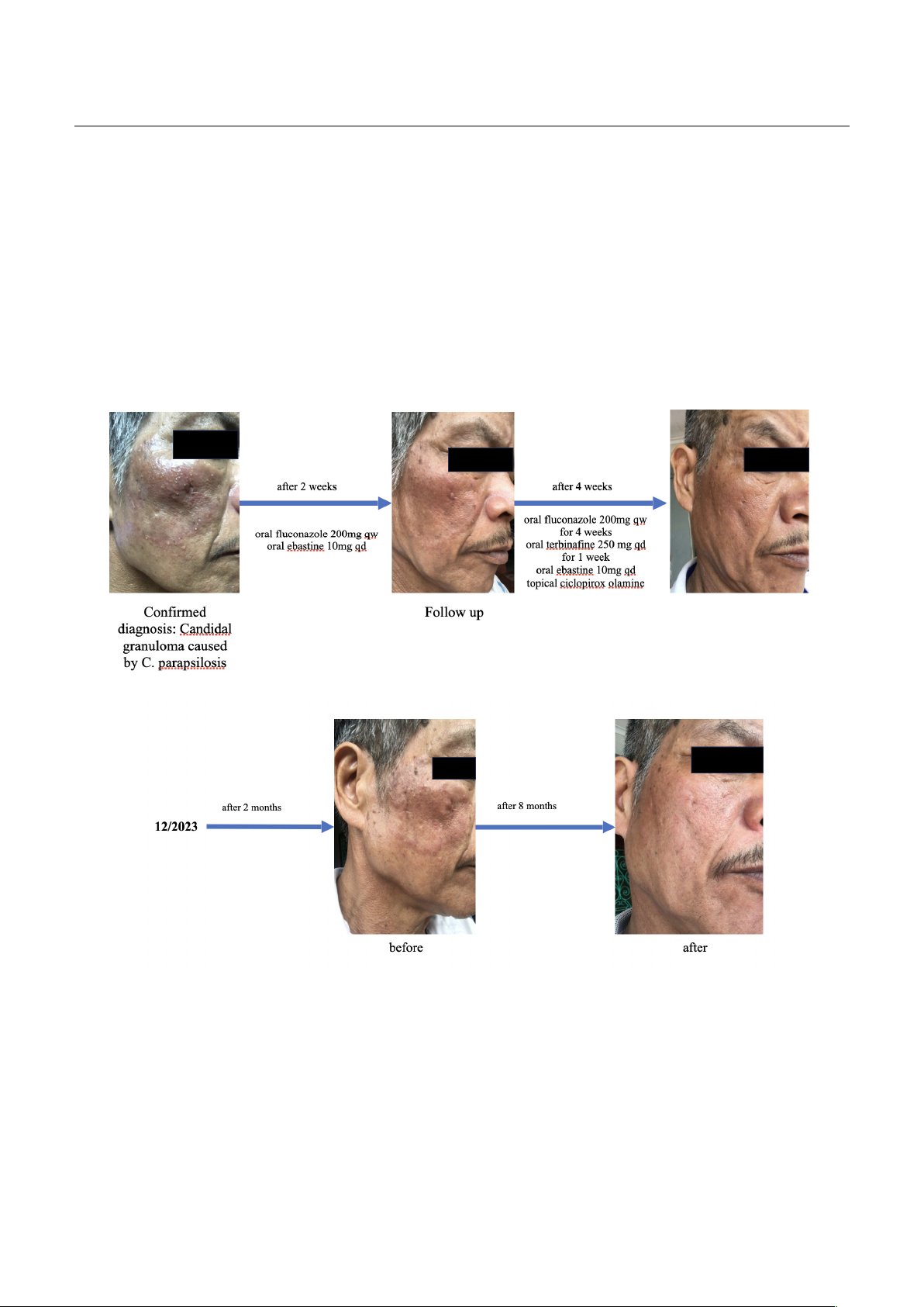
patient was subsequently treated with oral
fluconazole 200mg once weekly and oral ebastine
once daily. Follow-up after 2 weeks showed mild
improvement, thus the decision was made to add
oral terbinafine 250mg once daily for 1 week to the
treatment, as well as oral ebastine 10mg once daily
and topical ciclopirox olamine twice daily for 2
weeks. A complete resolution of skin lesions was
observed after 4 weeks, and there were no signs of
recurrence for the subsequent 6 months (figure 1, 2).
Figure 1. Detailed time course of the patient’s clinical course and therapeutic regimen
Figure 2. Skin lesions before and after treatment with follow-up
III. DISCUSSION
Candida spp. is an opportunistic pathogen,
having been isolated from domestic animals, soil,
insects and marine environment17. However it can
also be found in human skin, mucosa and nails,
causing local infections of these sites, and possibly
systemic infection with dissemination of the
disease7. The SENTRY Antimicrobial Surveillance
Programme reported that during the last few
decades, the spectrum of infections has undergone
a drastic change12. Although Candida albicans is the

JOURNAL OF 108 - CLINICAL MEDICINE AND PHARMACY Vol. 19 - Dec./2024 DOI: https://doi.org/10.52389/ydls.v19ita.2515
80
most common pathogenic fungal type of infection,
non-albicans Candida have emerged as an
important cause of infections. The prevalence of
Candida albicans infections is decreasing by year,
while the prevalence of non-albicans infections,
including Candida parapsilosis, is increasing in a
proportional way11. Despite the fact that the
incidence of candida infection, including C.
parapsilosis, is rising dramatically along with other
pathogens due to the wide use of broad-spectrum
antibiotics, glucocorticoids, immunosuppressive
drugs and invasive medical devices such as central
venous catheter, C. parapsilosis cutaneous infection
is considered to be rare1. Wu J, et al., 2021 reviewed
10 cases of C. parapsilosis cutaneous infection18. In
which onychomycosis is the most prevalent clinical
type of C. parapsilosis manifestation3,4,8,10,14,15,18. Three
cases reported subcutaneous candidiasis, which
include deep-seated subcutaneous ulcer on the
right shoulder, wart-like nodules on the planta and
subcutaneous ulcer on the right leg6,18,19.
Hauser and Rothman (1950) reported the first
case of Candida granuloma, which is a rare type of
deep cutaneous candidiasis, characterized by the
presence of papules, nodules, blisters, pustules,
abscesses and plaques covered with thick yellow-
brown scabs, commonly affected the face, scalp and
trunk of infants, children and immunocompromised
individuals or those using immunosuppressants5. C.
parapsilosis mainly causes superficial infections such
as onychomycosis, vulvovaginitis, and systemic
infections such as fungemia, endocarditis
meningitis, and peritonitis16. Candidal granuloma
caused by C. parapsilosis is consider to be an
uncommon disease and it is hard to diagnose and
treat in clinical practice. The most common cause of
granuloma is infection, such as bacterial or fungal. In
our case, the patient was uncorrectly diagnosed
twice as allergic contact dermatitis and granuloma
annulare, leading to the wrong treatment strategy.
Firstly, when there was no adequate response to
antibiotics, we need to think about other etiology
that cause the disease, such as fungal infection.
Thus, it is important to know differential diagnosis
for granuloma-like lesions.
Díaz-García J et al (2022) reported that
echinocandin susceptibility in C. parapsilosis
remained consistent, however, a notable increase in
fluconazole resistance was observed, particularly in
C. parapsilosis isolates, with rates rising from 2.6% in
2019 to 36.6% in 20222. Consequently, the treatment
of C. parapsilosis infections is still significantly
challenging. First-choice treatment of Candidal
granuloma is systemic antifungal medication.
However, as said above, fluconazole resistance was
observed in C. parapsilosis isolates, therefore
administering a combination of antifungal drugs
was a safe choice. Combination of fluconazole with
terbinafine acted synergistically on the ergosterol
biosynthetic enzymes, which enhanced the efficacy
of mycosis treatment by combining these two
medications13. In this case, the patient had good
response to fluconazole 200mg once weekly for 6
weeks combined with terbinafine 250mg once daily
for 1 week, resulting in complete resolution of skin
lesion and no recurrence after 6 months.
IV. CONCLUSION
In conclusion, candidal granuloma caused by C.
parapilaris is a rare disease. The incidence of
fluconazole resistance in Candida spp. is rising,
therefore the combination of oral fluconazole,
terbinafine and topical antifungal medication seems
to be a better choice over antifungal monotherapy
for better response to the treatment. The patient’s
skin lesions subsided without any complication
using the treatment combination and long-term
follow-up for this patient showed no relapse.
REFERENCES
1. Asogan M, Kim HY, Kidd S, Alastruey-Izquierdo A,
Govender NP, Dao A, Shin JH, Heim J, Ford NP,
Gigante V, Sati H, Morrissey CO, Alffenaar JW,
Beardsley J (2024) Candida parapsilosis: A
systematic review to inform the World Health
Organization fungal priority pathogens list. Med
Mycol 62(6):myad131.
2. Díaz-García J, Machado M, Alcalá L, Reigadas
E,Pérez-Ayala A, Gómez-García de la Pedrosa E,
Gónzalez-Romo F, Cuétara MS, García-Esteban

JOURNAL OF 108 - CLINICAL MEDICINE AND PHARMACY Vol. 19 - Dec./2024 DOI: https://doi.org/10.52389/ydls.v19ita.2515
81
C,Quiles-Melero I, Zurita ND, Muñoz-Algarra M,
Durán-Valle MT, Sánchez-García A, Muñoz P,
Escribano P, Guinea J (2023) Trends in antifungal
resistance in Candida from a multicenter study
conducted in Madrid (CANDIMAD study):
fluconazole-resistant C. parapsilosis spreading has
gained traction in 2022. Antimicrob Agents
Chemother 67(11):e0098623. doi:
10.1128/aac.00986-23.
3. Ge G, Li DM, Mei H, Lu GX, Zheng HL, Liu WD, Shi
DM (2019) Different toenail onychomycosis due to
Rhodotorula mucilaginosa and Candida parapsilosis
in an immu-nocopetent young adult. Med Mycol
Case Rep 28(24): 69-71.
4. Ge G, Yang ZY, Li DM, Hoog GS, Shi DM (2019)
Onychomycosis with greenish-black discolorations
and recurrent onycholysis caused by Candida
parapsilosis. Medical Mycol Case Rep 16(24): 48-50.
5. Hauser FV, Rothman S (1950) Monilial granuloma;
report of a case and review of the literature. Arch
Dermatol Syphilol 61(2): 297-310.
6. Honda Y, Hattori Y, Treashima T, Manabe T, Tanabe
H, Miyachi Y (2017) Plantar wart-like Candida
granuloma in a patient with myelodysplastic
syndrome. J Dermatol 44(11): 298-912.
7. Kang S, Amagai M, Bruckner AL, Enk AH, Margolis
DJ, McMichael AJ, Orringer JS. eds (2019)
Fitzpatrick's Dermatology, 9e. McGraw-Hill
Education: 2952.
8. Koklu E, Gunes T, Kurtoglu S, Gokoglu S, Koklu S
(2007) Onychomycosis in a premature infant caused
by Candida parapsilosis. Pediatr Dermatol 24(2):
155-156.
9. Mesini A, Mikulska M, Giacobbe DR, Del Puente F,
Gandolfo N, Codda G, Orsi A, Tassinari F,
Beltramini S, Marchese A, Icardi G, Del Bono V,
Viscoli C (2020) Changing epidemiology of
candidaemia: Increase in fluconazole-resistant
Candida parapsilosis. Mycoses 63(4): 361-368.
10. Noguchi H, Matsumoto T, Kimura U, Hiruma M,
Kano R, Yaguchi T, Fukushima S, IHN H (2019)
Fungal melanonychia caused by Candida
parapsilosis successfully treated with oral
fosravucanazole. J Dermatol 1(3): 1-3.
11. Qin J, Yang H, Shan Z, Jiang L, Zhang Q (2021)
Clinical efficacy and safety of antifungal drugs for
the treatment of Candida parapsilosis infections: A
systematic review and network meta-analysis. J Med
Microbiol 70(10): 001434.
12. Quindos G (2014) Epidemiology of candidaemia and
invasive candidiasis. A changing face. Rev Iberoam
Micol 31(1):42-8. doi: 10.1016/j.riam.2013.10.001.
13. Sayed MA, Abdel-Rahman TMA, Hassaneen HM, El
Kholy AA and Naguib RM (2014) Effect of
Fluconazole and Terbinafine on the Activities of
Lanosterol 14 α-Demethylase and Squalene
Epoxidase in Ergosterol Biosynthetic Pathway.
Egypt. J. Bot 54(2): 247-262.
14. Subramanya SH, Hamal DP, Nayak NR, Gokhale SS
(2016) Onychomycosis due to Candida parapsilosis
in a child with ventricular septal defect: An unusual
predisposition. Case Rep Pediatr. 2016:7026068.
doi: 10.1155/2016/7026068.
15. Subramanya SH, Subedi SJ, Metok Y, Kumar A,
Prakash PY, Nayak NR (2019) Distal and lateral
subungual onychomycosis of the finger nail in a
neonate: a rare case. BMC Pediatr 19(1):168.
16. Trofa D, Gácser A, Nosanchuk JD (2008) Candida
parapsilosis, an emerging fungal pathogen. Clin
Microbiol Rev 21(4): 606-625.
17. Weems JJ Jr (1992) Candida parapsilosis:
epidemiology, pathogenicity, clinical manifestations,
and antimicrobial susceptibility. Clin. Infect. Dis.
14:756–766.
18. Wu J, Tan J, Yang H, Gao Z, Yang L (2021) Two
cases of primary cutaneous candidiasis caused by
Candida parapsilosis: A report and literature review.
J Mycol Med 31(3): 101158.
19. Xi LY, Li XQ, Zhang JM, Lu CM, Xie T, Yin RF (2007)
Good response in a patient with deep-seated
subcutaneous ulcer due to Candida species.
Mycopathologia 164(2): 77-80.


![Bài giảng Cập nhật vấn đề hồi sức bệnh tay chân miệng nặng [mới nhất]](https://cdn.tailieu.vn/images/document/thumbnail/2025/20250920/hmn03091998@gmail.com/135x160/23301758514697.jpg)