doi:10.1046/j.1432-1033.2003.03440.x
Eur. J. Biochem. 270, 1089–1101 (2003) (cid:1) FEBS 2003
Enhanced expression of Mcm proteins in cancer cells derived from uterine cervix
Yukio Ishimi1, Isao Okayasu2, Chieko Kato1, Hyun-Ju Kwon1, Hiroshi Kimura3, Kouichi Yamada4 and Si-Young Song1 1Mitsubishi Kagaku Institute of Life Sciences, Machida, Tokyo; 2Department of Pathology, Kitasato University School of Medicine, Sagamihara, Kanagawa; 3Department of Functional Genomics, Medical Research Institute, Tokyo Medical and Dental University, Tokyo; 4National Institute of Health and Nutrition, Tokyo Japan
Consistently, Mcm2 mRNA was enriched in HeLa cells to approximately four times the level in WI-38 cells, and the synthesis of Mcm4, 6 and 7 proteins was accelerated in HeLa cells. Immunohistochemical studies of surgical materials from human uterine cervix showed that Mcm3 and 4 are ubiquitously expressed in cancer cells. Further, the positive rate and level of Mcm3 and 4 expression appeared to be higher in cancer cells than in normal proliferating cells of the uterine cervix and dysplastic cells, suggesting that they can be useful markers to distinguish these cells.
Keywords: cancer cells; DNA replication; Mcm; protein expression; uterine cervix.
Minichromosome maintenance proteins (Mcm) 2–7 play essential roles in eukaryotic DNA replication. Several reports have indicated the usefulness of Mcm proteins as markers of cancer cells in histopathological diagnosis. However, their mode of expression and pathophysiological significance in cancer cells remain to be clarified. We com- pared the level of expression of Mcm proteins among human HeLa uterine cervical carcinoma cells, SV40-transformed human fibroblast GM00637 cells and normal human fibro- blast WI-38 cells. All the proteins examined were detected in HeLa and GM cells at 6–10 times the level found in WI-38 cells on average. This increase was observed both in total cellular proteins and in the chromatin-bound fraction.
activated form of the Mcm2–7 complex is a Mcm4/6/7 hexamer.
Mcm proteins were identified as a component of the DNA replication licensing system by which a single round of DNA replication in a cell cycle is ensured [16–18]. It has been shown that cyclin-dependent kinase plays a central role in preventing over-replication [19]. Cdc6, involved in loading Mcm proteins onto chromatin, is one of the targets of regulation by the kinase [3]. Recently it has been shown that deregulation of Cdc6, ORC (origin recognition com- plex) and Mcm, all of which are targets of phosphorylation by cyclin-dependent kinase, leads to over-replication in Saccharomyces cerevisiae [20]. These findings indicate that Mcm proteins play a role in regulating the replication of DNA. The gene amplification that has been detected in various cancer cells [21] is probably generated by the over- replication of a genomic locus containing replication origins [22,23]. These notions suggest that the deregulation of DNA replication contributes to the development of malignant transformation of cells.
The entire Mcm family (Mcm2–7) is essential for eukaryotic DNA replication [1–4], playing roles in the initiation and elongation of DNA replication [5]. Mcm2–7 proteins constitute the prereplicative complex that is formed at the replication origin [6,7]. Among several Mcm complexes, only the Mcm2–7 hexamer has the ability to induce DNA replication in Xenopus egg extracts [8]. All family members have a DNA-dependent ATPase motif in the central domain [9]. However, it has been reported that Mcm4, 6, and 7 form a hexameric complex and function as a DNA helicase in vitro [10–13], suggesting that the Mcm4/6/7 complex acts as a DNA-unwinding enzyme in the replica- tion. The exact biochemical function of Mcm2, 3, and 5 remains to be determined, but it has been shown that these proteins can inhibit the helicase activity of Mcm4/6/7 by disassembling this hexamer [12,14,15], indicating a regula- tory role. In vivo findings suggest that Mcm2–7 proteins act is responsible for fork as a replicative helicase that movement [5,7]. Thus, it is likely that the Mcm2–7 complex is involved in DNA replication as a DNA helicase, and an
Recently, several groups have reported that Mcm proteins are more frequently detected in cells from malig- nant tissues than those from normal tissues [24–33]. This phenomenon was also observed in dysplastic cells, suggest- ing that Mcm proteins are a good indicator of proliferative or cancer cells in malignant tissues [28]. Elucidation of how the expression of Mcm protein changes relates to malignant transformation of cells and the pathophysiological signifi- cance of those changes awaits further studies. In this paper, we report that Mcm proteins are expressed at higher levels in the transformed cell lines than in normal fibroblasts. We also found more frequent and higher-level expression of
Correspondence to Y. Ishimi, Mitsubishi Kagaku Institute of Life Sciences, 11 Minamiooya, Machida, Tokyo 194–8511, Japan. Fax: + 81 42 724 6314, Tel.: + 81 42 724 6266, E-mail: yukio@libra.ls.m-kagaku.co.jp Abbreviations: BrdU, bromodeoxyuridine; CIS, carcinoma in situ; Mcm, minichromosome maintenance protein; ORC, origin recognition complex. (Received 24 September 2002, revised 16 December 2002, accepted 20 December 2002)
1090 Y. Ishimi et al. (Eur. J. Biochem. 270)
(cid:1) FEBS 2003
Mcm3 and 4 in cancer cells than in normal proliferating cells of human uterine cervix and dysplastic cells. The results suggest that enhanced expression of Mcm proteins plays a role in the malignant transformation of cells.
Materials and methods
Antibodies
Millipore). The membrane was incubated at 37 (cid:3)C for 1 h with primary antibodies in a blocking solution (Blockace, Dai-nippon Pharmaceuticals). After being washed with Tris buffered saline (TBS; 50 mM Tris/HCl, pH 7.5, and 0.15 M NaCl) plus 0.1% Triton X-100, the membrane was incu- bated with peroxidase-conjugated anti-rabbit or anti-mouse secondary antibodies (Bio-Rad). The immunoreacted pro- teins were detected using a chemiluminescence system (SuperSignal West Pico or Femto Maximum Sensitivity Substrate, Pierce), and the level of reactivity was quantified (Cool Saver AE-6935, Atto).
Immunostaining of cells
Rabbit anti-Mcm2 serum was prepared using mouse Mcm2 protein as an antigen, and the antibodies were purified with Mcm2-beads prepared by fixing Mcm2 protein to CNBr- activated Sepharose (Pharmacia). After the loading of antiserum onto the beads, the antibodies were eluted with 0.2 M glycine (pH 2.5) and 0.15 M NaCl. The solution was neutralized by adding 2 M Tris/HCl (pH 8.0) to a final concentration of 100 mM. Rabbit anti-Mcm3 serum was obtained as reported [34] and affinity-purified for immuno- staining. Anti-Mcm4 Ig were affinity-purified using beads conjugated with the fragment of mouse Mcm4 (amino acids 683–862) that had been used for immunizing rabbits [34]. Rabbit anti-Mcm5 serum was obtained as reported [35] and specific antibodies were affinity-purified. Rabbit anti-Mcm6 (sc-9843), mouse anti-Mcm7 (sc-9966) and mouse anti- PCNA (sc-56) IgG were purchased from Santa Cruz Biotechnology Inc. Rabbit anti-Ki67 Ig were purchased from DAKO. Anti-ORC2 Ig were produced as reported [36].
Cells
Cells cultured on eight-well chambers (Falcon) were pulse- labeled with 20 lM bromodeoxyuridine (BrdU) for 15 min. After being washed with phosphate-buffered saline (Dul- becco’s NaCl/PBS–, Nissui), the cells were fixed by incuba- tion with 4% paraformaldehyde in NaCl/Pi for 5 min at room temperature. The cells were washed with NaCl/Pi, and then permeabilized and blocked by incubation with 0.1% Triton X-100, 0.02% SDS and 2% nonfat dried milk in NaCl/Pi for 1 h at 37 (cid:3)C. Incubation of the cells with anti- Mcm4 rabbit Ig was performed for 1 h at 37 (cid:3)C in the above blocking solution. The cells were washed with the same solution and then incubated with Cy3-conjugated anti-rabbit IgG (Jackson ImmunoResearch) for 1 h in the blocking solution. Then, they were re-fixed, treated with 4 M HCl for 30 min at room temperature and incubated with rat anti-BrdU Ig (Harlan Sera Laboratory, Clone BU1/75) followed by the incubation with FITC-conjugated anti-rat IgG (Cappel). Positive immunoreactivities were detected with fluorescence microscopy (AX-80, Olympus).
Cell labeling and immunoprecipitation
HeLa cells were cultured in DMEM (Dulbecco’s modified Eagle’s medium) supplemented with 10% calf serum. WI-38 cells obtained from RIKEN GenBank and SV40-trans- formed human fibroblasts (GM00637) purchased from Coriell Cell Repositories were cultured in DMEM supple- mented with 10% fetal calf serum. WI-38 cells at a population doubling level of 33–38 were used for experi- ments and had almost stopped proliferating at approxi- mately 41 population doubling level.
HeLa and WI-38 cells that had been plated in dishes (30 mm in diameter) were cultured for 30 min in medium depleted of methionine (Sigma). Ten microlitres of [35S]methionine (10 mCiÆmL)1) was added to the medium, and incubation was continued for given periods. For pulse and chase experiments, the cells labeled with [35S]methionine for 2 h were cultured further for different periods in normal growth medium. The cells were lysed in solution A, and the precipitate after centrifugation was re-suspended with solution A. DNase I (Takara) was added to the solution at a final concentration of 700 unitsÆmL)1 and the mixture was incubated at 30 (cid:3)C for 30 min. After centrifugation, the supernatant was combined with the first supernatant and incubated with 1 lg of anti-Mcm4 Ig for 1.5 h at 4 (cid:3)C. Protein G-Sepharose (30 lL) was added to the solution, and the incubation was continued overnight at 4 (cid:3)C. After being spun down, the Sepharose beads were washed four times with RIPA buffer (150 mM NaCl, 0.5% Nonidet P-40, 1% Na-deoxycholate, 0.1% SDS, and 50 mM Tris/ HCl, pH 7.5) containing proteinase inhibitors and then mixed with 30 lL of 2· concentrated SDS sample buffer.
Fractionation of cell extracts and Western-blot analysis of Mcm proteins Cells (4 · 106 cells) were lysed in 0.2 mL of CSK buffer (10 mM Pipes, pH 6.8, 100 mM NaCl, 1 mM MgCl2, 1 mM EGTA, 1 mM dithiothreitol and 1 mM phenylmethanesulfo- nyl fluoride) containing 0.1% Triton X-100, 1 mM ATP and proteinase inhibitors (Pharmingen; solution A) [37]. The suspension was mixed with 0.1 mL of 3· concentrated sample buffer for SDS gel electrophoresis and then sonicated for 20 s to shear chromosomal DNA before being loaded onto the SDS gel. Thus, it contained total cellular proteins. To obtain chromatin-bound proteins, cells (4 · 106 cells) were lysed with solution A as described above and placed on ice for 15 min. The cell suspension was centrifuged, and the recovered precipitate was washed once with solution A. The precipitate was suspended in 0.1 mL of solution A and then mixed with 0.05 mL of concentrated SDS sample buffer.
RT-PCR
Total cellular proteins and chromatin-bound proteins were electrophoresed on a 10% acrylamide gel contain- ing SDS and transferred to a membrane (Immobilon,
mRNA was purified from HeLa and WI-38 cells using a kit (QuickPrep Micro mRNA Purification Kit, Amersham
Mcm expression in cancer cells (Eur. J. Biochem. 270) 1091
(cid:1) FEBS 2003
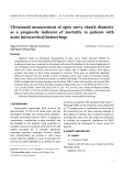
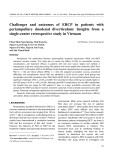
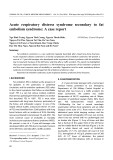
Fig. 1. Mcm proteins in total cell extracts and chromatin-bound fraction from HeLa and WI 38 cells. (A) Different volumes (0.7–12 lL) of total cellular proteins (equivalent to 2 · 104 cellsÆlL)1) were separated in SDS-polyacrylamide gel, transferred to a membrane and analyzed by immunoblotting using anti-Mcm and anti-PCNA Ig. Total proteins were analyzed on 15 : 25% gradient gel and stained with Coomassie Brilliant Blue. Core histones are indicated. (B) Chromatin-bound proteins (4 · 104 cellsÆlL)1) were analyzed by immunoblotting using anti-Mcm and anti- ORC2 Ig. Chromatin-bound proteins were analyzed on 15 : 25% gradient gel and stained with Coomassie Brilliant Blue.
1092 Y. Ishimi et al. (Eur. J. Biochem. 270)
(cid:1) FEBS 2003
Table 1. Quantitation and comparison of Mcm amounts. From the data in Fig. 1 and others, the concentrations of Mcm proteins, ORC2 and PCNA were compared between HeLa and WI-38 cells with respect to their two fractions, total cellular protein and chromatin-bound protein. After the band intensities were quantitated in each protein, the concentrations of these proteins in total and chromatin-bound fraction were determined. The relative ratio (HeLa/WI) in the concentration was calculated. When several experiments were performed in each Mcm, the average of the values (ratio) was presented with a range of the values (in parenthesis). ND, not determined. The total number of molecules in a single HeLa and WI-38 cell was calculated by determining the Mcm concentration in total cellular protein using a standard curve which was determined by immunoblotting human Mcm2, 4, 6, and 7 proteins purified from HeLa cells by histone-column chromatography.
Numbers of HeLa/WI Numbers of molecules (total)
Total Chromatin HeLa WI
1.6 · 106 1 · 105
2.5 · 106 5.2 · 105
12 11(7–15) 6(6–7) ND 10(4–14) 13(10–16) 2 · 106 1.5 · 106 1.8 · 105 2.1 · 105
14(13–16) 10(8–12) 5(4–7) 7 10(5–15) 9(6–10) 8 1.5(1.4–1.9) Mcm2 3 4 5 6 7 ORC2 PCNA Histones 1.5–2
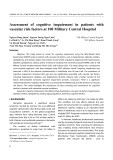
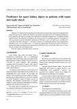
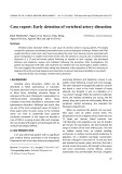
Fig. 2. Immunostaining of HeLa and WI-38 cells with anti-Mcm4 Ig. Logarithmically growing HeLa (A and B) and WI-38 cells (C and D) in one section that had been pulse-labeled with BrdU for 15 min were fixed and detected with anti-Mcm4 Ig (A and C) or anti-BrdU Ig (B and D).
Mcm expression in cancer cells (Eur. J. Biochem. 270) 1093
(cid:1) FEBS 2003
1094 Y. Ishimi et al. (Eur. J. Biochem. 270)
(cid:1) FEBS 2003
Table 2. Quantitation and comparison of proteins in GM and WI-38 cells. From the data in Fig. 2 and others, the concentrations of Mcm proteins, ORC2 and PCNA in total protein and chromatin-bound protein were compared between GM and WI-38 as described in Table 1.
GM/WI
Total Chromatin
12(5–20) 6(4–7) 5 10 6 5(5–6)
decreasing concentrations of ethanol, then with deionized water. They were boiled in 0.01 M citrate buffer (pH 6.0) at 95 (cid:3)C for 10 min using a microwave oven to facilitate antigen retrieval. Following washes in deionized water and NaCl/Pi (0.01 M phosphate buffer pH 7.2 with 0.9% NaCl), the endogenous peroxidase activity was quenched by incubation in 0.3% hydrogen peroxide in methanol for 30 min. Sections were then washed in NaCl/Pi, blocked with 10% normal swine serum in NaCl/Pi for 30 min and incubated with each of the following primary antibodies at 4 (cid:3)C overnight: rabbit anti-Mcm3, anti-Mcm4 and anti- Ki-67, and mouse anti-PCNA IgG. These antibodies were diluted in NaCl/Pi containing 2% normal swine serum (DAKO). The slides were washed in NaCl/Pi, and the subsequent immunostaining was performed by the labeled streptavidin biotin-peroxidase method using a kit (LSAB2 kit/HRP, DAKO) following the manufacturer’s protocol. The coloring reaction was performed using a ready-made substrate solution of diaminobenzidine (Stable DAB, Pharma). The slides were then lightly counterstained with methyl green, dehydrated in a graded series of ethanol, cleared in xylene and coverslipped.
6(5–7) 4(2–6) 3 9 4 4(3–7) 2 4 1 Mcm2 3 4 5 6 7 ORC2 PCNA Histones
Results
Pharmacia Biotech). The cDNA was prepared by RT-PCR using random primers (ThermoScript RT-PCR System, GibcoBRL). For the amplification of human Mcm2 cDNA, 5¢-AGACGAGATAGAGCTGACTG-3¢ as a forward primer and 5¢-CACCACGTACCTTGTGCTTG-3¢ as a reverse primer were used. Primers for the amplification of the human glyceraldehyde-3-phosphate dehydrogenase cDNA were purchased from Toyobo.
Northern blot analysis
Total RNA was extracted from HeLa and WI-38 cells with phenol, and electrophoresed on agarose gel containing formaldehyde [38]. The RNA on the gel was transferred to a nylon membrane (Hybond-N plus, Amersham). Mcm2 and glyceraldehyde-3-phosphate dehydrogenase probes ampli- fied by RT-PCR were labeled at the 5¢ end with poly- nucleotide kinase in the presence of [c-32P]ATP. The blotted membrane was incubated with the labeled probe in 1% dextran sulphate, 1% SDS and 1 M NaCl in the presence of salmon sperm DNA (50 lgÆmL)1). It was then washed twice with 2· NaCl/Cit at room temperature for 5 min, twice with 2· NaCl/Cit plus 1% SDS at 60 (cid:3)C for 30 min and finally once with 0.1 · NaCl/Cit at room temperature. The radioactivity on the membrane was detected and quantified with an Image Analyzer (FLA2000, Fuji).
Enhanced expression of Mcm proteins in cancer cells Total cellular proteins (2 · 107 cellsÆmL)1) and proteins bound to chromatin (4 · 107 cellsÆmL)1) were prepared from logarithmically growing HeLa uterine cervical carci- noma cells and human normal fibroblast WI-38 cells. They were analyzed by Western blotting using anti-Mcm2, 3, 4, 5, 6 and 7 Ig (Fig. 1). On comparing the amounts of Mcm in total cellular proteins and chromatin-bound proteins, approximately one-third of total Mcm protein was found to be recovered in the chromatin-bound fraction in HeLa cells (data not shown). Thus, a considerable portion of Mcm protein is present in the nucleoplasm and/or easily released from chromatin. Titration of these two fractions in the Western blot analysis shows that Mcm2–7 proteins are 5–14 times more abundant in HeLa cells than in WI-38 cells for the total cellular proteins and are 6–13 times for chromatin- bound proteins (Fig. 1 and Table 1). Using purified human Mcm proteins as a standard, it was calculated that approximately 1.5–2.5 · 106 molecules of Mcm2, 4, 6 and 7 proteins are present in a single HeLa cell on average and 0.1–0.5 · 106 molecules in a single WI-38 cell (Table 1).
Immunostaining of human tissues
Paraffin-embedded surgical material from uterine cervical cancer was cut 4-lm thick, and sections were placed on aminopropyltriethoxysilane-coated slides. These sections were dewaxed in xylene, and treated with a series of
While total cellular proteins from these cells appeared to be detected at a comparable level (Fig. 1), the amount of histone was slightly enriched in HeLa cells compared to WI-38 cells, which may be consistent with evidence of increased chromosomal ploidy in HeLa cells. The level of PCNA, a factor required for processivity of DNA poly- merase d and e, was comparable between HeLa cells and WI-38 cells. ORC2, a subunit of the ORC1-6 complex that is required for loading Mcm proteins onto chromatin, was detected at eight times the level in HeLa cells as in WI-38 cells. These results suggest that Mcm2–7 and ORC2 proteins are expressed at particularly high levels in HeLa cells compared to WI-38 cells. As the differences of Mcm expression between HeLa and WI-38 cells could be due to their different growth rates, we examined the percentage of
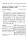
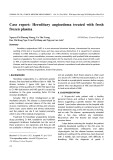
in SV40-transformed human fibroblast Fig. 3. Mcm proteins (GM00637) and WI-38 cells. Mcm2–7 proteins, ORC2 and PCNA were detected in total cellular proteins (A) and the chromatin-bound fraction (B) from GM and WI-38 cells as in Fig. 1. Total proteins including histones were also detected by Coomassie Brilliant Blue staining.
Mcm expression in cancer cells (Eur. J. Biochem. 270) 1095
(cid:1) FEBS 2003
intensity was relatively constant within BrdU-positive and -negative HeLa cells, more intense staining was observed in BrdU-positive WI-38 cells. These data suggest that the level of Mcm4 protein was maximized at the S phase in WI-38 cells and that the Mcm4 expression in HeLa cells is maintained irrespective of cell cycle.
Next, we compared the concentration of Mcm proteins in cell lysate prepared from SV40-transformed human fibro- blasts (GM00637) and normal human fibroblast WI-38 cells (Fig. 3). Mcm2–7 proteins were detected in GM cells at 3–9 times the level found in WI-38 cells for total cellular proteins and at 5–12 times the level for chromatin-bound proteins (Fig. 3 and Table 2). In contrast, the amounts of total
cell population in the S phase in logarithmically growing cells and doubling time. The percentage of BrdU-positive cells was approximately 32% for HeLa cells and 35% for WI-38 cells. Measurements of growth curves suggested that the doubling time was approximately 21 h for HeLa cells and 25 h for WI-38 cells. These results indicated that the difference in the level of Mcm expression between these two cells cannot be attributed to the difference in the growth rate. Then we performed double immunostaining of HeLa and WI-38 cells with anti-Mcm4 and anti-BrdU Ig (Fig. 2) to gain insight into the correlation between the expression of Mcm4 and the cell cycle. In both cells, clear nuclear staining was observed with anti-Mcm4 Ig. Although the staining
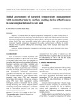
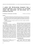
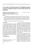
Fig. 4. Abundance of Mcm2 mRNA in HeLa and WI-38 cells. (A) Total mRNA was purified from HeLa and WI-38 cells, and the concentration of Mcm2 and glyceraldehyde-3-phosphate dehydrogenase (G3PDH) mRNA was determined by RT-PCR. Increasingly larger volumes of the mRNA fractions were added to the reaction as indicated. The amplified glyceraldehyde-3-phosphate dehydrogenase cDNA fragment was detected by staining with ethidium bromide, and the Mcm2 cDNA fragment was detected by hybridizing with the same labeled fragment. These fragments are indicated. (B) Total RNA was purified from HeLa and WI-38 cells and analyzed by Northern blot analysis. Increasing volumes (0.7, 1.5 and 3 lL each) of the total RNA were loaded onto the gel. Mcm2 and G3PDH mRNA were detected with specific probes using the same filter. The electrophoresed RNA (2 lL each) was stained with ethidium bromide (EtBr) to detect ribosomal RNAs.
1096 Y. Ishimi et al. (Eur. J. Biochem. 270)
(cid:1) FEBS 2003
Expression of the human Mcm2gene
To understand the mechanism behind the enhanced expression of Mcm proteins in HeLa cells, the concentra- tions of Mcm2 mRNA in the mRNA fractions purified from HeLa and WI-38 cells was compared by RT-PCR (Fig. 4A). A 300-base fragment of expected size was amplified from both mRNA fractions. Titration of these two fractions indicated that Mcm2 mRNA is 4–8 times more abundant in the HeLa mRNA fraction than in the WI-38 fraction. In contrast, the concentration of glyceral- dehyde-3-phosphate dehydrogenase mRNA (1000 nucleo- tides), which is known to be relatively constant among various cells, was at most doubled in the HeLa mRNA fraction. Thus, the RT-PCR experiment suggests that the level of mRNA is enriched in HeLa cells 2–4 times more than in WI-38 cells. Next, the abundance of Mcm2 mRNA was examined by Northern blot analysis. Total RNA was prepared from HeLa and WI-38 cells, and the RNA transferred to a membrane was probed with a labeled Mcm2 fragment (Fig. 4B). RNA of the expected size (approximately 3 kb) was detected on electrophoresis of the mRNA prepared from HeLa cells, but only a faint band was detected from the WI-38 mRNA. Titration of the samples loaded on the gel indicated that Mcm2 RNA is 4–8 times more abundant in HeLa cells than in WI-38 cells. In contrast, the concentrations of glyceraldehyde-3-phosphate dehydrogenase mRNA and ribosomal RNA were compar- able in these two fractions. These results indicate that Mcm2 mRNA is approximately four times more abundant in HeLa than in WI-38 cells.
Southern blot analysis of genomic DNA purified from HeLa and WI-38 cells was performed using a probe from an exon of the Mcm2 gene that codes for amino acids 214–287. Expected bands were detected from HeLa and WI-38 DNA, although the intensity of a BamH1 fragment decreased and an additional EcoRI fragment was detected in WI-38 DNA (data not shown). The results suggest that the Mcm2 gene is not amplified in HeLa cells but there is some alteration of the Mcm2 gene structure in WI-38 cells.
Protein synthesis of Mcm4
To further clarify why Mcm proteins are more abundant in HeLa cells than in WI-38 cells, we compared the synthesis of Mcm4 protein between the two cell lines. After pulse- labeling with [35S]methionine, Mcm4 protein was immuno- precipitated. The immunoprecipitates were stringently washed, and bound proteins were analyzed on SDS gel
Fig. 5. Synthesis and stability of Mcm4 protein. (A) HeLa and WI-38 cells were labeled with [35S]methionine for 0.5 or 2 h in medium depleted of methionine. The total cell extracts were prepared, immu- noprecipitated with anti-Mcm4 Ig and the immunoprecipitates were analyzed on SDS gel. (B) HeLa and WI-38 cells were labeled for 2 h as described above and incubated in the growth medium for 0, 2, 4, 7 and 22 h. Immunoprecipitation with anti-Mcm4 Ig was performed as described above. (C) The radioactivity in Mcm4 in (B) was quantitated and displayed as a course of chase periods. WI-38 (squares, solid line), HeLa (diamonds, dashed line).
cellular proteins and core histones were comparable between these two cells (Fig. 3), in spite of evidence that half of the GM cells were tetraploids. ORC2 and PCNA were detected in GM cells at two and four times the levels of those in WI-38 cells, respectively. Thus, on comparing the two human fibroblasts, it was found that Mcm proteins are expressed at higher levels in transformed GM cells than in normal WI-38 cells. We also observed a higher level of Mcm expression in SV40-transformed WI-38 cells (VA-13 cells) (2–3 times the level), human osteosarcoma (approximately four times), human Burkitt’s lymphoma (Raji cells) and human colorectal COLO 320 DM cells than in WI-38 cells (data not shown). In contrast, we observed a comparable level of Mcm expression in normal human fibroblasts from umbilical cord (HUC-F cells) and WI-38 cells.
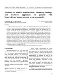
Fig. 6. Immunohistochemical detection of Mcm4, PCNA and Ki67 in normal, dysplastic and malignant cells of uterine cervix. The cancer-free squamous cell epithelium layer (left), carcinoma in situ (CIS; middle) and a region with deep invasion (right) were selected from adjacent regions in the same section to avoid staining artifacts. Four consecutive sections were stained by hematoxylin and eosin (HE; first), anti-Mcm4 (second), anti-PCNA (third) and anti-Ki67 (fourth) Ig. (B) Sections containing part of the boundary of CIS (left portion) and dysplasia (CIN1 of FIGO classification, right portion) were stained with HE, anti-Mcm4, anti-PCNA and anti-Ki67 Ig as indicated.
Mcm expression in cancer cells (Eur. J. Biochem. 270) 1097
(cid:1) FEBS 2003
1098 Y. Ishimi et al. (Eur. J. Biochem. 270)
(cid:1) FEBS 2003
(Fig. 5A). In addition to Mcm4 protein, Mcm6 and Mcm7 proteins tightly bound to Mcm4 were coimmunoprecipi- tated from HeLa cell extracts. Newly synthesized Mcm4 was also detected in the immunoprecipitates from WI-38 cells, but the intensity of the band was much weaker (approximately one tenth) than that from HeLa cells. Thus, the synthesis of Mcm4 was accelerated in HeLa cells in comparison to WI-38 cells.
Next, a pulse and chase experiment was performed to compare the protein stability of Mcm4 in these two cells. After 2-h pulse labeling, the cells were chased in normal medium for 2, 4, 7 and 22 h. The cell extracts were prepared and immunoprecipitated using anti-Mcm4 Ig, and bound proteins were analyzed on a SDS gel (Fig. 5B). Constant amounts of Mcm4 protein were recovered from different cell extracts (data not shown). The intensity of the Mcm4 band was clearly decreased at 22 h chase in both HeLa and WI-38 cells, indicating the presence of turnover of the protein. Quantitation of the Mcm4 band suggested that the protein stability of Mcm4 does not greatly differ between HeLa and WI-38 cells (Fig. 5C).
Expression of Mcm3 and 4 proteins in malignant tissues
epithelial cells, carcinoma in situ (CIS) and invasive cancer, all of which were observed in the same section. In the cancer-free squamous cell epithelial layer, the expression of Mcm4, PCNA and Ki67 was observed mainly in the basal cell layer (Fig. 6A). The immunoreactivity varied among the cells, and strongly immunopositive cells were scattered along the epithelial layer. Ki67-immunopositive cells were scarce compared with Mcm4- or PCNA-immunopositive cells. In cells of the CIS lesion, the expression of Mcm4 and PCNA was diffuse, and almost all cancer cells were immunopositive. These results are consistent with the observation that Mcm2, 3, 5 and 7 proteins are more frequently detected in cells from malignant tissues than those from normal tissues [24–33]. Mcm4-immunoreactivity was enhanced in the larger nuclei of cells of CIS compared with the cancer-free squamous cell epithelial layer. The increase in PCNA- or Ki67-immunoreactivity in cancer cells was not so marked, and Ki67-immunopositivity was detected in some of the cancer cells. All these findings were also observed in cancer cells with deep invasion. To further characterize the expression of Mcm4, PCNA and Ki67 in cancer cells, a section that contains a boundary region of CIS (CIN3 of FIGO classification) and dysplasia (CIN1) was immunostained (Fig. 6B). Mcm4- and PCNA-immu- noreactivity showed a more diffuse distribution in dysplasia than in the normal squamous cell epithelial layer, but was still localized compared with the distribution in CIS. Mcm4- immunoreactivity was stronger in cancer cells (CIS) than in cells with dysplasia. PCNA-immunoreactivity was slightly increased, but Ki67-immunoreactivity did not significantly change in the cancer cells. The results of anti-Mcm3 immu- nostaining was similar to those attained with anti-Mcm4 Ig: more intense Mcm3- and Mcm-4 immunoreactivities are
Next, we compared the expression of Mcm4 protein in cells from malignant tissues (Fig. 6). The expression of Mcm4 as well as two other proliferation marker proteins, PCNA and Ki67 [39], was examined in five cases of human uterine cervical cancer by immunohistochemical techniques, and the results were compared with the findings obtained by hematoxylin and eosin staining. We examined the expres- sion of these proteins in a cancer-free layer of squamous
Fig. 7. Immunohistochemical detection of Mcm3 and Mcm4 in normal, dysplastic and malignant cells of uterine cervix. Mcm3- and Mcm-4 immu- noreactivities were examined in the normal squamous cell epithelial layer (Normal) and invasive cancer lesion (Cancer), both of which were found in adjacent regions in the same section. Mcm3- and Mcm-4 immunoreactivities were also examined in a section containing the boundary of CIS (left portion) and dysplasia (right portion, Cancer/dysplasia).
Mcm expression in cancer cells (Eur. J. Biochem. 270) 1099
(cid:1) FEBS 2003
findings on uterine cervical cancer are consistent with an enhanced expression in transformed cell lines. It should be noted that the results suggest that Mcm proteins are expressed at high levels in cancer cells derived from uterine cervix and in HeLa cells.
ubiquitously expressed in the larger nuclei of cancer cells compared with those in basal cells of the normal squamous cell epithelial layer (Fig. 7). The ubiquitous nature of the expression of both Mcm proteins in cancer cells was also clear compared with the localized expression in dysplastic lesions. These results suggest that Mcm3 and 4 proteins are expressed more ubiquitously in cancer cells than in prolif- erative cells from normal uterine cervix or cells with dysplasia. Their expression is probably more abundant in cancer cells than in normal proliferative cells, although it is difficult to compare these results quantitatively.
Discussion
The expression of Mcm genes in growth-arrested cells is induced by addition of serum [43]. There are several E2F binding sites in the promoter region of the Mcm5, 6, and 7 genes, and transcription of the genes seems to be regulated by an E2F transcription factor [44–46]. In HeLa cells, the human papilloma E7 oncogene product binds to hyper- phosphorylated Rb to destabilize the Rb/E2F complex ([47] and references therein), and thereby several genes, including Mcm, whose expression is dependent on E2F may be activated [43]. Thus, it is possible that the human papilloma oncogene products are involved in the higher level of Mcm expression in HeLa cells and cancer cells from human uterine cervix.
Although it remains to be determined how enhanced expression of Mcm proteins affects DNA replication in cancer cells, the following findings suggest a possible involvement of Mcm in the malignant transformation. The human Mcm5 gene has been identified as one of the cancer-related genes linked to hepatitis B virus-induced carcinogenesis [48]. The Mcm7 gene has also been identified as a gene whose expression is up-regulated in colon cancer metastasis [49]. Recently, it has been reported that Mcm7 in neuroblastoma is a direct target of the MYCN transcription factor that binds to an E-box element in the Mcm7 promoter [50]. In conclusion, the present study as well as previous reports shed light on the biological significance of Mcm proteins in the aberrant proliferation of cancer cells. In addition, Mcm3 and 4 proteins seem to be useful as a marker to discriminate cancer cells and dysplastic cells in the uterine cervix, as was revealed in the present work.
Acknowledgements
We thank Yuki Komamura-Kohno for assistance. This work is in part supported by a grant-in-aid for scientific research on priority area from the Ministry of Education, Science, Sports and Culture of Japan.
References
We showed that Mcm2–7 proteins were expressed at higher levels in HeLa cells and SV40-transformed human fibro- blasts than in normal human fibroblast WI-38 cells, albeit the total proteins and histone were present at comparable levels. The higher level of Mcm expression was detected not only in total cellular proteins but also in chromatin-bound proteins. The Mcm proteins bound to chromatin probably function in DNA replication, but the role of the proteins not tightly bound to chromatin remains to be determined. Consistent with the results at the protein level, semiquan- titative PCR and Northern blot analyses suggest that Mcm2 mRNA is approximately four times more abundant in HeLa cells than in WI-38 cells. Synthesis of Mcm4, 6 and 7 proteins was accelerated in HeLa cells compared to WI-38 cells, but the turnover rate of the Mcm4 protein within 22 h did not differ between these two cells. Thus, the enhanced expression of Mcm proteins in HeLa cells may be explained by the abundance of mRNA. However, it should also be noted that the level of Mcm4 protein varies among logarithmically growing WI-38 cells (Fig. 2), suggesting the presence of regulated turnover of Mcm4 protein in WI-38 cells. A higher level of Mcm expression was observed in several other transformed cell lines, although the extent of the enhancement varied among the cells. The enhanced level of Mcm expression in these transformed cell lines would contribute to the growth of cancer cells by facilitating genome replication. As Mcm proteins bound to chromatin are present at higher levels in these cells, whether the number of replication initiation sites increases and/or the rate of the replication fork movement is accelerated, compared to WI-38 cells, deserves to be examined. It is also possible that the enhanced expression of Mcm proteins in these transformed cells contributes to cell growth by facilitating transcription [40,41] or through interaction with the Rb (retinoblastoma) protein [42].
It has been reported that
1. Kearsey, S.E. & Labib, K. (1998) MCM proteins: evolution, properties, and role in DNA replication. Biochim. Biophys. Acta 1398, 113–136. 2. Tye, B.K. (1999) MCM proteins in DNA replication. Annu. Rev. Biochem. 68, 649–686. 3. Kelly, T.J. & Brown, G.W. (2000) Regulation of chromosome replication. Annu. Rev. Biochem. 69, 829–880. 4. Bell, S.P. & Dutta, A. (2002) DNA replication in eukaryotic cells. Annu. Rev. Biochem. 71, 333–374.
5. Labib, K., Tercero, J.A. & Diffley, J.F. (2000) Uninterrupted MCM2-7 function required for DNA replication fork progression. Science 288, 1643–1647.
6. Tanaka, T., Knapp, D. & Nasmyth, K. (1997) Loading of an Mcm protein onto DNA replication origins is regulated by Cdc6p and CDKs. Cell 90, 649–660.
the frequency of Mcm expression is much higher in malignant tissues than normal tissues [24–33]. Consistent with these findings we observed that Mcm3 and 4 are ubiquitously expressed in cancer cells from human uterine cervix (Figs 6 and 7). It has also been indicated that Mcm5 protein is expressed at a similar level in normal and cancer cells from uterine cervix, breast and large intestine [28]. However, we suggest that the amounts of Mcm3 and 4 protein were increased in cancer cells compared to proliferating cells from normal tissue. Further quantitative experiments are required to confirm whether Mcm proteins are expressed at higher levels in cancer cells, but the present immunohistochemical
7. Aparicio, O.M., Weinstein, D.M. & Bell, S.P. (1997) Components and dynamics of DNA replication complexes in S. cerevisiae: redistribution of MCM proteins and Cdc45p during S phase. Cell 91, 59–69.
1100 Y. Ishimi et al. (Eur. J. Biochem. 270)
(cid:1) FEBS 2003
8. Thommes, P., Kubota, Y., Takisawa, H. & Blow, J.J. (1997) The RLF-M component of the replication licensing system forms complexes containing all six MCM/P1 polypeptides. EMBO J. 16, 3312–3319. 27. Todorov, I.T., Werness, B.A., Wang, H.-Q., Buddharaju, L.N., Todorova, P.D., Slocum, H.K., Brooks, J.S. & Huberman, J.A. (1998) HsMCM2/BM28: a novel proliferation marker for human tumors and normal tissues. Laboratory Invest. 78, 73–78.
28. Freeman, A., Morris, L.S., Mills, A.D., Stoeber, K., Laskey, R.A., (1999) Minichromosome Williams, G.H. & Coleman, N. maintenance proteins as biological markers of dysplasia and malignancy. Clin. Cancer Res. 5, 2121–2132. 9. Koonin, E.V. (1993) A common set of conserved motifs in a vast variety of putative nucleic acid-dependent ATPases including MCM proteins involved in the initiation of eukaryotic DNA replication. Nucleic Acids Res. 21, 2541–2547.
10. Ishimi, Y. (1997) A DNA helicase activity is associated with an MCM4,-6, and -7 protein complex. J. Biol. Chem. 272, 24508– 24513.
29. Stoeber, K., Halsall, I., Freeman, A., Swinn, R., Doble, A., Morris, L., Coleman, N., Bullock, N., Laskey, R.A., Hales, C.N. & Williams, G.H. (1999) Immunoassay for urothelial cancers that detects DNA replication protein Mcm5 in urine. Lancet 354, 1524–1525. 11. You, Z., Komamura, Y. & Ishimi, Y. (1999) Biochemical analysis of the intrinsic Mcm4-Mcm6-Mcm7 DNA helicase activity. Mol. Cell. Biol. 19, 8003–8015.
30. Stoeber, K., Tisty, T.D., Happerfield, L., Thomas, G.A., Romanov, S., Bobrow, L., Williams, E.D. & Williams, G.H. (2001) DNA replication licensing and human cell proliferation. J. Cell Sci. 114, 2027–2041.
12. Lee, J.-K. & Hurwitz, J. (2000) Isolation and characterization of various complexes of the minichromosome maintenance proteins of Schizosaccharomyces pombe. J. Biol. Chem. 275, 18871–18878. 13. Lee, J.-K. & Hurwitz, J. (2001) Processive DNA helicase activity of the minichromosome maintenance proteins 4, 6, and 7 complex requires forked DNA structures. Proc. Natl Acad. Sci. USA 98, 54–59. 31. Wharton, S.B., Chan, K.K., Anderson, J.R., Stoeber, K. & Williams, G.H. (2001) Replicative Mcm2 protein as a novel proliferation marker in oligodendrogliomas and its relationship to Ki67 labelling index, histological grade and prognosis. Neuropath. Appl. Neurobiol. 27, 305–313.
14. Ishimi, Y., Komamura, Y., You, Z. & Kimura, H. (1998) Biochemical function of mouse minichromosome maintenance 2 protein. J. Biol. Chem. 273, 8369–8375.
32. Endl, E., Kausch, I., Baak, M., Knippers, R., Gerdes, J. & Scholzen, T. (2001) The expression of Ki-67, MCM3, and p27 defines distinct subsets of proliferating, resting, and differentiated cells. J. Pathol. 195, 457–462.
33. Tan, D.-F., Huberman, J.A., Hyland, A., Loewen, G.M., Brooks, J.S.J., Beck, A.F., Todorov, I.T. & Bepler, G. (2001) MCM2- a promising marker for premalignant lesions of the lung: a cohort study. BMC Cancer 1, 6.
34. Kimura, H., Ohtomo, T., Yamaguchi, M., Ishii, A. & Sugimoto, (1996) Mouse MCM proteins: complex formation and 15. Sato, M., Gotow, T., You, Z., Komamura-Kohno, Y., Uchiyama, Y., Yabuta, N., Nojima, H. & Ishimi, Y. (2000) Electron microscopic observation and single-stranded DNA binding activity of the Mcm4,6,7 complex. J. Mol. Biol. 300, 421–431. 16. Kubota, Y., Mimura, S., Nishimoto, S.I., Takisawa, H. & Nojima, H. (1995) Identification of the yeast MCM3-related protein as a component of Xenopus DNA replication licensing factor. Cell 81, 601–609. K. transportation to the nucleus. Genes Cells 1, 977–993.
35. Kimura, H., Takizawa, N., Nozaki, N. & Sugimoto, K. (1995) Molecular cloning of cDNA encoding mouse Cdc21 and CDC46 the products: physical homologs and characterization of interaction between P1 (MCM3) and CDC46 proteins. Nucleic Acids Res. 23, 2097–2104. 17. Chong, P.J., Mahbubani, H.M., Khoo, C.Y. & Blow, J.J. (1995) Purification of an MCM-containing complex as a component of the DNA replication licensing system. Nature 375, 418–421. 18. Madine, M.A., Khoo, C.Y., Mills, A.D. & Laskey, R.A. (1995) MCM3 complex required for cell cycle regulation of DNA replication in vertebrate cells. Nature 375, 421–425.
19. Lygerou, Z. & Nurse, P. (2000) Controlling S-phase onset in fission yeast. Biological responses to DNA damage. Symposium on Quantitative Biology, 65, 323–332.
20. Nguyen, Y.Q., Co, C. & Li, J.J. (2001) Cyclin-dependent kinases prevent DNA re-replication through multiple mechanisms. Nature 411, 1068–1073. 36. Fujita, M., Ishimi, Y., Nakamura, H., Kiyono, T. & Tsurumi, T. (2002) Nuclear organization of DNA replication initiation proteins in mammalian cells. J. Biol. Chem. 277, 10354–10361. 37. Fujita, M., Kiyono, T., Hayashi, Y. & Ishibashi, M. (1997) In vivo interaction on of human MCM heterohexameric complexes with chromatin. Possible involvement of ATP. J. Biol. Chem. 272, 10928–10935.
21. Brodeur, G.M. & Hogarty, M.D. (1998) Gene amplification in human cancers: biological and clinical significance. In The Genetic Basis of Human Cancer (Vogelstein, B. & Kinzler, K.W., eds), pp 161–172. McGraw-Hill, Health Professions Division. 38. Sambrook, J., Fritsch, E.F. & Maniatis, T. (1989) Molecular Cloning: A Laboratory Manual, Vol. 3, 2nd edn. Cold Spring Harbor Laboratory Press, Cold Spring Harbor, New York. 39. Scholzen, T. & Gerdes, J. (2000) The Ki-67 protein: from the 22. Schimke, R.T. (1988) Gene amplification in cultured cells. J. Biol. known and the unknown. J. Cell Physiol. 182, 311–322. Chem. 263, 5989–5992.
23. Stark, G.R., Debatisse, M., Giulotto, E. & Wahl, G.M. (1989) Recent progress in understanding mechanisms of mammalian DNA amplification. Cell 57, 901–908.
24. Hiraiwa, A., Fujita, M., Nagasaka, T., Adachi, A., Ohashi, M. & Ishibashi, M. (1997) Immunolocalization of hCDC47 protein in normal and neoplastic human tissues and its relation to growth. Int. J. Cancer 74, 180–184. 40. DeFonseca, C.J., Shu, F. & Zhang, J.J. (2001) Identification of two residues in MCM5 critical for the assembly of MCM complexes and Stat1-mediated transcription activation in response to IFN-c. Proc. Natl Acad. Sci. USA 98, 3034–3039. 41. Yankulov, K., Todorov, I., Romanowski, P., Licatalosi, D., Cilli, K., McCracken, S., Laskey, R. & Bentley, D.L. (1999) MCM proteins are associated with RNA polymerase II holoenzyme. Mol. Cell Biol. 19, 6154–6163.
25. Hiraiwa, A., Fujita, M., Adachi, A., Ono, H., Nagasaka, T., Matsumoto, Y., Ohashi, M., Tomita, Y. & Ishibashi, M. (1998) Specific distribution patterns of hCDC47 expression in cutaneous diseases. J. Cutan. Pathol. 25, 285–290. 42. Sterner, J.M., Dew-Knight, S., Musahl, C., Kornbluth, S. & Horowitz, J.M. (1998) Negative regulation of DNA replication by the retinoblastoma protein is mediated by its association with Mcm7. Mol. Cell Biol. 18, 2748–2757.
43. Leone, G., DeGregori, J., Yan, Z., Jakoi, L., Ishida, S., Williams, R.S. & Nevins, J.R. (1998) E2F3 activity is regulated during the cell cycle and is required for the induction of S phase. Genes Dev. 12, 2120–2130. 26. Williams, G.H., Romanowski, P., Morris, L., Madine, M., Mills, A.D., Stoeber, K., Marr, J., Laskey, R.A. & Coleman, N. (1998) Improved cervical smear assessment using antibodies against proteins that regulate DNA replication. Proc. Natl Acad. Sci. USA 95, 14932–14937.
Mcm expression in cancer cells (Eur. J. Biochem. 270) 1101
(cid:1) FEBS 2003
44. Tsuruga, H., Yabuta, N., Hosoya, S., Tamura, K., Endo, Y. & Nojima, H. (1997) HsMCM6: a new member of the human MCM/P1 family encodes a protein homologous to fission yeast Mis5. Genes Cells 2, 381–399. 48. Gozuacik, D., Murakami, Y., Saigo, K., Chami, M., Mugnier, C., Lagorce, D., Okanoue, T., Urashima, T., Brechot, C. & Paterlini- Brechot, P. (2001) Identification of human cancer-related genes by naturally occurring hepatitis B virus DNA tagging. Oncogene 20, 6233–6240.
45. Suzuki, S., Adachi, A., Hiraiwa, A., Ohashi, M., Ishibashi, M. & Kiyono, T. (1998) Cloning and characterization of human MCM7 promoter. Gene 216, 85–91.
49. Saha, S., Bardelli, A., Buckhaults, P., Velculescu, V.E., Rago, C., Croix, B.S., Romans, K.E., Choti, M.A., Lengauer, C., Kinzler, K.W. & Vogelstein, B. (2001) A phosphatase associated with metastasis of colorectal cancer. Science 294, 1343–1346.
46. Ohtani, K., Iwanaga, R., Nakamura, M., Ikeda, M., Yabuta, N., Tsuruga, H. & Nojima, H. (1999) Cell growth-regulated expression of mammalian MCM5 and MCM6 genes mediated by the transcription factor E2F. Oncogene 18, 2299–2309.
50. Shohet, J.M., Hicks, M.J., Plon, S.E., Burlingame, S.M., Stuart, (2002) S., Chen, S.-Y., Brenner, M.K. & Nuchtern, J.G. Minichromosome maintenance protein MCM7 is a direct target of the MYCN transcription factor in neuroblastoma. Cancer Res. 62, 1123–1128. 47. Goodwin, E.C. & DiMaio, D. (2000) Repression of human papillomavirus oncogenes in HeLa cervical carcinoma cells causes the orderly reactivation of dormant tumor suppressor pathways. Proc. Natl Acad. Sci. USA 97, 12513–12518.