
BioMed Central
Page 1 of 12
(page number not for citation purposes)
Respiratory Research
Open Access
Research
Arsenic trioxide, a potent inhibitor of NF-κB, abrogates
allergen-induced airway hyperresponsiveness and inflammation
Lin-Fu Zhou*1,2,3, Yi Zhu1, Xue-Fan Cui1, Wei-Ping Xie1, Ai-Hua Hu3 and Kai-
Sheng Yin*1
Address: 1Department of Respiratory Medicine, The First Affiliated Hospital, Nanjing Medical University, Nanjing, China, 2Global Health
Programs, University of Pennsylvania School of Medicine, Philadelphia, USA and 3Division of Pulmonary Medicine, Joseph Stokes Jr. Research
Institute, The Children's Hospital of Philadelphia, University of Pennsylvania School of Medicine, Philadelphia, USA
Email: Lin-Fu Zhou* - lfzhou@njmu.edu.cn; Yi Zhu - zhuyi2000@citiz.net; Xue-Fan Cui - xuefancui@njmu.edu.cn; Wei-
Ping Xie - wpxie@njmu.edu.cn; Ai-Hua Hu - hua@email.chop.edu; Kai-Sheng Yin* - yinks@126.com
* Corresponding authors
Abstract
Background: Overactivation of nuclear factor κB (NF-κB) orchestrates airway
eosinophilia, but does not dampen airway hyperresponsiveness in asthma. NF-κB repression
by arsenic trioxide (As2O3) contributes to apoptosis of eosinophils (EOS) in airways. Here
we provide evidence that As2O3 abrogates allergen (OVA)-induced airway eosinophilia by
modulating the expression of IκBα, an NF-κB inhibitory protein, and decreases the airway
hyperresponsiveness.
Methods: Using a murine model of asthma, the airway hyperresponsiveness was conducted
by barometric whole-body plethysmography. Airway eosinophilia, OVA-specific IgE in
serum, and chemokine eotaxin and RANTES (regulated upon activation, normal T cell
expressed and secreted) in bronchoalveolar lavage fluid were measured by lung histology,
Diff-Quick staining, and ELISA. Chemokine-induced EOS chemotactic activity was evaluated
using EOS chemotaxis assay. Electrophoretic mobility shift assay and Western blot analysis
were performed to assess pulmonary NF-κB activation and IκBα expression, respectively.
Results: As2O3 attenuated the allergen-induced serum IgE, chemokine expression of
eotaxin and RANTES, and the EOS recruitment in bronchoalveolar lavage fluid, which is
associated with an increased IκBα expression as well as a decreased NF-κB activation. Also,
As2O3 suppressed the chemotaxis of EOS dose-dependently in vitro. Additionally, As2O3
significantly ameliorated the allergen-driven airway hyperresponsiveness, the cardinal
feature underlying asthma.
Conclusion: These findings demonstrate an essential role of NF-κB in airway eosinophilia,
and illustrate a potential dissociation between airway inflammation and
hyperresponsiveness. As2O3 likely exerts its broad anti-inflammatory effects by suppression
of NF-κB activation through augmentation of IκBα expression in asthma.
Published: 20 December 2006
Respiratory Research 2006, 7:146 doi:10.1186/1465-9921-7-146
Received: 19 July 2006
Accepted: 20 December 2006
This article is available from: http://respiratory-research.com/content/7/1/146
© 2006 Zhou et al; licensee BioMed Central Ltd.
This is an Open Access article distributed under the terms of the Creative Commons Attribution License (http://creativecommons.org/licenses/by/2.0),
which permits unrestricted use, distribution, and reproduction in any medium, provided the original work is properly cited.

Respiratory Research 2006, 7:146 http://respiratory-research.com/content/7/1/146
Page 2 of 12
(page number not for citation purposes)
Background
Asthma is now accepted as a T-helper type 2 (Th2) lym-
phocyte-mediated chronic inflammatory disorder, charac-
terized by airway eosinophilia and airway
hyperresponsiveness (AHR) [1]. Eosinophils (EOS)
appear to play a crucial role in the ongoing inflammation
due to either an impaired clearance or a delayed apoptosis
in the airways, where accumulation of a number of EOS
cytotoxic proteins including major basic protein, cationic
proteins and peroxidase could occur [2]. Existing data
support the notion that morphologic changes in airway
tissue to the development and severity of AHR in asthma
correlates with the presence of activated airway inflamma-
tory cells, in particular EOS [3].
The molecular regulatory pathways in induction of
chronic cytokine expression and recruitment/activation of
inflammatory cells in asthma remain elusive. However,
there is growing recognition that these processes involve
increased transcription of inflammatory genes via tran-
scription factors [4]. One such transcription factor,
nuclear factor κB (NF-κB), is abundant of p50 (NF-κB1)/
p65 (RelA) heterodimer. In a latent state, NF-κB is seques-
tered as an inactive trimer by complexing with IκBα, a 37
kDa inhibitory protein, which promotes cytoplasmic
retention and maintains a low basal transcriptional activ-
ity. IκBα consists of an N-terminal domain containing
specific phosphorylation sites, five ankyrin repeat
sequences, and a C-terminal domain of Pro-Glu-Ser-Thr
polypeptides [5]. Upon stimulation, IκBα is phosphor-
ylated by the IκB kinase, ubiquitinated and degraded
through the 26S proteasome pathway [6]. Subsequently,
the nuclear localization sequence of NF-κB is unmasked
to allow its translocation into the nucleus, where it binds
to DNA and initiates transcription of a wide range of NF-
κB-dependent genes in association with immune and
inflammatory responses [7].
Arsenic compound has long been considered as a proto-
plasmic poison that can bind to human sulfydryl-contain-
ing proteins with high affinity. Arsenic trioxide (As2O3),
extracted from arsenic compound, is a powerful ancient
medication for a variety of ailments with the principle of
"using a toxic against another toxic" in traditional Chi-
nese medicine. Strikingly, As2O3 treatment in a regime of
10 mg/d of intravenous infusion for 28 to 60 days is effec-
tive in patients with acute promyelocytic leukemia (APL)
without viable toxicity in refractory to the all-trans retin-
oic acid (ATRA) and the conventional chemotherapy by
inducing apoptosis of APL cells [8]. Many studies have
demonstrated that NF-κB overactivation underlines the
chronicity of airway inflammation characteristic of
asthma [9-12]. Recently, we have reported that As2O3-
mediated NF-κB repression in airways facilitated EOS
apoptosis in a dose-dependent manner, contributing to
the resolution of airway eosinophilic inflammation [13].
In this study, we investigated the effects of As2O3 on aller-
gen-induced AHR and NF-κB-mediated airway inflamma-
tion in a murine model of asthma. Our data indicate that
inhibition of NF-κB activation through induction of IκBα
expression may account for the broad anti-inflammatory
action of As2O3.
Methods
Asthma modeling
Specified pathogen-free female BALB/c mice, aged 6 to 8
weeks, were provided by the Chinese Academy of Medical
Sciences (Beijing, China). The animal experiment was
approved by Nanjing Medical University according to the
guidelines of the Institutional Animal Care and Use Com-
mittee. A murine asthma model was established as
described previously [14] with minor modifications.
On days 0 and 7, mice received intraperitoneal injection
of 20 µg of chicken ovalbumin (OVA, Grade V, Sigma-
Aldrich, St. Louis, MO) adsorbed to 20 mg of aluminum
hydroperoxide gel (Pierce, Rockford, IL). On days 14,
mice were randomized to receive aerosol challenge with
either 6% OVA in phosphate-buffered saline (PBS) or PBS
alone via a nebula (1–5 µM particles, Bohringer Ingel-
heim, Germany) for 40 min per day up to 7 days. During
the treatment period, As2O3 (Yida Pharmaceutics, Harbin,
China) at dose of 0.5–4.5 mg/kg, dexamethasone (Dex,
Phoenix Pharmaceutics, Belmont, CA) at dose of 2.5 mg/
kg or PBS alone was injected into the peritoneum 30 min
before each airway challenge. After the last aerosol expo-
sure, mice were sacrificed at designated timepoints.
Airway physiology
Baseline resistance and AHR induced by nebulized meth-
acholine (Sigma-Aldrich, St. Louis, MO) at dose of 12.5–
100 mg/ml in conscious unrestrained-mice were assessed
using barometric whole-body plethysmography (Buxco
Electronics Inc., Troy, NY) as described previously [15].
Airway resistance is expressed as: Penh = [(Te/0.3 Tr)-1] × [2
Pef/3 Pif], where Penh = enhanced pause, Te = expiratory
time (sec), Tr = relaxation time (sec), Pef = peak expiratory
flow (ml/sec), and Pif = peak inspiratory flow (ml/sec).
Bronchoalveolar lavage
Four hours after the last airway challenge, mice underwent
euthanasia and were cannulated in the trachea. The lungs
were washed twice with 1 ml aliquots of PBS to collect the
bronchoalveolar lavage fluid (BALF). Subsequently, the
lungs were removed, quickly frozen in liquid nitrogen,
and stored at -70°C. Additionally, the lungs were col-
lected at 1, 12, and 24 hrs post the last airway challenge to
study the kinetics of pulmonary NF-κB activation.

Respiratory Research 2006, 7:146 http://respiratory-research.com/content/7/1/146
Page 3 of 12
(page number not for citation purposes)
Lung histology
Paraffin embedded lung sections (5 µm) collected 24 hrs
after airway challenge were stained with hemotoxylin &
eosin (Sigma-Aldrich, St. Louis, MO) for examination of
histology.
Diff-Quick staining
Diff-Quick staining is a modified Wright's staining [16].
Centrifuged at 300 × g for 10 min, the pelleted cells of
BALF were suspended in a serum-free RPMI 1640
medium. The cell viability, evaluated by the trypan blue
exclusion method, was over 95%. Total and differential
cell counts were enumerated on cytospins (Thermo Shan-
don, Pittsburgh, PA) in compliance with the Diff-Quick
staining profile (Merck, Germany) by counting at least
200 to 500 cells in cross-section.
Enzyme-linked immunosorbant assay (ELISA)
Serum levels of OVA-specific immunoglobulin E (IgE)
were analyzed by ELISA using samples collected 24 hrs
after the last OVA challenge. Briefly, 96-well plates were
coated with either purified anti-mouse IgE (5 µg/ml, BD
PharMingen, San Diego, CA) or OVA (100 µg/ml). After
addition of serum samples, OVA-specific IgE was detected
using horseradish peroxidase (HRP)-conjugated sheep
anti-IgG (Calbiochem, La Jolla, CA). Arbitrary units (AU)
were calculated according to OD50 of the standard curve.
Murine chemokines, eotaxin and RANTES (regulated
upon activation, normal T cell expressed and secreted), in
the BALF samples were measured by utilizing paired anti-
bodies following the manufacturer's recommendations.
The ELISA kits were purchased from R&D Systems (Min-
neapolis, MN) with a minimum detectable levels of 3 and
5 pg/ml for eotaxin and RANTES, respectively.
EOS chemotaxis assay (ECA)
Interleukin (IL)-5 transgenic mice (CBA/CaH-TnN) were
provided by the Institute of Chemistry and Cell Biology,
Chinese Academy of Sciences (Shanghai, China). EOS
(~98% purity) were derived from spleen of IL-5 transgenic
mice with depletion of B, T, and antigen-presenting cells
using anti-B220, anti-CD4, anti-CD8 and anti-class II, as
well as rat anti-mouse Ig-conjugated magnetic beads
(Miltenyi Biotec, Auburn, CA) as described previously
[17]. EOS were seeded at 5 × 10⁴ density in triplicate and
preincubated for 15 min at room temperature with 0.25–
2 µM of As2O3 prior to chemotaxis measurement.
Chemotaxis was assessed in 48-well micro-Boyden cham-
bers using polyvinylpyrrolidone-free polycarbonate mem-
branes (NeuroProbe, Bethesda, MD). Cell suspension and
diluted chemokines of eotaxin or RANTES (PeproTech,
London, UK) were added into the chamber with RPMI
1640 containing 25 mM N-2-hydroxyethylpiperazine-N'-
2-ethanesulfonic acids (HEPES, pH 7.4) and 0.05%
bovine serum albumin. The plates were incubated for 60
min at 37°C under 5% CO2. The migrated cells were
counted in five randomly selected high-power fields
(magnification was × 1,000). Spontaneous migration was
evaluated in the absence of chemoattractant.
Extraction of nuclear and total proteins
Nuclear and total proteins of lung tissue were collected as
described previously [18]. Briefly, aliquots of liquid nitro-
gen-frozen tissue were pulverized and lysed in 200 µl of
cold Buffer A [10 mM Tris-HCl (pH7.5), 150 mM NaCl,
1.5 mM MgCl2, 0.65% Nonidet P-40, 0.5 mM phenyl-
methylsulfonyl fluoride (PMSF) and 0.5 mM dithiothrei-
tol (DTT)] for 3 min. After centrifugation at 10,000 × g for
1 min at 4°C, the nuclear pellets were extracted with 20 µl
of Buffer B [20 mM HEPES (pH7.9), 1.5 mM MgCl2, 420
mM NaCl, 0.5 mM DTT, 0.2 mM ethylenediamine-
tetraacetic acid (EDTA), 0.5 mM PMSF and 25% glycerol]
for 30 min with intermittent mixing on ice. The superna-
tant containing nuclear proteins was collected by centrif-
ugation at 12,000 × g for 5 min.
The total proteins were prepared by addition of Buffer A
to the lung powder and subjected to two freeze/thaw
cycles to fracture the nuclear membranes. After centrifuga-
tion, the supernatant was collected. The nuclear and total
proteins were quantitated using the Bradford assay (Bio-
Rad, Hercules, CA), aliquoted and stored at -70°C until
use.
Electrophoretic mobility shift assay (EMSA)
EMSA analysis was performed using a commercial kit
(Promega, Madison, WI). Double-stranded oligonucle-
otide probe (5'-AGTTGAGGGGACTTTCCCAGGC-3')
containing a consensus NF-κB sequence (underlined) was
end-labelled with [γ-32P]-adenosine triphosphate (Furui
Biotechnology, Beijing, China) by T4 polynucleotide
kinase and purified by chromatography. The binding reac-
tion was conducted in a final volume of 20 µl containing
5 µg of nuclear proteins and 30 fmol of 32P-labelled oligo-
nucleotide probe. Protein-DNA complexes were separated
by electrophoresis on a 5% native polyacrylamide gel
(37:1 acrylamide:bis-acrylamide) in a 0.5 × Tris-borate-
EDTA running buffer. The dried gel was exposed to Phos-
phorImager (Molecular Dynamics) using ImageQuant
software (Amersham Life Science, Arlington Heights, IL).
For competition assay, a 100-fold excess of unlabelled NF-
κB or activator protein 1 (AP-1) oligonucleotide probe
was added to the reaction mixture 10 min before addition
of the labelled probe. For supershift assay, a 0.5 µg of anti-
p50 or anti-p65 antibody (Santa Cruz Biotechnology,
Santa Cruz, CA) was added to the reaction mixture prior
to the labelled probe for 30 min.

Respiratory Research 2006, 7:146 http://respiratory-research.com/content/7/1/146
Page 4 of 12
(page number not for citation purposes)
Western blot analysis
Denatured samples (100 µg of total proteins) were frac-
tionated by 10% sodium dodecyl sulfate polyacrylamide
gel eletrophoresis (SDS-PAGE) and transferred to nitrocel-
lulose membranes. Blots were blocked with 5% milk con-
taining 1 × TBST [40 mM Tris-HCl (pH7.6), 300 mM NaCl
and 0.1% Tween-20] at 4°C overnight. Thereafter the blot
was probed with primary antibodies of anti-IκBα (1:1,000
dilution) or anti-β-actin antibody (1:800 dilution) for 1
hr. After an HRP-conjugated goat anti-rabbit IgG (1:5,000
dilution, Santa Cruz Biotechnology, Santa Cruz, CA) incu-
bation, the immunoblots were visualized by an enhanced
chemiluminescence (ECL) kit (Pierce, Rockford, IL)
according to the manufacture's instructions.
Data analysis
Statistical analysis was performed by one-way analysis of
variance (ANOVA) and q test with SPSS 11.0 software
package (SPSS Inc., Chicago, IL). The negative relation-
ship was evaluated by Pearson correlation analysis. Data
were expressed as mean ± SEM, and p < 0.05 was consid-
ered statistically significant.
Results
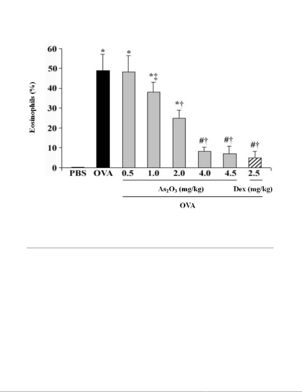
Attenuation of airway EOS recruitment by As2O3
OVA-challenged mice in response to 0.5–4.5 mg/kg of
As2O3 reduced the number of EOS in BALF in a dose-
dependent manner (Fig. 1). Since the anti-inflammatory
effects of As2O3 were similar at the doses of 4 and 4.5 mg/
kg, and it was comparable to the effect of 2.5 mg/kg of Dex
(p > 0.05), the 4 mg/kg of As2O3 was herein chosen as the
effective dosage in the rest of experiments. This dosage
was also proved to be relatively safe based on our previous
experiments [13,14]. Histological analysis of the OVA-
challenged mice lung revealed an enhanced airway eosi-
nophilia as compared to the naïve control mice that were
treated with PBS (Fig. 2A). Conversely, pretreatment of
As2O3 protected mice from developing the allergen-
induced peribronchial inflammation (Fig. 2A). Examina-
tion of BALF collected from mice at 24 hrs after OVA chal-
lenge showed a marked influx of inflammatory cells into
the airways, including EOS, lymphocytes, macrophages
and neutrophils (Fig. 2B–C). The increased EOS in the
BALF was correlated with an increase of EOS recruitment
by the Diff-Quick analysis in OVA-challenged mice (Fig.
2B). The number of EOS in BALF from naïve mice was less
than 1%, whereas that of OVA-challenged mice was about
49% (p < 0.01). Pretreatment of As2O3 dramatically atten-
uated the airway eosinophilia in the OVA-challenged
mice (p < 0.01; Fig. 2A–C; Table 1).
Amelioration of AHR by As2O3
Penh, relative to the measured airway resistance, was
obtained as an index and was normalized to the postsa-
line – Penh. This readout was used as a measure of AHR.
Mice previously sensitized and challenged with OVA
developed a dose-dependent methacholine-induced
bronchospasm as compared to the naïve mice that were
treated with PBS. As2O3 treatment significantly reduced
the effect (p < 0.01; Fig. 3).
Reduction of serum IgE and BALF chemokines by As2O3
IgE can augment allergic airway responses in a high affin-
ity receptor-dependent manner. Serum levels of OVA-spe-
cific IgE were elevated in OVA-challenged mice compared
with the naïve control mice (p < 0.01), whereas pretreat-
ment with As2O3 resulted in a 4.8-fold decrease to the lev-
els of the OVA mice (p < 0.01; Fig. 4A). Eotaxin and
RANTES play a critical role in inducing chemotaxis of EOS
[19]. ELISA analysis showed that levels of eotaxin and
RANTES in BALF were markedly increased in OVA-chal-
lenged mice in comparison with the control mice (p <
0.01). However, these chemokine levels were largely
reduced by pretreatment with As2O3 (p < 0.05 or 0.01; Fig.
4B).
Ablation of EOS chemotaxis by As2O3
Eotaxin and RANTES with respective concentrations of 1
(100) and 103 nM reached a maximal chemotaxis
response indicating that eotaxin is a more active chemo-
taxin to EOS than RANTES (Fig. 5A). As2O3 significantly
inhibited the EOS chemotaxis mediated by eotaxin or
RANTES in a dose-dependent manner (p < 0.05 or 0.01;
Fig. 5B).
Inhibition of pulmonary NF-
κ
B activation by As2O3
The OVA challenged mice showed a sharp increase in the
pulmonary DNA binding activity of NF-κB at various
timepoints as compared to the unchallenged mice lung.
Indeed, NF-κB activity was increased within 1 hr (p <
0.01), peaked at 4 hrs (p < 0.01), and decreased by 12 (p
< 0.01) to 24 hrs (p < 0.05). This effect of OVA challenge
was clearly ameliorated by pretreatment with As2O3 (p <
0.01; Fig. 6, lane 6 as compared to lane 3; Table 1). In the
competition assay, addition of 100-fold excess of unla-
belled NF-κB, but not AP-1, oligonucleotide probe com-
peted away the NF-κB-DNA complexes, verifying the
specificity of NF-κB binding. In the supershift assay, addi-
tion of antibodies against p50 and p65 resulted in retarda-
tion of supershifted bands, with reciprocal decreases in
the intensity of the NF-κB bands, confirming the classic
subunits of NF-κB heterodimer (Fig. 6).
Augmentation of pulmonary I
κ
B
α
expression by As2O3
The pulmonary IκBα expression in the lung lysate was rel-
atively decreased in OVA-challenged mice (p < 0.01; Fig.
7; Table 1) compared to the control lung. In contrast, pre-
treatment of As2O3 accumulated the pulmonary IκBα (p <
0.01). Furthermore, there was a tight negative correlation
between EOS recruitment in the BALF or the pulmonary
Respiratory Research 2006, 7:146 http://respiratory-research.com/content/7/1/146
Page 5 of 12
(page number not for citation purposes)
NF-κB activation and IκBα expression (r = -0.82 and -
0.94, respectively; p < 0.01).
Discussion
Multiple upstream signal events converge on the NF-κB-
inducing kinase (NIK) [20]. Activation of NIK results in
phosphorylation of IκB kinases, which render the phos-
phorylation of IκBα at N-terminal serines 32 and 36
(Ser32 and Ser36) residues, leading to a proteolytic degra-
dation of IκBα. Consequently, the activated NF-κB trans-
locates to the nucleus, where it bonds to specific κB sites
to facilitate the transcription of target genes. This results in
expression of numerous pro-inflammatory cytokines,
chemokines and adhesion molecules [21]. These pro-
inflammatory mediators are essential in the recruitment
of airway inflammatory cells, including EOS and CD4+ T
lymphocytes, which in turn secret Th2 cytokines [22].
Therefore, NF-κB repression in airways via suppression of
IκBα degradation or augmentation of IκBα synthesis
would decrease the transcription of a myriad of NF-κB-
dependent genes. This strategy proved to be more effective
than that of blocking a single downstream inflammatory
or an immune gene among the inflammatory cascade
[23,24].
Several lines of evidence suggest a central role of NF-κB in
the pathogenesis of asthma. Activated NF-κB has been
identified in sputum-induced macrophages and bronchial
biopsy specimens of asthmatic patients [25]. Agents such
as allergens, ozone and viral infections, which are associ-
ated with exacerbation of asthma, stimulate activation of
NF-κB [26]. As the major effective treatment for asthma,
As2O3 decreases EOS recruitment in BALF in a dose-dependent manner
Figure 1
As2O3 decreases EOS recruitment in BALF in a dose-dependent manner. Intraperitoneal administration of OVA-
challenged mice with As2O3 (0.5–4.5 mg/kg) reduced the EOS in BALF, in which both 4, 4.5 mg/kg of As2O3 and 2.5 mg/kg of
Dex achieved the similar anti-inflammatory effects. BALF EOS, stained with Diff-Quick solution, were counted using a hemato-
cytometer, and expressed as a percentage in total leukocytes. Data represent the mean ± SEM of four separate experiments (n
= 6 per group). # p < 0.05, *p < 0.01, vs the control mice; ‡ p < 0.05, † p < 0.01, vs the OVA-challenged mice.


%20--%3e%3cdefs%3e%3cstyle%3e%20.st0%20{%20fill:%20%23fff;%20}%20.st1%20{%20fill:%20%237800fa;%20}%20%3c/style%3e%3c/defs%3e%3cpath%20class='st1'%20d='M117.78,12.18H43.11c2.9,3.47,4.65,7.94,4.65,12.82,0,5.6-2.3,10.66-6.01,14.29h76.02l7.22-13.56-7.22-13.56Z'/%3e%3cg%3e%3cpath%20class='st0'%20d='M53.58,26.17h-.59v-1.46h.59v-4.96h2.83c1.78,0,2.67.94,2.67,2.82v5.76c0,1.87-.89,2.81-2.67,2.81h-2.83v-4.96ZM55.36,21.37v3.34h1.1v1.46h-1.1v3.34h1.01c.61,0,.91-.37.91-1.1v-5.93c0-.74-.3-1.1-.91-1.1h-1.01Z'/%3e%3cpath%20class='st0'%20d='M65.99,31.14h-1.8l-.31-2.07h-2.19l-.31,2.07h-1.64l1.82-11.39h2.62l1.82,11.39ZM65.28,18.04c-.25.46-.51.77-.75.94-.21.15-.47.22-.79.22-.26,0-.57-.07-.92-.22l-.38-.15c-.14-.05-.26-.07-.37-.07-.3,0-.53.18-.71.54l-.91-.68c.25-.46.51-.77.75-.94.21-.14.48-.21.79-.21.26,0,.57.07.92.21l.38.15c.14.05.26.07.37.07.3,0,.53-.18.71-.54l.91.68ZM61.91,27.52h1.73l-.87-5.76-.87,5.76Z'/%3e%3cpath%20class='st0'%20d='M74.53,26.89v1.52c0,1.91-.89,2.86-2.67,2.86s-2.67-.95-2.67-2.86v-5.93c0-1.91.89-2.86,2.67-2.86s2.67.95,2.67,2.86v1.11h-1.69v-1.22c0-.75-.31-1.12-.93-1.12s-.93.37-.93,1.12v6.15c0,.74.31,1.11.93,1.11s.93-.37.93-1.11v-1.63h1.69Z'/%3e%3cpath%20class='st0'%20d='M81.4,31.14h-1.8l-.31-2.07h-2.19l-.31,2.07h-1.64l1.82-11.39h2.62l1.82,11.39ZM75.9,19.2l1.52-1.91h1.71l1.51,1.91h-1.61l-.76-.95-.75.95h-1.61ZM77.32,27.52h1.73l-.87-5.76-.87,5.76ZM83.1,15.99l-1.76,1.91h-1.26l1.17-1.91h1.86Z'/%3e%3cpath%20class='st0'%20d='M84.86,19.75c1.78,0,2.67.94,2.67,2.82v1.48c0,1.87-.89,2.81-2.67,2.81h-.85v4.28h-1.79v-11.39h2.64ZM84.01,21.37v3.86h.85c.58,0,.87-.36.87-1.08v-1.71c0-.71-.29-1.07-.87-1.07h-.85Z'/%3e%3cpath%20class='st0'%20d='M93.51,19.75c1.78,0,2.67.94,2.67,2.82v1.48c0,1.87-.89,2.81-2.67,2.81h-.85v4.28h-1.79v-11.39h2.64ZM92.66,21.37v3.86h.85c.58,0,.87-.36.87-1.08v-1.71c0-.71-.29-1.07-.87-1.07h-.85Z'/%3e%3cpath%20class='st0'%20d='M98.8,31.14h-1.79v-11.39h1.79v4.88h2.03v-4.88h1.83v11.39h-1.83v-4.88h-2.03v4.88Z'/%3e%3cpath%20class='st0'%20d='M105.36,24.55h2.46v1.62h-2.46v3.34h3.09v1.63h-4.88v-11.39h4.88v1.63h-3.09v3.18ZM108.17,17.29l-1.76,1.91h-1.26l1.17-1.91h1.86Z'/%3e%3cpath%20class='st0'%20d='M112.2,19.75c1.78,0,2.67.94,2.67,2.82v1.48c0,1.87-.89,2.81-2.67,2.81h-.85v4.28h-1.79v-11.39h2.64ZM111.35,21.37v3.86h.85c.58,0,.87-.36.87-1.08v-1.71c0-.71-.29-1.07-.87-1.07h-.85Z'/%3e%3c/g%3e%3ccircle%20class='st1'%20cx='25'%20cy='25'%20r='20'/%3e%3cpath%20class='st0'%20d='M32.78,19.27c2.92,0,4.43,2.55,5.28,5.33l.71,2.17c.14.38-.33.75-.71.75h-5.61c.19-.33.24-.71.09-1.08l-.75-2.45c-.43-1.32-.99-2.64-1.79-3.77.75-.57,1.65-.94,2.78-.94h0ZM25,18.38c3.25,0,4.9,2.78,5.89,5.89l.76,2.45c.14.42-.33.8-.8.8h-11.69c-.42,0-.94-.38-.8-.8l.75-2.45c.99-3.11,2.64-5.89,5.89-5.89h0ZM25,11.35c1.74,0,3.11,1.37,3.11,3.11s-1.37,3.11-3.11,3.11-3.11-1.41-3.11-3.11,1.41-3.11,3.11-3.11h0ZM17.27,19.27c1.08,0,1.98.38,2.73.94-.8,1.13-1.37,2.45-1.74,3.77l-.8,2.45c-.14.38-.05.75.09,1.08h-5.56c-.42,0-.9-.38-.75-.75l.71-2.17c.9-2.78,2.41-5.33,5.33-5.33h0ZM17.27,12.91c1.51,0,2.78,1.27,2.78,2.83s-1.27,2.83-2.78,2.83-2.83-1.27-2.83-2.83,1.27-2.83,2.83-2.83h0ZM32.78,12.91c1.56,0,2.78,1.27,2.78,2.83s-1.23,2.83-2.78,2.83-2.83-1.27-2.83-2.83,1.27-2.83,2.83-2.83h0ZM27.07,28.56v.09c0,.57-.24,1.08-.61,1.46h0v.05c-.38.33-.9.57-1.46.57s-1.08-.24-1.46-.61h0c-.38-.38-.61-.9-.61-1.46v-.09h1.41v.09c0,.19.05.38.19.47v.05c.09.09.28.19.47.19s.38-.09.47-.19v-.05c.14-.09.24-.28.24-.47t-.05-.09h1.41ZM30.99,28.56v.09c0,1.65-.66,3.16-1.74,4.24-1.08,1.08-2.59,1.79-4.24,1.79s-3.16-.71-4.24-1.79l-.05-.05c-1.04-1.08-1.7-2.55-1.7-4.2v-.09h1.41v.09c0,1.27.47,2.4,1.27,3.25h.05c.85.85,1.98,1.37,3.25,1.37s2.4-.52,3.25-1.37c.85-.8,1.37-1.98,1.37-3.25v-.09h1.37ZM34.99,28.56v.09c0,2.78-1.13,5.28-2.92,7.07-1.79,1.79-4.29,2.92-7.07,2.92s-5.23-1.13-7.07-2.92c-1.79-1.79-2.92-4.29-2.92-7.07v-.09h1.41v.09c0,2.4.94,4.53,2.5,6.08,1.56,1.56,3.72,2.5,6.08,2.5s4.52-.94,6.08-2.5c1.56-1.56,2.5-3.68,2.5-6.08v-.09h1.41Z'/%3e%3c/svg%3e)