Biological characteristics of two lysines on human serum albumin in the high-affinity binding of 4Z,15Z-bilirubin-IXa revealed by phage display Ai Minomo1, Yu Ishima1, Ulrich Kragh-Hansen2, Victor T. G. Chuang3, Makiyo Uchida4, Kazuaki Taguchi1, Hiroshi Watanabe1, Toru Maruyama1, Hiroshi Morioka4 and Masaki Otagiri1,5
1 Department of Biopharmaceutics, Graduate School of Pharmaceutical Sciences, Kumamoto University, Japan 2 Department of Medical Biochemistry, University of Aarhus, Denmark 3 School of Pharmacy, Curtin Health Innovation Research Institute, Curtin University, Perth, Australia 4 Department of Analytical and Biophysical Chemistry, Graduate School of Pharmaceutical Sciences, Kumamoto University, Japan 5 Drug Delivery System Research Institute, Faculty of Pharmaceutical Sciences, Sojo University, Kumamoto, Japan
Keywords bilirubin; domain II; high-affinity binding; human serum albumin; phage display
Correspondence M. Otagiri, Faculty of Pharmaceutical Sciences, Sojo University, 1-22-4 Ikeda, Kumamoto-shi, Kumamoto 860-0082, Japan Fax: +81 96 362 7690 Tel: +81 96 371 4150 E-mail: otagirim@ph.sojo-u.ac.jp
(Received 12 April 2011, revised 30 June 2011, accepted 24 August 2011)
doi:10.1111/j.1742-4658.2011.08316.x
Database Model data are available in the PMDB database under accession numbers 2VUE and 2BXA.
4Z,15Z-bilirubin-IXa (4Z,15Z-BR), an endogenous compound that is spar- ingly soluble in water, binds human serum albumin (HSA) with high affin- ity in a flexible manner. A phage library displaying recombinant HSA domain II was constructed, after three rounds of panning against immobi- lized 4Z,15Z-BR, and eight clones with high affinity for the pigment were found to contain conserved basic residues, such as lysine or arginine, at positions 195 and 199. The wild type and two mutants, K195A and K199A, of whole HSA as well as stand-alone domain II were expressed in Pichia pastoris for ligand-binding studies. The binding of 4Z,15Z-BR to the K195A and K199A mutants was decreased in both whole HSA and the domain II proteins. The P-helicity conformer (P-form) of 4Z,15Z-BR was found to preferentially bind to the wild types and the K195A mutants, whereas the M-form bound to the K199A mutants. Photoconversion experiments showed that the P-form of 4Z,15Z-BR was transformed into highly water-soluble isomers at a much faster rate than the M-form. In addition, the M-form of 4Z,15Z-BR showed higher affinity for domain I than for domain II. The present findings suggest that, whereas both Lys195 and Lys199 in subdomain IIA are important for the high-affinity binding of 4Z,15Z-BR, Lys199 plays a more prominent role in the elimination of 4Z,15Z-BR.
Introduction
Abbreviations 4Z,15E-BR, 4Z,15E-bilirubin-IXa; 4Z,15Z-BR, 4Z,15Z-bilirubin-IXa; BR, bilirubin; CMPF, 3-carboxy-4-methyl-5-propyl-2-furanpropanoic acid; HRP, horseradish peroxidase; HSA, human serum albumin; TU, transducing unit.
FEBS Journal 278 (2011) 4100–4111 ª 2011 The Authors Journal compilation ª 2011 FEBS
4100
Bilirubin (BR), which is produced during the catabo- lism of hemoglobin, can exist in several isomeric forms [1]. 4Z,15Z-BR-IXa (4Z,15Z-BR) is the main isomer in the body, and can cause kernicterus in newborns. The neurotoxicity of 4Z,15Z-BR is most probably related to its extremely low solubility in water at neu- tral pH [2]. The neurotoxicity of the pigment is dimin- ished and its solubility in aqueous media is increased
A. Minomo et al.
Bilirubin-binding site on human serum albumin
on binding to human serum albumin (HSA), to which it binds with a high-affinity constant in the order of )1. Hydrogen bonds and aromatic p–p and 107–108 M hydrophobic interactions are important for binding [3,4]. On the other hand, the conversion of 4Z,15Z-BR to 4Z,15E-BR-IXa (4Z,15E-BR) by photosentization results in the formation of a geometric isomer that has a lower binding affinity for HSA but higher water sol- ubility, which permits it to be excreted more easily than the precursor 4Z,15Z-BR [5].
HSA is the most abundant protein in plasma, and is responsible for most of the oncotic pressure. It is a simple protein without prosthetic groups; it does not contain any glycan chains, and hence is not a glyco- protein. It is a heart-shaped protein composed of three homologous domains, each of which is further divided into two subdomains, A and B [6]. HSA binds a vari- ety of different endogenous substances, such as fatty acids, BR, various uremic toxins, and thyroxins. Two discrete drug-binding sites, designated Sudlow site I and site II, have also been found on subdomains IIA and IIIA, respectively [6].
Affinity binding experiments on 4Z,15Z-BR with albumin fragments point to a fragment composed of amino acids from about 180 to 250, which roughly correspond to subdomain IIA [7]. Petersen et al. [8] studied the binding of 4Z,15Z-BR to eight different HSA mutants in which residues assumed to be impor- tant for binding had been mutated. Surprisingly, none of the single-residue substitutions had any significant effect on the binding of 4Z,15Z-BR. This result was taken to indicate the existence of a dynamic, unusually flexible high-affinity binding site for 4Z,15Z-BR in subdomain IIA. In addition, the bisignate CD spec- trum of 4Z,15Z-BR becomes inverted when 4Z,15Z- BR binds to various mammalian albumins with more than 80% homology with HSA [9]. mode exists, but multiple interactions are ongoing between 4Z,15Z-BR and HSA [11–17]. In order to conduct a binding site topology analysis, it is necessary to simultaneously introduce multiple mutations into domain II. However, one of the inherent limitations of point mutation techniques is that they require the selection of a reasonably small number of residues as targets. To overcome this limitation, phage display technology, which can be used to efficiently perform multiple mutations, represents an alternative and potentially useful approach. Phage display is a power- ful technology for identifying polypeptides with novel properties, and altering the properties of existing ones [18–22]. Phage display has been used in epitope map- ping and studies of protein–protein interactions. This technique involves the insertion of a foreign DNA fragment into the structural gene of a bacteriophage, which leads to the expression of a peptide or protein at the surface of the viral particle. The rapid isolation of specific ligands by phage display is advantageous in many applications, including the selection of inhibitors for active and allosteric sites of enzymes, and receptor agonists and antagonists, and specific ligands isolated from phage libraries can also be used in therapeutic target validation, drug design, and vaccine develop- ment. The use of this technology to identify residues that are involved in ligand binding is not uncommon. The purpose of this study was to identify the func- tional residues in HSA that play a role in high-affinity 4Z,15Z-BR binding by constructing a phage display library with the M13 phage. We selected domain II, which contains the binding site of 4Z,15Z-BR, as the display protein. As electrostatic interactions have been shown to be important in the binding of 4Z,15Z-BR to HSA, we screened the role of two positively charged residues, namely lysine and arginine, which could potentially participate in electrostatic interactions with the carboxylate groups of 4Z,15Z-BR.
Results
Construction of a phage library displaying HAS domain II
FEBS Journal 278 (2011) 4100–4111 ª 2011 The Authors Journal compilation ª 2011 FEBS
4101
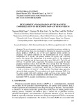
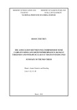
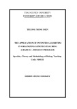
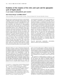
In thermodynamic studies of interactions of 4Z,15Z- BR and HSA, DH(cid:2) and DS(cid:2) were found to be ) 13.5 kcalÆmol)1 and ) 0.0085 kcalÆmol)1ÆK)1, respec- tively [3]. The large negative free energy for the inter- action, which is mainly the result of a large enthalpy change, suggests the importance of electrostatic rather than hydrophobic interactions. Earlier studies pro- posed that the bound BR interacts with histidines, arginines, tyrosines [10], and lysines [11–14], especially buried lysines [15], but not with Lys240 [16]. Hence, salt linkages between the carboxyl groups of BR and e-NH2 groups of lysines of HSA appear to be neces- sary for the binding of BR [10,17]. Therefore, it is highly probable that the multiple pattern binding mode is attributable to the formation of a flexible 4Z,15Z- BR–HSA complex, and that no single crucial binding We (resi- initially displayed wild-type domain II dues 187–385, 22 kDa) on the M13KO7 phage, as described in Experimental procedures. The expression of domain II on the phage was confirmed by ELISA. The domain II-displaying phage and the M13KO7 helper phage were equally reactive to the antibody against M13KO7, whereas only the domain II-display- ing phage showed specific activity for the polyclonal antibody against HSA (Fig. 1). These results confirm
A. Minomo et al.
Bilirubin-binding site on human serum albumin
the phage library from round 1 to 3
Helper phage
Table 1. Enrichment of expressed as recovery rate.
3
1.5
2
1
Rounds
Input phage (TU)
Output phage (TU)
Recovery rate (per 105 phages)
1
0.5
1 2 3
4.0 0.63 2.4
3.0 · 1013 2.2 · 1012 5.0 · 1011
1.2 · 109 1.4 · 107 1.2 · 107
A m n 0 9 4 t a e c n a b r o s b A
Domain II display phage B m n 0 9 4 t a e c n a b r o s b A
0 0.001 0.01 0.1
1
100
0 0.001 0.01 0.1
1
100
10 Phage volume (%)
10 Phage volume (%)
Fig. 1. Detection of domain II display phage. (A) Various concentra- tions of phage solutions were plated on 96-well microtiter plates at 100 lL per well. After an overnight incubation, the wells were washed and blocked with 0.5% gelatin, and treated with an HRP- conjugated antibody against M13KO7. (B) Various concentrations of phage solutions were plated on 96-well microtiter plates at 100 lL per well. After an overnight incubation, the coated wells were washed and blocked with gelatin, and then reacted with polyclonal antibody against HSA. Color development was performed with 5-amino-2-hydroxybenzoic acid, and 0.02% H2O2 was used as a chromogen.
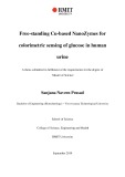
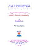
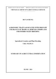
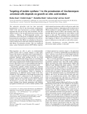
their binding affinities for 4Z,15Z-BR were evaluated with a horeradish peroxidase (HRP) assay. Among these 111 clones, 15 were found to bind 4Z,15Z-BR with the same or higher affinity as wild-type domain II (data not shown). DNA sequencing analysis of these clones showed that seven clones had frameshifts caused by nucleotide deletions or mutations and did not retain a complete domain II sequence. These deletions and mutations are the result of the facts that the phagemid vector contains an ssDNA, which is relatively unstable, and ⁄ or that the HSA sequence has a stretch of five adenines that is a spontaneous mutational ‘hot spot’ [23]. The remaining eight clones retained the sequence of domain II and were numbered serially (Fig. 2A). As can be seen from Fig. 2A, one of the clones (no. 7) had the same amino acid sequence as wild-type domai- n II. It is noteworthy that a DNA sequencing analysis of these eight clones showed that seven of them con- tained a basic residue at position 195, and that all of the eight clones contained a basic residue at posi- tion 199 (Fig. 2A).
The above findings were confirmed by the following experiments. First, the proteins of the eight selected clones were expressed with the Pichia pastoris protein expression system and purified. Their 4Z,15Z-BR- binding capabilities were then evaluated with the HRP assay. As can be seen in Fig. 2B, all of the mutated domains bound 4Z,15Z-BR equally well and with an affinity comparable to that of wild-type domain II (no. 7). These results suggest that Lys195 and Lys199 in domain II are important residues in the binding of 4Z,15Z-BR via electrostatic interactions. that domain II is displayed on the M13 phage. In the second phase of this study, we constructed a phage library displaying mutated versions of recombinant domain II. Because 4Z,15Z-BR contains two carboxyl groups, previous researchers have proposed that basic residues, lysine and ⁄ or arginine, are key residues that interact with the carboxyl groups of 4Z,15Z-BR via salt bridge formation [11–16]. To test this hypothesis and to identify the positions of such key residues, the eight lysines and four arginines located in subdo- main IIA were randomly replaced with basic lysine or arginines, neutral glycines, or acidic glutamic acids. To produce these substitutions, the lysine and arginine codons were replaced with RRA (where R represents A ⁄ G) in the cDNA library. Because RRA has only four codons (AAA, Lys; AGA, Arg; GAA, Glu; GGA, Gly), the bias of this library can be assumed to be insignificant. As shown in Fig. S1, the sequences of unselected 24 phages displayed little or no bias. The library contained (cid:2) 1.7 · 107 phage clones, a size that covers all potential clones that can be expressed.
Biopanning and evaluation of the 4Z,15Z-BR-binding capacity of selected clones Effects of mutations of Lys195 and Lys199 to alanine on the binding of 4Z,15Z-BR to domain II and HSA
FEBS Journal 278 (2011) 4100–4111 ª 2011 The Authors Journal compilation ª 2011 FEBS
4102
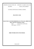
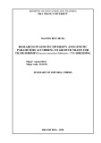
In order to identify the recombinant domain II-dis- playing phages with high 4Z,15Z-BR binding affinities, the phage library was subjected to three rounds of panning against 4Z,15Z-BR immobilized on an EAH Sepharose 4E gel. The recovery rates of the different rounds were calculated, and are listed in Table 1. After the third panning, 111 phage clones were isolated, and Wild-type domain II and domains with the K195A or K199A mutations were produced, and their 4Z,15Z- BR-binding activities were determined with an HRP assay. Both mutants, but especially domain II-K199A, had a significantly lower affinity for 4Z,15Z-BR than wild-type domain II (Fig. 3A). Consistent with the domain II studies, both point mutations, K195A and
A. Minomo et al.
Bilirubin-binding site on human serum albumin
190
205 209 212 218 222 225 233
195197199
240 DEGKASSAKQRLKCASLQKFGERAFKAWAVARLSQRFPKAEFAEVSKLVTDLTK
A WT
*
*
1 DEGGASSAKQRLRCASLQGFGEGAFEAWAVAGLSQEFPKAEFAEVSGLVTDLTE 2 DEGEASSARQELRCASLQRFGEEAFKAWAVAGLSQRFPRAEFAEVSGLVTDLTG 3 DEGRASSAGQRLRCASLQEFGEEAFGAWAVAELSQGFPEAEFAEVSRLVTDLTK 4 DEGKASSARQGLRCASLQEFGEGAFRAWAVAELSQGFPRAEFAEVSELVTDLTE 5 DEGEASSARQGLKCASLQRFGEGAFRAWAVAGLSQRFPGAEFAEVSELVTDLTR 6 DEGKASSARQRLRCASLQGFGEGAFRAWAVAGLSQGFPRAEFAEVSELVTDLTE 7 DEGKASSAKQRLKCASLQKFGERAFKAWAVARLSQRFPKAEFAEVSKLVTDLTK 8 DEGKASSARQKLRCASLQGFGEGAFGAWAVAELSQGFPRAEFAEVSGLVTDLTK
40
B
30
20
10
) M µ ( . c n o c R B d n u o b n U
0
1
2
3
4
5
6
7
8
Fig. 2. Selection and confirmation of domain II clones that bind 4Z,15Z-BR. (A) Partial amino acid sequences of domain II clones selected after three rounds of panning on 4Z,15Z-BR. (B) 4Z,15Z-BR binding to domain II clones as determined by an HRP assay. The molar ratio between 4Z,15Z-BR and protein was 2 : 1, and the concentration of unbound ligand is shown. Protein solutions were plated on 96-well mi- crotiter plates at 100 lL per well. After a 2-h incubation, the coated wells were washed, and 60 lM 4Z,15Z-BR was added to each well. Then, 10 lL of 1.75 mM H2O2 and 10 lL of 1 ngÆmL)1 HRP were added, and the absorbance at 450 nm was recorded. The free 4Z,15Z-BR concentrations were estimated as described in Experimental procedures. Data are expressed as means ± standard errors of the mean (n = 3). When the results obtained for the mutated domain II clones were compared with those obtained for the wild-type domain II clone (no. 7), the P-values were, in all cases, > 0.05. WT, wild type.
K199A, resulted in significantly weaker 4Z,15Z-BR binding to full-length HSA mutants (Fig. 3B).
)1) determined by Minchiotti et al.
for
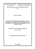
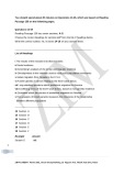
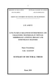
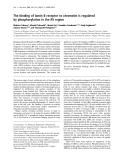
The interaction of 4Z,15Z-BR with full-length HSA and mutants was also investigated by fluorescence spectroscopy. The binding constant (Ka) for wild-type )1 HSA was determined to be (5.03 ± 0.53) · 107 M (Table 2). This value is reasonably consistent with the constant of the high-affinity binding value )1 reported by Petersen et al. 1.56 · 107 M [8], who used a peroxidase protection assay, and to that (1.10 · 108 M [16], who used fluorescence quenching titration. The Ka-val- ues for binding to the K195A and K199A mutants )1 and were calculated to be (1.10 ± 0.32) · 107 M )1, respectively (Table 2). Thus, (1.01 ± 0.15) · 107 M the two point mutations both resulted in a five-fold reduction in binding affinity for 4Z,15Z-BR. CD spectrum that is typical for the P-enantiomer of 4Z,15Z-BR. Introduction of the K195A mutation had little effect on the spectrum. In contrast, in the case of the K199A mutation, the 4Z,15Z-BR CD spectrum was altered, and resembled the form typical for the M-enantiomer. As shown in Fig. 4C, essentially the same observations were made for full-length HSA; that is, the replacement of Lys195 with an alanine had a small effect on the CD spectrum, whereas the same type of substitution for Lys199 had a dramatic, invert- ing effect on the 4Z,15Z-BR CD spectrum. The rela- tively small amplitudes observed for the wild-type and K195A domain II preparations can be attributed to the weaker binding of the P-form of 4Z,15Z-BR to these domain preparations and ⁄ or to the fact that the bend angle between the two dipyrrole chromophores of 4Z,15Z-BR had been modified.
Conformation of 4Z,15Z-BR in binding to wild-type and mutant HSA domain II and HSA examined with CD spectroscopy
FEBS Journal 278 (2011) 4100–4111 ª 2011 The Authors Journal compilation ª 2011 FEBS
4103
Figure 4B shows that the binding of 4Z,15Z-BR to wild-type domain II resulted in an induced 4Z,15Z-BR A similar mutation-induced trend was found for the domain II preparations as for the full-length HSAs, both with respect to changes in binding (Fig. 3A,B) and with respect to changes in the CD spectra for bound 4Z,15Z-BR (Fig. 4B,C). These findings indicate the high-affinity 4Z,15Z-BR site is contained that within domain II. However, because the changes were
A. Minomo et al.
Bilirubin-binding site on human serum albumin
Domain II
HSA
A
B
30
30
4Z,15Z-BR binding to albumins of different species
**
**
**
25
25
20
20
**
15
15
10
10
5
5
) M µ ( . c n o c R B d n u o b n U
) M µ ( . c n o c R B d n u o b n U
0
0
WT
K195A K199A
WT
K195A
K199A
HSA showed the highest binding affinity for 4Z,15Z- BR, followed by albumins from bovine and sheep. Dog albumin showed the weakest binding affinity for 4Z,15Z-BR (Fig. 6A). As shown in Figs 6B and S4, the preferred binding conformation for dog and rat al- bumins was the P-form, whereas the M-form was the preferred binding conformation for bovine and sheep albumins.
Photoconversion of 4Z,15Z-BR bound to HSA, albumins of different species, or HSA mutants
Fig. 3. 4Z,15Z-BR-binding properties of recombinant domain II and HSA (wild type, K195A mutant, and K199A mutant). The concentra- tion of unbound 4Z,15Z-BR in the presence of recombinant domai- n II proteins (A) or recombinant HSA proteins (B) as determined by the HRP assay. 4Z,15Z-BR at 60 lM was incubated with 30 lM domain II proteins or 15 lM HSA proteins, each dissolved in 67 mM phosphate buffer (pH 7.4). The free 4Z,15Z-BR concentrations were estimated as described in Experimental procedures. Data are expressed as means ± standard errors of the mean (n = 3). **P < 0.01, as compared with the corresponding wild-type poly- peptide, WT, wild type.
4Z,15Z-BR bound to wild-type HSA and the K195A mutant showed a higher photoconversion rate than that for the K199A mutant (Fig. 7A). On the other hand, HSA and dog albumin showed a higher photo- conversion rate than bovine and sheep albumins (Fig. 7B).
Discussion
Table 2. BR affinity binding constants of recombinant domain II and HSA (wild type and the K195A and K199A mutants). BR bind- ing to different recombinant domain II and HSA preparations was studied with the fluorescence enhancement titration technique. The protein solutions were excited at 460 nm, and the emission was measured at 512 nm in the case of wild-type HSA and the K199A mutant, and at 507 nm in the case of the K195A mutant. The protein concentration was kept constant at 3 lM, whereas the concentration of BR was varied from 0 to 3.0 lM; the medium was 67 mM sodium phosphate buffer (pH 7.4). Data are expressed as means ± standard errors of the mean (n = 3).
M
M
)1)
)1)
HSA Ka (· 107
Domain II Ka (· 107
5.03 ± 0.53 1.10 ± 0.32 1.01 ± 0.15
1.93 ± 0.26 0.327 0.209
Wild type K195A K199A
Identification of the key 4Z,15Z-BR-binding residues in domain II of HSA with phage display
quantitatively different, the detailed structure of the 4Z,15Z-BR–domain II complex is most probably dif- ferent from that of those formed with full-length HSA. The use of a phage display permitted the presentation of (poly)peptides as fusion bodies to capsid proteins on the surface of bacterial viruses or phage particles. The novel display formats, in vitro selection via inno- vative library designs and screening strategies, enabled the rapid identification and optimization of proteins on the basis of their structural or functional properties. techniques have been developed to Recombinant increase the diversity of the library. Phage display technology has been used to develop polypeptides with novel properties and small antibodies such as scFv. There are some examples of ‘nonantibody’ proteins that are macromolecular proteins displayed on a phage, such as protective antigen protein domain 4 (24 kDa) [19], insulin-like growth factor I (14.5 kDa) [20], human growth hormone (22 kDa) [21] and gluta- thione-S-transferase (25 kDa) [22].
4Z,15Z-BR binding to recombinant HSA domain I
FEBS Journal 278 (2011) 4100–4111 ª 2011 The Authors Journal compilation ª 2011 FEBS
4104
et al. study, Dockal [25] HSA is a single-stranded polypeptide that folds to form a heart-shaped protein containing three homolo- gous domains. An HSA fragment composed mainly of domain II has been shown to house the primary bind- ing site for 4Z,15Z-BR [24]. The aim of this study was to identify residues in domain II that are of impor- tance for the high-affinity binding of 4Z,15Z-BR. In a previous successfully expressed and characterized each individual HSA The binding affinity of 4Z,15Z-BR for wild-type domain I (residues 1–186) was significantly lower than that for wild-type domain II. Furthermore, 4Z,15Z- BR, when bound to domain I, adopts an unusual M-conformation that is quite different from the typical P-conformation that it adopts when binding to domain II and full-length HSA (Fig. 5A,B).
A. Minomo et al.
Bilirubin-binding site on human serum albumin
A
O
O
H O
H O
N H
N H
O
O
H
H
N
N
N
N
H
H
O
O
H
H
N
N
O
O
Ridge-tile
H
H
O
O
M-form
P-form
WT
K199A
Domain II
HSA
K195A C
B
80
300
200
40
P
) 1 – l o m d · 2
) 1 – l o m d · 2
P
100
0
0
M
–40
M
–100
m c . g e d ( 3 – 0 1 × ] [
m c . g e d ( 3 – 0 1 × ] [
–80
–200
550
350
550
350
450 Wavelength (nm)
450 Wavelength (nm)
Fig. 4. Induced CD spectra of 4Z,15Z-BR bound to recombinant domain II and HSA (wild type, K195A mutant, and K199A mutant). (A) Structures of the M-enantiomer and P-enantiomer of 4Z,15Z-BR. The ridge- tile conformations are indicated. (B) CD spectra of 4Z,15Z-BR bound to different versions of domain II. (C) CD spectra of 4Z,15Z-BR bound to different HSAs. In both (B) and (C), 800 lL of a 40 lM domain II solution or a 20 lM HSA solution in 67 mM sodium phosphate buffer (pH 7.4) was placed in a 1.0-cm-pathlength cell and scanned from 350 to 550 nm. 4Z,15Z-BR was subsequently added to a final concen- tration of 60 or 30 lM, respectively. After being mixed and then allowed to stand for 10 min, the samples were rescanned from 350 to 550 nm. WT, wild type.
ing results with the phage library indicated that Lys195 and Lys199 in native subdomain IIA appeared to play an important role in the binding of BR, which contains two free carboxyl groups. On the basis of the phage display results, the role of Lys195 and Lys199 in binding 4Z,15Z-BR was further examined by site- directed mutagenesis. domain. Therefore, taking advantage of the fact that each of the three HSA domains can be independently produced with recombinant techniques without signifi- cant disruption of the secondary and tertiary struc- tures, it was possible to display a large number of domain II mutants on the phage and screen for candi- date residues for 4Z,15Z-BR binding.
Binding affinity of 4Z,15Z-BR for HSA
)1
the wild-type investigation of
the sequences of
FEBS Journal 278 (2011) 4100–4111 ª 2011 The Authors Journal compilation ª 2011 FEBS
4105
Libraries with various biases can be created for spe- cific purposes when the sequence of the peptide that binds to the target is known, and its affinity can be selectively increased by screening libraries created with limited mutagenesis of the peptide. In this study, the lysine and arginine codons located in subdomain IIA were replaced with RRA (where R represents A ⁄ G) in the cDNA library. Because RRA has only four codons (AAA, Lys; AGA, Arg; GAA, Glu; GGA, Gly), the bias of this library is considered to be insignificant. Figure S3 shows the unselected library phages that display little or no bias. This phage library was composed of (cid:2) 1.7 · 107 clones. A reason- ably sized, diverse library ensures that every possible clone is represented in the library. In fact, the input phage number at round 1 was 3.0 · 1013 transducing units (TUs) which is sufficient to cover the diversity requirement of a phage library (Table 1). The screen- The findings reported so far indicate that mutations of Lys195 and Lys199 to alanines in subdomain IIA caused an increase in the concentration of unbound 4Z,15Z-BR, indicating that both lysines are involved in the binding of 4Z,15Z-BR (Table 2). However, a similar stand-alone domain II showed the binding affinity to be lower, only (1.93 ± 0.26) · 107 M (Table 2). A possible explanation for the weaker binding to the stand-alone domain II is that the BR binding site is more exposed to the solvent in the absence of domains I and III. A similar observation has been reported by Dockal et al. [25] for the binding of the site I ligand, warfarin, where the primary warfarin-binding site is centered around subdomain IIA, with indispensable structural
A. Minomo et al.
Bilirubin-binding site on human serum albumin
3
P < 0.01
A
A
25
2.5
20
2
1.5
15
1
10
0.5
) M µ ( . c n o c R B d n u o b n U
5
) M µ ( . c n o c R B d n u o b n U
0
Human
Dog
Bovine
Sheep
0
Domain I
Domain II
B
Human Bovine
Dog Sheep
HSA
200
Domain I
Domain II
B
300
100
) 1 – l o m d · 2
P
200
0
) 1 – l o m d · 2
100
M
.
P
–100
0
m c . g e d ( 3 – 0 1 × ] [
–200
–100
350
550
m c g e d ( 3 – 0 1 × ] [
450 Wavelength (nm)
–200
350
550
450 Wavelength (nm)
Fig. 6. 4Z,15Z-BR-binding properties of albumins of different spe- cies. (A) The concentration of unbound 4Z,15Z-BR in the presence of albumins of different species was determined with the HRP assay. 4Z,15Z-BR at 15 lM was incubated with 30 lM albumin in 67 mM phosphate buffer (pH 7.4). The free 4Z,15Z-BR concentra- tions were estimated as described in Experimental procedures. Data are expressed as means ± standard errors of the mean (n = 3). (B) Induced CD spectra of 4Z,15Z-BR bound to albumin of different species. The CD spectra were initially obtained by placing 800 lL of a 20 lM albumin solution in 67 mM sodium phosphate buffer (pH 7.4) in a 1.0-cm-pathlength cell and scanning from 350 to 550 nm. 4Z,15Z-BR was then added to a final concentration of 20 lM. After being mixing and allowed to stand for 10 min, the samples were scanned from 350 to 550 nm.
Fig. 5. 4Z,15Z-BR-binding properties of recombinant domain I as compared with domain II and HSA. (A) The concentration of unbound 4Z,15Z-BR in the presence of domain I or II was deter- mined with the HRP assay. 4Z,15Z-BR at 60 lM was incubated with 30 lM recombinant HSA, domain I or domain II in 67 mM phosphate buffer (pH 7.4). The free 4Z,15Z-BR concentrations were estimated as described in Experimental procedures. Data are (B) expressed as means ± standard errors of the mean (n = 3). Induced CD spectra of 4Z,15Z-BR bound to wild-type domain I or II or HSA. First, the CD spectra were obtained by placing 800 lL of a 40 lM domain I or II solution or a 20 lM HSA solution in 67 mM sodium phosphate buffer (pH 7.4) in a 1.0-cm-pathlength cell and scanning from 350 to 550 nm. 4Z,15Z-BR was then added to a final concentration of 60 or 30 lM, respectively. After being mixed and allowed to stand for 10 min, the samples were scanned from 350 to 550 nm.
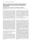
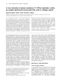
contributions from subdomain IIB and domain I. We also reported previously that the recombinant albumin domain II protein binds warfarin and dansyl L-asparagine only negligibly [26]. This indicates that the integrity of the binding pocket in domain II is affected by interdo- main interactions between domains I and III. Goncharova and Urbanova´ crystallographic study, 3-carboxy-4-methyl-5-propyl-2- furanpropanoic acid (CMPF), a dicarboxylic acid- containing renal toxin, has been shown to undergo five hydrogen bond or salt bridge interactions with Tyr150, Lys199, Arg222, His242, and Arg257 [28]. In a previ- ous study, we found that CMPF competitively inhib- 4Z,15Z-BR to HSA [11]. ited the binding of Inspection of the CMPF–HSA X-ray crystallographic structure revealed that CMPF interacts electrostatically with Lys199, but that obvious interactions with Lys195 are lacking (Fig. 8B). These findings provide further support for the essential role of Lys199 in the binding of 4Z,15Z-BR.
FEBS Journal 278 (2011) 4100–4111 ª 2011 The Authors Journal compilation ª 2011 FEBS
4106
[27] proposed that ligands with a carboxylate group may interact with the positively charged Lys199, Arg222 and Arg257 in in an X-ray subdomain IIA. On the other hand, The importance of Lys199 over Lys195 was clearly observed from the results of the third panning, where
A. Minomo et al.
Bilirubin-binding site on human serum albumin
0.5
0.4
0.3
i
·
0.2
) 1 – n m m n (
0.1
A n o i s r e v n o c o t o h p f o e t a R
be attributed to hydrophobic interactions. In a study using molecular dynamics modeling, Dı´ az et al. reported that Lys195 and Lys199 could play a com- bined and comparable chemical role in maintaining a positive charge, whereby a potential Lys195 fi Lys199 proton-transfer process via water bridges has been pro- posed [15]. The model illustrates the importance of the ionization state of Lys199, a key residue, and the nearby Lys195 residue on the structure and dynamics of the IIA binding site.
0
HSA
K195A
K199A
Enantioselective binding of 4Z,15Z-BR to HSA
0.4
0.3
i
0.2
·
) 1 – n m m n (
0.1
B n o i s r e v n o c o t o h p f o e t a R
0
Human
Dog
Bovine
Sheep
Fig. 7. Rate of photoconversion of 4Z,15Z-BR into other, more water-soluble, forms of BR. (A) Recombinant HSA and its K195A and K199A mutants. (B) Albumin from different species.
The fully protonated unconjugated 4Z,15Z-BR diacid and the dianion adopt a folded biplanar ridge-tile structure, analogous to a half-opened book, which is stabilized through intramolecular hydrogen bonding of each of the two carboxylate groups to the oxygen and nitrogen of the opposing dipyrrole (Fig. 4A). This explains the considerable lipophilicity of the pigment as compared with its biogenetic precursor biliverdin, and is a dominant factor in its metabolism, antioxidant behavior, and toxicity [1]. The right-handed configura- tion is denoted as P (plus) and the left-handed one as M (minus); these are referred to as the P-helicity folded conformer (P-form) and the M-helicity folded conformer (M-form) [29]. In solution at 37 (cid:2)C, the two mirror-image forms interconvert rapidly, forming a 50 : 50 mixture of the P-form and the M-form. Never- theless, when a chiral complexation agent such as serum albumin is added, the equilibrium shifts towards either the M-form or the P-form. In such cases, one observes typically intense bisignate CD cotton effects. The conformational structure of the bound pigment, and hence the shape and nature of the binding site on HSA, can be inferred from the CD data [13].
clone 3 retained an arginine at position 199 but a gly- cine was located at position 195. In addition, the con- centration of free 4Z,15Z-BR for K199A was higher than that for K195A and wild-type HSA in this study. In an X-ray crystallographic study of drug–HSA com- plexes, Ghuman reported that the polar groups of Lys199 and Lys195 were clustered with other polar residues at the entrance of the binding pocket of sub- domain IIA, whereas only the aliphatic portion of Lys199, which contributes to the delineation of the subchamber, protruded from the front of the pocket [28]. This shows that Lys199 plays more than one role in the binding of 4Z,15Z-BR.
FEBS Journal 278 (2011) 4100–4111 ª 2011 The Authors Journal compilation ª 2011 FEBS
4107
Petersen et al. [8] studied the binding of 4Z,15Z-BR to HSA preparations with several single-residue substi- tutions, including K195M and K199M, and found that none of the mutants had a significantly altered affinity for 4Z,15Z-BR. This is in contrast to the present study, in which we found that the binding of 4Z,15Z- BR to the K195A and K199A mutants was diminished (Table 2). A comparison of the binding results for methionine and alanine mutants illustrates the impor- tance of having the spaces at positions 195 and 199 occupied by an aliphatic side chain, in order to retain 4Z,15Z-BR at the binding pocket. This can most likely As shown by the 4Z,15Z-BR-induced CD spectra, mutations of Lys195 and Lys199 to alanine effectively changed the binding microenvironment of the 4Z,15Z- BR-binding site to different extents, depending on the location of the mutation, and thus the preferred bind- ing conformation of 4Z,15Z-BR. The preferred confor- mation of BR for binding by wild-type HSA is the P-form, as shown in Fig. 4C. The mutation of K195A resulted in only a slight decrease in the intensities of the spectrum, and the P-form of 4Z,15Z-BR remains the preferred binding conformation at the binding site (Fig. 4C). However, when Lys199 is replaced with an alanine, the spectrum for 4Z,15Z-BR changed dramat- ically, indicating that the M-form was the preferred binding conformation in the absence of a lysine ali- phatic side chain at position 199. A similar binding chirality preference was observed for the wild type and
A. Minomo et al.
Bilirubin-binding site on human serum albumin
A
4Z,15E-BR
Table 3. Amino acids at positions 195 and 199 of albumins of dif- ferent species and the corresponding BR binding helicity form.
Mammalian
BR helicity
195
199
222
Homology (%)
100 80 73 75 75
Human Dog Rat Bovine Sheep
P P P (Fig. S2 [9]) M M
K K R R R
K K K R R
R R R K K
B
CMPF
CMPF
K195
R222
K199
R218
Fig. 8. Binding regions of 4Z,15E-BR and CMPF on HSA. Magnifi- cation of the binding regions of 4Z,15E-BR (A) and CMPF (B), with the four residues lining the entrance of the site I binding pocket in subdomain IIA, namely Lys195, Lys199, Arg218, and Arg222, shown as ball and stick models; CPK color style. Domain I (gray) and domain II (green) are displayed in backbone style, and domain III (gray) is displayed in strand style. Both diagrams were generated with RASMOL, using the X-ray crystallographic structure data files from the Protein Data Bank: PDB ID 2VUE for 4Z,15E-BR–HSA (A), and PDB ID 2BXA for CMPF–HSA (B). The insert in (A) shows the whole HSA structure.
M-form is the preferred binding conformation for bovine and sheep albumins. When the amino acid sequences of the albumins were compared, it was found that the preferred P-forms of albumins of humans and dogs contain lysines at positions 195 and 199, the rat albumin contains an arginine at position 195 (Fig. S2) [9], and bovine and sheep albumins, with the M-form preferred, contain arginines at positions 195 and 199 (Table 3). Hence, Lys199 is a key residue responsible for the preferred P-form conformation for binding of 4Z,15Z-BR by albumin. As the mutation of lysine to methionine at position 199 had no effect on the P-form binding preference of HSA in the point mutation study reported by Petersen et al. [8], this provides a demon- stration of the role of an aliphatic side chain at posi- tion 199 in determining the helicity of 4Z,15Z-BR in the binding preference of albumin.
K195A and K199A recombinant domain II proteins (Fig. 4B). the Interestingly, albumins of different
Albumin has been found to facilitate a conforma- tional change in 4Z,15Z-BR resulting in a more water-soluble form during phototherapy [5]. The physi- ological significance of the preference for a particular 4Z,15Z-BR binding conformation by albumin was evident in a further investigation of the effect of photo- irradiation on 4Z,15Z-BR and albumin mixtures. In general, the P-form-preferring albumins facilitated a faster transformation of the low-solubility 4Z,15Z-BR into more water-soluble forms, as shown in Fig. 7B for human and dog. A similar observation was made when both wild-type and K195A mutant HAS, which prefer- entially bind the P-form, exhibited faster transforma- tion of 4Z,15Z-BR. In contrast, the mutation of Lys199 to alanine effectively changed the binding preference to the M-form, leading to a slower photo- low-solubility 4Z,15Z-BR conversion rate of (Fig. 7A). This observation also points to the impor- tance of an aliphatic side chain at position 199 for the efficient photoconversion of 4Z,15Z-BR. Zunszain et al.
FEBS Journal 278 (2011) 4100–4111 ª 2011 The Authors Journal compilation ª 2011 FEBS
4108
species also showed such a preferred binding conformation of 4Z,15Z-BR, depending on the species. Pistolozzi et al. [9] reported that the induced CD spectrum of 4Z,15Z- BR bound to rat albumin showed the P-form, which is in agreement with our findings. As shown in Figs 6B and S4, the P-form is the preferred binding conforma- tion for human, dog and rat albumins, whereas the [30] recently published a crystallo- graphic analysis of HSA complexed with 4Z,15E-BR (Fig. 8A). The analysis revealed that this isomeric form of the pigment binds with high affinity to a pocket in subdomain IB, and that the carboxylate groups form
A. Minomo et al.
Bilirubin-binding site on human serum albumin
the P-form of 4Z,15Z-BR, which affects the binding preference of albumin, thus leading to more rapid photoconversion of 4Z,15Z-BR into a more water-solu- ble molecule for subsequent renal elimination.
Experimental procedures
Construction of a phage library displaying recom- binant HSA domain II
salt bridges with arginines at positions 117 and 186, but not with lysine. Our results show that 4Z,15Z-BR binds to domain I with a lower affinity than to domai- n II (Fig. 5A), and in an atypical M-helicity conforma- tion instead of the usual P-form (Fig. 5B). Thus, it is possible that subdomain IB is a binding site for 4Z,15E-BR, but may not be the primary high-affinity binding site for 4Z,15Z-BR. However, further investi- gations will be needed to investigate the possible photoconversion of 4Z,15Z-BR to 4Z,15E-BR that leads to a shift in the binding site from subdomain IIA to subdomain IB.
Domain II cDNA containing sites for restriction enzymes SfiI and NotI, located at the N-terminus and C-terminus, respectively, was constructed. The plasmid pCANTAB 5E also contained SfiI and NotI sites, and both domain II cDNA and pCANTAB 5E were digested with SfiI–NotI and ligated by T4 DNA ligase. A DNA sequence analysis confirmed that, in the case of the plasmid pCANTAB 5E, which encodes for the HSA-domain II, the C terminus of the HSA-domain II was fused to the N terminus of the M13 phage gene 3 signal sequence.
Oligonucleotides 1–6 were designed to have the sequence RRA (or TYY in the complementary strand), where R and Y represent G ⁄ A and C ⁄ T, respectively, at the subdo- main IIA codons for Lys190, Lys195, Arg197, Lys199, Lys205, Arg209, Lys212, Arg218, Arg222, Lys225, Lys233, and Lys240. The sequence RRA can randomly encode for Lys, Arg, Gly, and Glu, without bias (Fig. S3). The sequences were: oligonucleotide 1, 5¢-CCGGCCATGTCCGATGAAGGG RRAGCTTCGTCGTCTGCC-3¢; oligonucleotide 2, 5¢-TT GGAGACTGGCACATYYGAGTYYCTGTYYGGCAGA CGAAGC-3¢; oligonucleotide 3, 5¢-GTGCCAGTCTCCAA RRATTTGGAGAARRAGCTTTCRRAGCATGGGCAG TAGCT-3¢; oligonucleotide 4, 5¢-ACTTCTGCAAACTCA GCTYYGGGAAATYYCTGGCTCAGTYYAGCTACTG CCCATGC-3¢; oligonucleotide 5, 5¢-GCTGAGTTTGCA GAAGTTTCCRRATTAGTGACAGATCTT-3¢; oligonu- cleotide 6, 5¢-GCAGCATTCCGTGTGGACTYYGGTAAGAT GTGTCACTAA-3¢; HSA_d II_SfiI, 5¢-CCTTTCTATGCG GCCCAGCCGGCCATGTCCGATGAA-3¢; HSA_d II_120r, 5¢-GGAAACTTCTGCAAACTCAGC-3¢; and HSA_d II_NcoI, 5¢-CAGATCTCCATGGCAGCATTCCGTGTGGAC-3¢.
Potential clinical usefulness of domain II in hyperbilirubinemia
Mutagenic dsDNAs were synthesized from these oligonu- (TaKaRa, cleotides with the use of Klenow fragment Japan). Prepared dsDNAs with oligonucleo- Kyoto, tides 1–2, 3–4 and 5–6 are referred to as fragments 1, 2 and 3, respectively (Fig. S4).
The present work laid the groundwork for develop- ment of new therapeutic agents for treating newborns with jaundice. The major treatment for 4Z,15Z-BR- induced encephalopathy in newborns, and especially in premature infants, is phototherapy, which converts 4Z,15Z-BR, which primarily binds to its high-affinity HSA binding site, into more soluble nontoxic struc- tural isomers that are more easily eliminated from the circulatory system. The rate of formation of isomers and the isomeric composition of the reaction products have been shown to be highly dependent on the con- formation and electronic environment of 4Z,15Z-BR bound to HSA [8,31]. The results of this study show that the P-form of 4Z,15Z-BR is more readily con- verted into photoisomers with higher water solubility than the M-form (Fig. 7). In a previous study, we reported that there was an (cid:2) 50-fold difference in total clearance between HSA and individual recombinant domains, including domain II [26]. Similar findings have been made by Sheffield et al. [32], using recombi- nant domains of rabbit serum albumin. This indicates that domain II with preserved Lys199 could be useful in treating newborns with jaundice without being dependent on a premature liver, because it preferen- tially binds the P-form of 4Z,15Z-BR with a compara- bly high affinity, and more importantly it would be cleared from the body via the kidney at a rate 50-fold higher than whole albumin.
To identify key residues for the binding of BR to HSA, we prepared an HSA display phage library. Because it is most likely that the present isomer of BR binds to domain II, we proceeded to display that domain. As seen in Fig. S4, the synthesized dsDNA fragments were elongated and amplified by PCR with PfuUltra High-Fidelity DNA polymerase (Stratagene, La Jolla, CA, USA). Finally, mutagenized subdomain IIA cDNA was ligated into pCANTAB 5E.
Concluding remarks
FEBS Journal 278 (2011) 4100–4111 ª 2011 The Authors Journal compilation ª 2011 FEBS
4109
Using a phage display system, we have successfully dis- played domain II of HSA on M13 phages and identified two key lysines, Lys195 and Lys199, that are involved in the binding of 4Z,15Z-BR. Further investigations indi- cated that Lys199 plays additional roles, as compared with Lys195, in that it is a determinant of the helicity of
A. Minomo et al.
Bilirubin-binding site on human serum albumin
11 Tsutsumi Y, Maruyama T, Takadate A, Goto M,
Matsunaga H & Otagiri M (1999) Interaction between two dicarboxylate endogenous substances, bilirubin and an uremic toxin, 3-carboxy-4-methyl-5-propyl-2-furan- propanoic acid, on human serum albumin. Pharm Res 16, 916–923.
12 Tayyab S, Ahmad B, Kumar Y & Khan MM (2002)
Salt-induced refolding in different domains of partially folded bovine serum albumin. Int J Biol Macromol 30, 17–22.
Two hundred microliters of BR coupling gel was blocked with 1 mL of 0.5% gelatin overnight at room temperature. One milliliter of the phage library was added to the gel, which was then allowed to bind for 2 h at 37 (cid:2)C. After binding, the gel was washed three or 10 times with NaCl ⁄ Pi containing 0.005% Tween-20, and eluted with 1 mL of (pH 2.2) containing 1 mgÆmL)1 BSA. 0.1 M glycine ⁄ HCl The eluate was collected and neutralized with 60 lL of 2 M Tris ⁄ HCl. The eluted phages were amplified and used for next-round selection.
13 Khan MM & Tayyab S (2001) Understanding the role of internal lysine residues of serum albumins in confor- mational stability and bilirubin binding. Biochim Biophys Acta 1545, 263–277.
Biopanning
Acknowledgements
14 Dı´ az N, Sua´ rez D, Sordo TL & Merz KMJ (2001) Molecular dynamics study of the IIA binding site in human serum albumin: influence of the proton- ation state of Lys195 and Lys199. J Med Chem 44, 250–260.
15 Mir MM, Fazili KM & Qasim MA (1992) Chemical
This work was supported in part by Grants-in-Aid from the Japan Society for the Promotion of Science (JSPS). Thanks are due to the members of the Gene Technology Center at Kumamoto University for their important contributions to the experiments.
References
modification of buried lysine residues of bovine serum albumin and its influence on protein conformation and bilirubin binding. Biochim Biophys Acta 1119, 261–267.
1 Brodersen R (1980) Binding of bilirubin to albumin.
CRC Crit Rev Clin Lab Sci 11, 305–399.
2 Brodersen R (1979) Bilirubin. Solubility and interaction with albumin and phospholipid. J Biol Chem 254, 2364– 2369.
16 Minchiotti L, Galliano M, Zapponi MC & Tenni R (1993) The structural characterization and bilirubin- binding properties of albumin Herborn, a [Lys240– >Glu] albumin mutant. Eur J Biochem 214, 437–444. 17 Lightner DA, Wijekoon WMD & Zhang MH (1988)
3 Jacobsen J (1977) Studies of the affinity of human
serum albumin for binding of bilirubin at different tem- peratures and ionic strength. Int J Pept Protein Res 3, 235–239.
Understanding bilirubin conformation and binding. Circular dichroism of human serum albumin complexes with bilirubin and its esters. J Biol Chem 263, 16669– 16676.
18 Smith GP (1985) Filamentous fusion phage: novel
expression vectors that display cloned antigens on the virion surface. Science 228, 1315–1317.
4 Berde CB, Hudson BS, Simoni RD & Sklar LA (1979) Human serum albumin. Spectroscopic studies of bind- ing and proximity relationships for fatty acids and bilirubin. J Biol Chem 254, 391–400.
19 Chen KH, Liu S, Bankston LA, Liddington RC &
5 Maisels MJ & McDonagh AF (2008) Phototherapy for
neonatal jaundice. N Engl J Med 358, 920–928.
6 Peters T Jr (1981) The albumin molecule: its structure
Leppla SH (2007) Selection of anthrax toxin protective antigen variants that discriminate between the cellular receptors TEM8 and CMG2 and achieve targeting of tumor cells. J Biol Chem 282, 9834–9845.
and chemical properties. In All About Albumin: Biochemistry, Genetics, and Medical Applications (Peters T Jr, ed.), pp. 9–75. Academic Press, San Diego, CA.
20 Dubaquie´ Y & Lowman HB (1999) Total alanine-scan- ning mutagenesis of insulin-like growth factor I (IGF-I) identifies differential binding epitopes for IGFBP-1 and IGFBP-3. Biochemistry (Mosc) 38, 6386–6396.
7 Kragh-Hansen U (1981) Molecular aspects of ligand binding to serum albumin. Pharmacol Rev 33, 17–53. 8 Petersen CE, Ha CE, Harohalli K, Feix JB & Bhagavan
21 Lowman HB & Wells JA (1993) Affinity maturation of human growth hormone by monovalent phage display. J Mol Biol 234, 564–578.
NV (2000) A dynamic model for bilirubin binding to human serum albumin. J Biol Chem 275, 20985–20995.
22 Widersten M & Mannervik B (1995) Glutathione trans- ferases with novel active sites isolated by phage display from a library of random mutants. J Mol Biol 250, 115–122.
9 Pistolozzi M & Bertucci C (2008) Species-dependent stereoselective drug binding to albumin: a circular dichroism study. Chirality 20, 552–558.
23 Streisinger G, Okada Y, Emrich J, Newton J, Tsugita
10 Jacobsen C (1972) Chemical modification of the high-
A, Terzaghi E & Inouye M (1966) Frameshift mutations and the genetic code. This paper is dedicated to
affinity bilirubin-binding site of human-serum albumin. Eur J Biochem 27, 513–519.
FEBS Journal 278 (2011) 4100–4111 ª 2011 The Authors Journal compilation ª 2011 FEBS
4110
A. Minomo et al.
Bilirubin-binding site on human serum albumin
Professor Theodosius Dobzhansky on the occasion of his 66th birthday. Cold Spring Harbor Symp Quant Biol 31, 77–84.
31 Khan MM, Muzammil S & Tayyab S (2000) Role of salt bridge(s) in the binding and photoconversion of bilirubin bound to high affinity site on human serum albumin. Biochim Biophys Acta 1479, 103–113.
24 Geisow MJ & Beaven GH (1977) Physical and binding properties of large fragments of human serum albumin. Biochem J 163, 477–484.
25 Dockal M, Carter DC & Ru¨ ker F (1999) The three
32 Sheffield WP, Marques JA, Bhakta V & Smith IJ (2000) Modulation of clearance of recombinant serum albumin by either glycosylation or truncation. Thromb Res 99, 613–621.
recombinant domains of human serum albumin. Struc- tural characterization and ligand binding properties. J Biol Chem 274, 29303–29310.
Supporting information
26 Matsushita S, Isima Y, Chuang VTG, Watanabe H,
Tanase S, Maruyama T & Otagiri M (2004) Functional analysis of recombinant human serum albumin domains for pharmaceutical applications. Pharm Res 21, 1924– 1932.
27 Goncharova I & Urbanova´ M (2008) Stereoselective bile pigment binding to polypeptides and albumins: a circular dichroism study. Anal Bioanal Chem 392, 1355–1365.
28 Ghuman J, Zunszain PA, Petitpas I, Bhattacharya AA, Otagiri M & Curry S (2005) Structural basis of the drug-binding specificity of human serum albumin. J Mol Biol 353, 38–52.
29 Boiadjiev SE, Person RV, Puzicha G, Knobler C, Mav- erick E, Trueblood KN & Lightner DA (1992) Absolute configuration of bilirubin conformational enantiomers. J Am Chem Soc 114, 10123–10133.
30 Zunszain PA, Ghuman J, McDonagh AF & Curry S
The following supplementary material is available: Fig. S1. Probability of codon appearance in 24 clones in the unselected phage library. Fig. S2. Induced CD spectra of 4Z,15Z-BR bound to albumin of different species. Fig. S3. Substitution sites of a phage library displaying recombinant HSA domain II. Fig. S4. Construction of a phage library displaying recombinant HSA domain II. Doc. S1. Supplementary materials and methods. This supplementary material can be found in the online version of this article.
(2008) Crystallographic analysis of human serum albu- min complexed with 4Z,15E-bilirubin-IXalpha. J Mol Biol 381, 394–406.
FEBS Journal 278 (2011) 4100–4111 ª 2011 The Authors Journal compilation ª 2011 FEBS
4111
Please note: As a service to our authors and readers, this journal provides supporting information supplied by the authors. Such materials are peer-reviewed and may be re-organized for online delivery, but are not copy-edited or typeset. Technical support issues arising from supporting information (other than missing files) should be addressed to the authors.