
105
HNUE JOURNAL OF SCIENCE
Natural Sciences 2024, Volume 69, Issue 3, pp. 105-113
This paper is available online at http://hnuejs.edu.vn/ns
DOI: 10.18173/2354-1059.2024-0040
DEVELOPMENT AND VALIDATION OF THE MAGNETIC
IMMOBILIZATION TO DETERMINE PAPP-A IN HUMAN SERUM
Nguyen Bich Ngan1,*, Nguyen Thi Kim Lien1, Ta Van Thao2 and Bui Thi Bao3
1Faculty of Chemistry, Hanoi National University of Education, Hanoi city, Vietnam
2Department of Clinical Biochemistry, Hanoi Medical University, Hanoi city, Vietnam
3Chemedic Lab Centre, Chemedic Joint Stock Company, Hanoi city, Vietnam
Corresponding author: Nguyen Bich Ngan, e-mail: ngannb@hnue.edu.vn
Received October 5, 2024. Revised October 26, 2024. Accepted October 31, 2024.
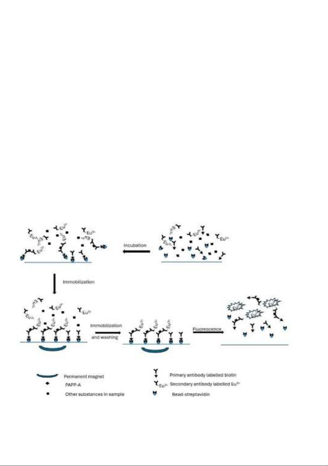
Abstract. The novel magnetic method used to immobilize the primary antibody in
96-well polystyrene plates was investigated to determine the pregnancy-associated
plasma protein-A (PAPP-A), one of the most important protein markers in
pregnancy. The optimal conditions for the immobilization are 20 μL magnetic
nanoparticles (0.72 mg/mL), 2μL primary antibody (30 μg/mL), and a one-step
process. The reaction was implemented in 40 minutes and 30 ºC. The calibration
curve was established and the linear range shows up to 2090 mU/L. The LOQ and
LOD are 7.2 and 24.0 mU/L, respectively. The reaction is non-specific for other
pregnancy hormones such as hCG and aFP even at a high level. The Passing &
Bablok regression showed the linear relationship and the agreement between new
and reference methods, y = a (95% CI)x + b(95% CI), a = 1.00 (0.971 to 1.019), b
= 22.07 (-73.104 to 117.244), R2 = 0.999, p<0.001. The Bland-Altman plot also
showed the high concordant. The new method can be used to determine PAPP-A in
the serum sample with mild conditions, simple and time-saving reaction, high
sensitivity and selectivity, and comparable results with the commercial method.
Keywords: PAPP-A, magnetic immobilization, one-step process.
I. Introduction
Pregnancy-associated plasma protein-A (PAPP-A) is a metalloprotease, produced
and secreted by the placental syncytiotrophoblast, the protein level increases from 5
weeks of gestation and continuously rises along with the age of the fetus. The main PAPP-
A function is to release insulin-like growth factor (IGF) from its binding protein (IGF-
binding protein). Therefore, the PAPP-A plays a critical role in placental invasion,
placental development, and maintenance of placental functions [1]-[3]. The level of
PAPP-A, especially in the first trimester was widely investigated and used as a biomarker
for many pregnancy complications. For instance, low serum PAPP-A is associated with
Nguyen BN*, Nguyen TKL, Ta VT & Bui TB
106
fetal growth restriction, fetal loss, intrauterine fetal demise, pre-eclampsia, gestational
diabetes, and T21 trisomy [1]-[2]. In addition, the low serum PAPP-A is also associated
with short stature in offspring and postpartum de-novo maternal diabetes mellitus [4].
Otherwise, the high serum PAPP-A level is related to a high risk of placenta accrete [5], [6].
A small quantity of PAPPA can be found in other tissues such as the breast, kidneys, bone
marrow, and colon, abnormal elevated PAPP-A serum in non-pregnancy is a marker for
relapse and prognostic clinical outcomes in malignant cancer such as breast cancer,
cardiac disease, and end-stage renal disease [2], [7]-[9].
Quantification PAPP-A is mainly based on the immunoassay. In the assay on 96-well
plates, the primary antibody is permanently immobilized by adsorption onto the well
surfaces [10]. The step is time-consuming and needs to be prepared before the
quantification reaction. This study aims to develop and validate the novel method for
primary antibody immobilization by magnetic to be used for immunoassay reaction
quantification of the PAPP-A in serum samples. In brief, the primary antibody is labelled
by biotin (Ab-biotin), and the secondary antibody, which has a different epitope compared
with the primary antibody is labelled with the fluorophore to obtain a fluorescent signal.
The primary antibodies, PAPP-As and secondary antibodies freely interact within the
solution. To wash non-specific binding, the immunocomplex is transiently immobilized
through the interaction of Ab-biotin and magnetic nanoparticles or beads labelled
streptavidin (MNP-S) in the presence of a magnet. Finally, the fluorescence intensity is
obtained to quantify the amount of PAPP-A in the samples (Figure 1).
Figure 1. The schematic of magnetic immobilization immunoassay reaction

Development and validation of the magnetic immobilization to determine PAPP-A in human serum
107
2. Content
2.1. Experiments
* Materials and equipment
Bead or magnetic nanoparticles-streptavidin (MNP-S) (0.72 mg/mL, Roche, USA),
primary antibody labelled biotin (Ab-biotin, 30 μg/mL, Perkin Elmer, Finland), secondary
antibody labelled europium (Ab-DTTA-Eu3+, Perkin Elmer, Finland), 1-(2-naphthoyl)-
3,3,3-trifluoroacetylacetone (NTAA) (Perkin Elmer, Finland), PAPP-A standard solutions
(0, 9.9, 39.7, 205, 820, 2090 mU/L) (Perkin Elmer, Finland), were within their expiration
date from … The standard 0 was a pseudo serum sample or blank sample. The washing
solution (0.9 % NaCl, 0.005% NaN3, 0.05% tween 20, 0.050M Tris-HCl, pH 7.8) was
freshly prepared before using. The first trimester pregnancy serum samples were provided
by Chemedic Vietnam JSC. with the permission of patients and were used only for
scientific purposes.
The equipment used included a permanent magnet (6x4x2 cm), a thermal incubator
(Perkin Elmer, Finland), a plate shaker incubator (Perkin Elmer, Finland), 96-well
polystyrene plates (Corning, USA), a fluorescence reader Victor-2D (Perkin Elmer, Finland).
* Development and validation method
- Development method:
Magnetic immobilization primary antibody. The procedure was investigated by two
methods.
In the one-step assay method, MNP-S, Ab-biotin, PAPP-A, and Ab-DTTA-Eu³⁺ were
simultaneously incubated in 96-well plates. In step 1, the reaction mixture, consisting of
20 μL of MNP-S, 2 μL of Ab-biotin, 10 μL of PAPP-A standard or sample, and 10 μL of
Ab-DTTA-Eu³⁺, was vortexed for 15 seconds and then incubated in a shaking thermos
reactor at 800 rpm. The concentrations of MNP-S and Ab-biotin, along with the
incubation time and temperature, were optimized for this reaction. In Step 2, the reaction
product was immobilized onto a permanent magnet for 3 minutes and washed three times
with 200 μL of washing solution in total. In step 3, NTAA solution was added to the
reaction well, and the fluorescence signal was measured within 5 minutes.
In the stepwise method, MNP-S and Ab-biotin were first incubated together (Step
1a). This was followed by immobilization using a permanent magnet, with subsequent
washing performed three times (Step 1b). Next, PAPP-A and Ab-DTTA-Eu^3+ were
added to the reaction, and the mixture was continuously incubated (Step 1c). Steps 2 and
3 were performed as described in the one-step method.
The reactions were performed with a blank sample and spiked samples at two PAPP-
A levels (250 and 2090 mU/L). The scheme of reaction is described in Figure 1.
The effect of time to reaction: The time of reaction was investigated in 20, 30, 40, 60,
and 120 minutes, other conditions are fixed.
The effect of temperature on reaction: The temperature incubation was investigated
at 20, 25, 30, and 35ºC, other conditions were fixed.
The effect of MNP-S concentration on reaction: The concentration of MNP-S was
investigated at 5, 10, 15, 20, and 25 μL, other conditions were fixed.
Nguyen BN*, Nguyen TKL, Ta VT & Bui TB
108
The effect of Ab-biotin concentration on reaction: The concentration of ab-biotin was
investigated at 2, 4, 6, 8, and 10 μL, other conditions were fixed.
All reactions were performed at the 2090 mU/L PAPP-A concentration.
- Validation method:
+ The repeatability: The reaction with 2090 mU/L PAPP-A concentration was
repeated five times to assess repeatability.
+ The specificity: The blank samples spiked with hCG, and AFP (9930 U/L and 1010
U/L, respectively) were measured to determine the reaction cross-activity.
+ The calibration curve instruction: Six standard samples (0 – 2090 mU/L) were
used to construct the calibration curve.
+ Low detection (LOD) and low quantification (LOQ) estimation: The LOD and
LOQ were estimated as the average intensity fluorescence signal plus 3 standard
deviations of 10 blanks and the average intensity fluorescence signal plus 10 standard
deviations of blanks, respectively.
Comparison with reference method: Twelve pregnancy serum samples were
measured using the new method and the reference method (PAPP-A DELFIA assay,
Perkin Elmer, Finland). The results obtained from the developed method were compared
with those from the reference method by using the Passing & Bablok regression
(y = a (95% CI)x + b(95% CI)) to determine the correlation and linear regression. The
Bland-Altman plot describes the agreement between the two methods [11].
2.2. Results and discussion
* Development method
- Magnetic immobilization primary antibody:
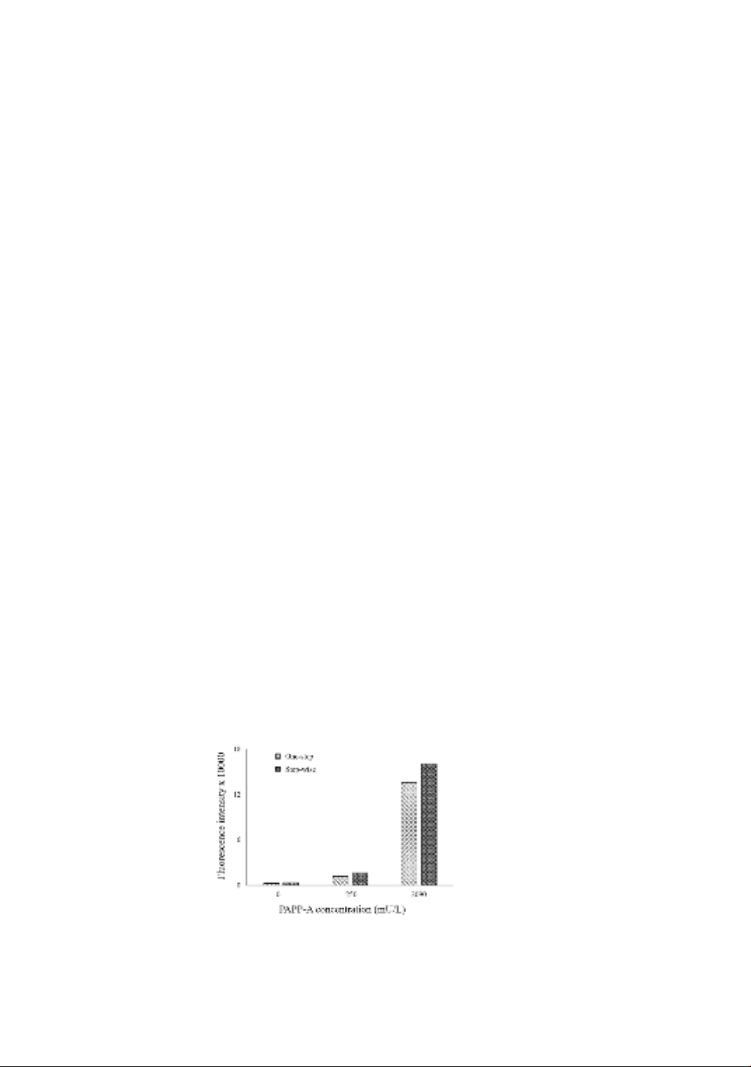
With the blank sample, at the 0 mU/L concentration of PAPP-A, the fluorescence
signal of the one-step method is slightly lower than the step-wise method. However, at
205 and 2090 mU/L concentrations, the one-step method is higher than the step-wise
method (Figure 2). The immobilization efficiency is a critical factor in the reaction, as it
directly influences the ability to capture PAPP-A in the sample. The results demonstrated
that the one-step method exhibited lower background noise and higher fluorescence
signals at both low and high concentrations. Furthermore, the one-step procedure is
simpler and more time-efficient compared to traditional or alternative enzyme-linked
immunosorbent assays (ELISA), which typically involve three steps and the use of pre-
coated primary antibodies.
Figure 2. The one-step and step-wise fluorescence signal
in different levels of PAPP-A
Development and validation of the magnetic immobilization to determine PAPP-A in human serum
109
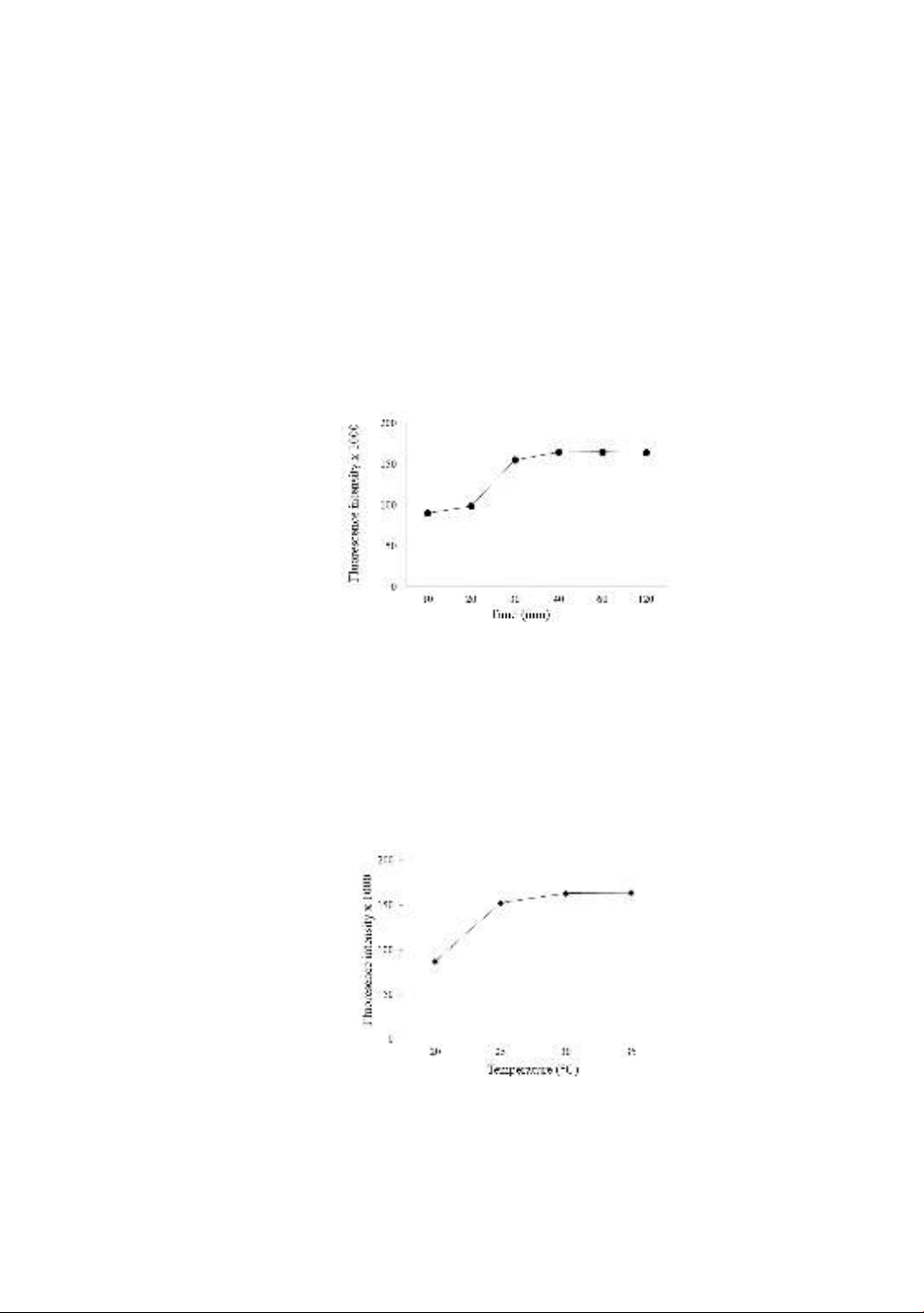
- The effect of time on the reaction:
The incubation time can affect the efficiency of binding immunocomplex. The
fluorescence signal slightly increases when incubation time increases from 10 to 20
minutes, however, significantly increases at 30 minutes and continuously slightly
increases at 40 minutes. The fluorescence signal remains unchanged and evenly extends
the incubation time to 120 minutes (Figure 3). The incubation time of 40 minutes is
appropriate for further reaction experiments. Instead of permanently immobilizing
primary antibodies on solid surfaces to form immunocomplexes, allowing free reactions
between primary antibodies, antigens, and secondary antibodies in solution offers
significant time advantages. This approach results in a reaction time considerably shorter
than that of traditional or alternative ELISA immunoassays, which typically took 1.5 to 2
hours, or even up to 3 hours. [12].
Figure 3. The effect of incubation time on fluorescence signal
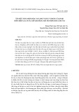
- The effect of temperature on the reaction:
The increased incubation reaction can accelerate the reaction rate. However, high
temperatures may cause instability in the protein and antibodies. The fluorescence signal
increases significantly when incubated from 20 ºC to 25 ºC and slightly increases
(approximately 7%) at 30 ºC and remains the intensity signal until 35 ºC (Figure 4). The
optimal temperature can be archived at 30 ºC. This range temperature is also widely used
on the immunoassay such as traditional or alternative ELISA immunoassay,
electrochemical luminescence, or chemiluminescent microparticle immunoassay [13].
Figure 4. The effect of temperature on the fluorescence signal
- The effect of MNP-S concentration on reaction:
The concentration of beads, when directly immobilized in the magnetic field, can
significantly affect the fluorescence intensity. The signal increases with the concentration


![Bài giảng Cộng hưởng từ EPR - NMR: Tổng hợp kiến thức [mới nhất]](https://cdn.tailieu.vn/images/document/thumbnail/2016/20160917/maiyeumaiyeu10/135x160/861474077701.jpg)


![Các công cụ kỹ thuật [mới nhất]](https://cdn.tailieu.vn/images/document/thumbnail/2011/20111014/cvbn123/135x160/cac_cong_cu_ky_thuat_378.jpg)
![Tài liệu giảng dạy Sinh học và di truyền [mới nhất]](https://cdn.tailieu.vn/images/document/thumbnail/2026/20260323/hoatudang2026/135x160/42181774414220.jpg)

![Giáo trình Công nghệ vi sinh (Nghề Công nghệ sinh học TC/CĐ) - Trường Cao đẳng Đà Lạt [Mới nhất]](https://cdn.tailieu.vn/images/document/thumbnail/2026/20260224/hoacattuong2026/135x160/87621772161812.jpg)


![Giáo trình Vi sinh vật học môi trường Phần 1: [Thêm thông tin chi tiết nếu có để tối ưu SEO]](https://cdn.tailieu.vn/images/document/thumbnail/2025/20251015/khanhchi0906/135x160/45461768548101.jpg)


