JOURNAL OF MILITARY PHARMACO-MEDICINE N04 - 2025
199
A CASE REPORT: CARCINOMA AFTER TRAUMATIC BRAIN INJURY
CAUSED BY INCENDIARY WEAPONS
Nguyen Xuan Phuong1, Nguyen Hai An2, Tran Manh Cuong1*
Abstract
Carcinoma in patients with traumatic brain injury having retained metal foreign
bodies does not commonly exist. Complications of local cancer by traumatic brain
injury, having retained metal foreign bodies can occasionally occur. Changes
mainly appear at the wound due to chronic inflammation and cell hyperplasia,
which leads to cancer. The clinical case the authors introduce is a patient
participating in the Resistance War for National Salvation who was shot by
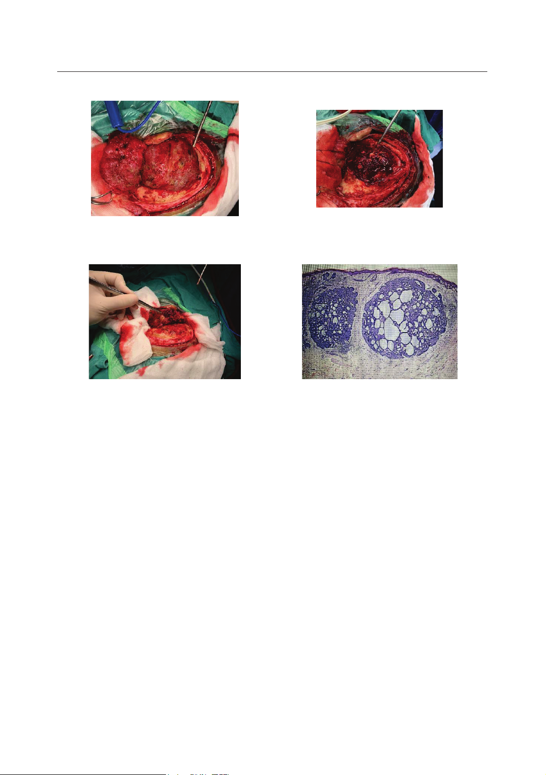
artillery pieces in the right temporal. The patient was admitted to the hospital and
had experienced surgery to remove a tumor from the right temporal. The
histopathology of the tumor was carcinoma. After surgery, the patient was awake,
the incision recovered well, and the patient could live normally.
Keywords: Carcinoma; Traumatic brain injury; Surgery.
INTRODUCTION
Cancer in chronic wounds was first
described by Jean-Nicolas Marjolin in
1828. Especially wounds with retained
foreign bodies in the head and neck area
can result in carcinoma, making up to
80%[1]. Early surgery to remove foreign
bodies is needed to prevent carcinoma
at the place. The authors present a rare
case with the aim to: Evaluate the status
of in situ cancer in a patient with a
metal foreign body from a resistance
war wound many years ago.
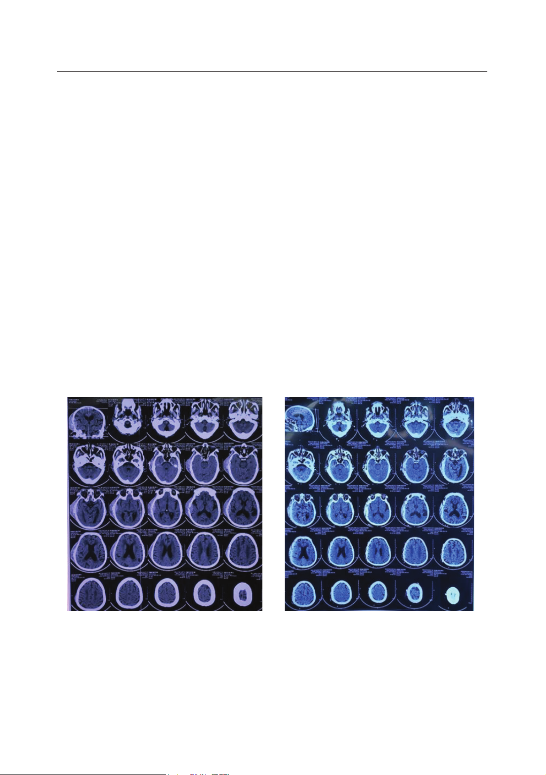
CASE REPORT
Patient Pham Van P, male, 72 years
old, was presented to the hospital due to
pain and swelling in his right temple
and a mild fever of 37.4°C. The patient
participated in the resistance war in
1972 and was hit by shell fragments in
the right temple area.
1Neurosurgery Department, Military Hospital 103, Vietnam Military Medical University
2War Surgery Department, Military Hospital 103, Vietnam Military Medical University
*Corresponding author: Tran Manh Cuong (tmcuongpttk@gmail.com)
Date received: 27/8/2024
Date accepted: 29/10/2024
http://doi.org/10.56535/jmpm.v50i4.994
JOURNAL OF MILITARY PHARMACO-MEDICINE N04 - 2025
200
Since then, the patient has been living
with a foreign metal fragment under the
scalp in the right temple area. The
wound is swelling, recurring many
times, and gradually getting bigger. The
patient had sleep disorders, dizziness,
and weight loss (lost 5kg/2 months).
He was admitted to the hospital with
a Glasgow score of 15 points, felt
pain in the right temporal region, had
no paralysis, no epilepsy, and no
meningeal syndrome. In the right
temple area, there is a shiny mass under
the scalp, about 8 x 10cm in size. A
computed tomography scan of the brain
shows the parenchymal window with
oval heterogeneous hyperdense mass
including the subcutaneous area, skull,
and the subscapular area adjacent to
dura mater, bone window with the
image of skull bone destruction in the
tumor area, metal foreign body image
under the skin in the tumor area. The
patient underwent surgery to remove
the tumor, skull bone, and invaded
dura mater. Dural reconstruction
with artificial dura mater and
cranioplasty with titanium brain mesh.
After surgery, the patient had no
complications, and the incision
recovered well. The patient was
recharged from the hospital and lives
normally. Histopathology results: Basal
cell carcinoma.
Figure 1. Preoperative computed
tomography image of the brain
-
metal
foreign body under the skin
of the right temple.
Figure 2. Post-operative computed
tomography image of the brain
-
the
tumor in the right temporal region
was completely removed.
JOURNAL OF MILITARY PHARMACO-MEDICINE N04 - 2025
201
Figure 3. Exposing tumor.
Figure 4. Tumor invades the cerebral
dura mater.
Figure 5. Tumor removal.
Figure 6. Histopathology.
DISCUSSION
Wounds resulting from retained
foreign objects are common in Vietnam.
Foreign bodies left on the body after
war can be anywhere-under the skin or
in the brain parenchyma or lung
parenchyma. Metal foreign bodies in
the head area can be in the brain
parenchyma, skull, or under the scalp.
We can meet patients who still have
metal foreign bodies in their bodies and
live peacefully with these. However,
authors around the world have mentioned
that cancer is due to lingering foreign
objects in the wound. Verifying the
relationship between cancer and lasting
foreign body injuries is still difficult.
Whether the retained metal foreign
body is a direct cause of carcinoma
remains controversial. However, some
hypotheses have been proposed that
the poor vascularity of the scar
tissue facilitates the development of
ulceration in the thin epidermis,
making it more susceptible to changes
and infection. The retained foreign
body will result in changes in
epithelial cells during the regeneration
process. Dysplasia will occur when
the repair process is repeated in
chronic wounds, leading to carcinoma
in local tissue [2, 3, 4].
JOURNAL OF MILITARY PHARMACO-MEDICINE N04 - 2025
202
The location of the wound by a
foreign body is also a crucial factor
resulting in carcinoma. The authors
believe that location exposure to
sunlight will increase the risk of the
disease [4, 5]. Ozyazgan I [6] studied
92 patients with basal cell carcinoma,
which is common in the elderly, poor
wound healing, and exposed skin
locations. These are all related to
ultraviolet rays and become factors
increasing the risk of cancer. Our
patient suffered a metal foreign body
wound under the scalp in the right
temple area. As this is a location that is
less covered and usually exposed to
sunlight, it will raise the risk of cancer
at the wound site.
Wozniak SE describes a patient with
a war-related foreign body wound in his
hand that had complications causing
basal cell carcinoma. The authors
believe that these wounds are related to
the Marjolin degeneration process of
epithelial cells and cause carcinoma in
the long term [2]. Ebrahimzadeh (2013)
described a case of a 44-year-old
male patient with a metal fragment in
the lower end of the femur while
participating in the Iran-Iraq war,
which, after 22 years, caused local bone
cancer without any special clinical
manifestations. The patient was
examined periodically, and found a
bone-destroying lesion and the biopsy
results showed local bone cancer. The
patient underwent surgery to remove
the tumor and replace the joint. The
authors believe that the cause may be
metal or lead poisoning which resulted
in changes in the cells around the
foreign body [6]. Our patient suffered
from a fire injury for 50 years - long
enough for the wound to change,
causing local carcinoma.
CONCLUSION
Wounds retaining foreign objects
can be found anywhere on the body.
Foreign metal objects under the skin
can cause carcinoma. Surgery to remove
foreign bodies is necessary, especially
those in skin areas exposed to sunlight.
Surgery to remove foreign bodies prevents
cancer and prolongs the patient's life.
Ethics: Research strictly complies
with the regulations on research ethics
of the Ministry of Health. Patients were
on operations following the surgical
procedures of Military Hospital 103 and
the Ministry of Health. The patient's
database has been approved for use and
publication by Military Hospital 103.
The authors commit that the research is
carried out objectively and without
conflicts of interest.
JOURNAL OF MILITARY PHARMACO-MEDICINE N04 - 2025
203
REFERENCES
1. Noodleman FR, Pollack SV.
Trauma as a possible etiologic factor in
basal cell carcinoma. J Dermatol Surg
Oncol. 1986; 12:841-846.
2. Wozniak SE, Zuo S, Khan K,
Coleman J. Old war scar revisited.
J Am Coll Clin Wound Spec. 2016 Oct 3;
7(1-3):35-39. DOI: 10.1016/j.jccw.
2016.09.001. PMID: 28053867; PMCID:
PMC5197046.
3. Koga Y, Sawada Y. Basal cell
carcinoma developing on a burn scar.
Burns. 1997 Feb; 23(1):75-77. DOI:
10.1016/s0305-4179(96)00077-0. PMID:
9115617.
4. Kandamany N, Monk B. Basal cell
carcinoma presenting late in a shotgun
scar. Am J Clin Dermatol. 2009;
10(4):271-272. DOI: 10.2165/00128071-
200910040-00009. PMID: 19489662.
5. Ozyazgan I, Kontaş O. Previous
injuries or scars as risk factors for the
development of basal cell carcinoma.
Scand J Plast Reconstr Surg Hand
Surg. 2004; 38(1):11-15. DOI: 10.1080/
02844310310005883. PMID: 15074717.
6. Ebrahimzadeh MH, Vahedi E,
Ganji R, Bozorgnia S. Skeletal sarcoma
on the site of retained war bullet
fragments and a literature review on
long-term complications of retained
war shells. Arch Bone Jt Surg. 2013
Dec; 1(2):107-111. Epub 2013 Dec 15.
PMID: 25207300; PMCID: PMC4151411.