JOURNAL OF MILITARY PHARMACO-MEDICINE N04 - 2025
51
A RARE CASE REPORT: LUNG ADENOCARCINOMA
WITH DIFFUSE LESIONS IN A YOUNG MALE PATIENT
Dao Ngoc Bang1*, Ta Ba Thang1, Nguyen Tien Dung1
Abstract
The images of diffuse lung involvement are seen in various respiratory diseases,
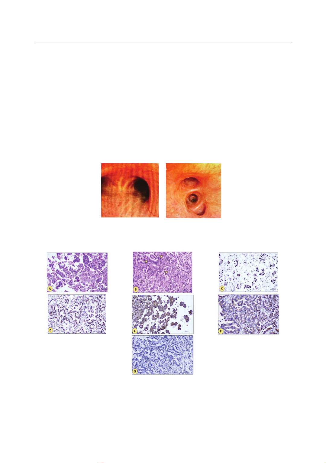
including lung adenocarcinoma (ADC). After ruling out acute infectious causes,
lung biopsy is valuable for a definitive diagnosis, with transbronchial biopsy via
flexible bronchoscopy being an effective and safe diagnostic approach. However,
the widespread lung damage in these patients poses a challenge for transbronchial
biopsy. Chemotherapy is often difficult due to the overall poor health of the patient.
Targeted therapies, specifically tyrosine kinase inhibitors (TKIs), have shown
efficacy in lung cancer treatment.
Keywords: Adenocarcinoma; Lung cancer; Tyrosine kinase inhibitors (TKIs).
INTRODUCTION
Lung cancer is a malignancy with
increasing incidence and mortality rates.
Its slow progression and nonspecific
clinical symptoms contribute to low
early-stage diagnosis rates, often leading
to misdiagnosis or confusion with other
respiratory diseases, especially in cases
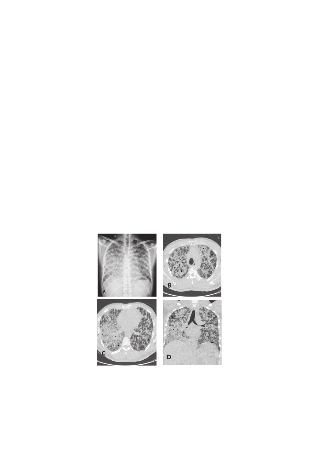
with atypical X-ray or computed
tomography (CT) findings. ADC with
diffuse lesions (named bronchioloalveolar
carcinoma - BAC before) is rare in
clinical practice in Vietnam, making
diagnosis challenging [1, 2, 3]. In
patients with epidermal growth factor
receptor (EGFR) mutation and low PS
[3, 4], TKIs are the first choice for
treatment, in which Afatinib is a
suitable indication for ADC with a
G719x mutation in exon 18. With this
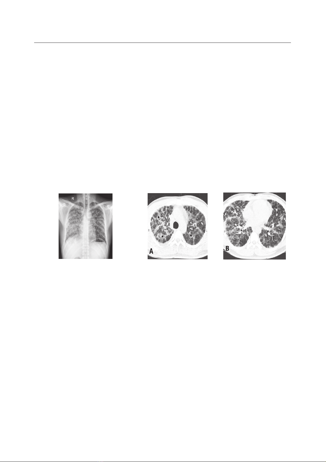
case study, we would like to: Present
the clinical characteristics of a rapidly
progressed ADC case with diffuse
lesions, having G719x mutation in exon
18, and the results of treatment by
Afatinib as first-line therapy.
1Respiratory Medicine Center, Military Hospital 103, Vietnam Military Medical University
*Corresponding author: Dao Ngoc Bang (bsdaongocbang@gmail.com)
Date received: 08/7/2024
Date accepted: 17/9/2024
http://doi.org/10.56535/jmpm.v50i4.896
JOURNAL OF MILITARY PHARMACO-MEDICINE N04 - 2025
52
CASE REPORT
Male patient, 39 years old, non-
smoker, no history of tobacco use, no
exposure to cancer risk factors, and no
family history of cancer. Occupation:
Military officer. The symptoms have
been present for about 4 months,
including occasional cough with
minimal sputum, dull chest pain, mild
progressive dyspnea, and mild fever.
The patient was admitted to the Internal
Department of a regional general
hospital, where chest X-rays, chest CT,
acid-fast bacillus (AFB) testing, and
GeneXpert/MTB testing of negative
sputum were conducted. Following
consultation, the patient was diagnosed
with miliary tuberculosis and received a
2RHZE/6RH regimen. After 3 weeks of
tuberculosis treatment, the patient's
symptoms were unresponsive, and
respiratory distress worsened, leading
to admission to the Respiratory Medicine
Center, Military Hospital 103. On
admission, the patient exhibited signs of
respiratory failure (persistent dyspnea,
cyanosis, respiratory muscle retractions;
SpO2 80%), no fever, no palpable
peripheral lymph nodes, and reduced
breath sounds in both lungs without
rales. Imaging findings on chest X-ray
and CT revealed hazy opacities
interspersed with diffuse reticular
patterns in both lungs, concentrated
in the mid and lower zones of both
lungs, with no hilar lymphadenopathy,
involvement of the mediastinum, or
pleural effusion (Figure 1).
Figure 1. Initial chest X-ray (A) demonstrates bilateral diffuse reticular opacities
with consolidation in the right lung predominant. Axial CT scan (B, C)
and coronal reconstructed CT image (D) showed bilateral extensive septal
thickening and reticular opacities (arrowhead) with consolidation (star).
JOURNAL OF MILITARY PHARMACO-MEDICINE N04 - 2025
53
Arterial blood gas (ABG) results:
PaO2 = 57 mmHg, PaCO2 = 37 mmHg,
pH = 7.38. The patient underwent treatment
for respiratory failure (high-flow nasal
cannula oxygen therapy - HFNC),
symptom management, and bronchoscopy.
Bronchoscopy images show normal
findings with no increased mucus secretion
(Figure 2). Selective bronchial lavage
technique is applied, collecting bronchial
lavage fluid for microbiological and
cytological examinations, and performing
transbronchial biopsy for pathological
examination. Pathological examination
reveals papillary ADC of the lung;
EGFR mutation testing indicates a
G719x mutation on exon 18 (Figure 3).
Microbiological tests, including AFB,
GeneXpert/MTB-Rif, bacterial and
fungal cultures of bronchial fluid, all
yield negative results. The patient
undergoes additional diagnostic imaging
tests (abdominal ultrasound, cranial
MRI) for staging.
A B
Figure 2. Bronchoscopy did not detect lesions in the main bronchus (A)
and segmental bronchus (B), so a transbronchial biopsy was indicated.
Figure 3. Initial chest X-ray (A) demonstrates bilateral diffuse reticular opacities
with consolidation in the right lung predominant. Axial CT scan (B, C)
and coronal reconstructed CT image (D) showed bilateral extensive septal
thickening and reticular opacities (arrowhead) with consolidation (star).
JOURNAL OF MILITARY PHARMACO-MEDICINE N04 - 2025
54
The patient is diagnosed with non-
mucinous ADC of the lung, stage IV
(T1miN0M1), with EGFR mutation, and
respiratory failure as a complication.
The patient is reviewed by a
multidisciplinary team and opts for
treatment with second-generation TKI:
Afatinib at a dose of 40 mg/day, taken 1
hour before lunch. Response assessment
after 1 month indicates a partial response:
Significant reduction in cough, sputum,
and chest pain; weight gain (BMI
21.3 kg/m2), no need for supplemental
oxygen, and improved ABG (PaO2 = 87
mmHg, PaCO2 = 38 mmHg). Chest X-
ray images show a partial reduction in
lung lesions after 10 days of treatment
(Figure 4), and CT scans demonstrate a
noticeable decrease in the extent of
spread compared to the patient's initial
presentation (Figure 5). Mild adverse
effects of TKIs are observed (mild skin
rash). After 6 months, the patient has
signs of disease progression. Afatinib
should be replaced by Osimertinib as a
recommendation of NCCN [7].
Figure 5. CT scan obtained 15 days after
treatment showed a decrease in consolidation,
but we still saw bilateral septal thickening, and
reticular opacities (arrow) with ground glass
opacities (star). (A) Axial section of apex
(B) Axial section of basal.
DISCUSSION
1. Diagnosis of diffuse lung ADC
Diffuse lung ADC is a type found at
a rate of 3 - 6% of all types of lung
cancer. It is more commonly found in
young, non-smoking females. However,
this type is rare in Vietnam. Key clinical
symptoms include chronic cough,
progressively increased difficulty in
breathing and a significant amount of
coughed-up sputum. In patients with
localized lesions visible on X-rays,
clinical symptoms may not be apparent,
and the diagnosis relies primarily on the
JOURNAL OF MILITARY PHARMACO-MEDICINE N04 - 2025
55
technique of invasive specimen collection
for pathological examination. Patients
with rare cases of bilateral spread of
lesions present more significant diagnostic
challenges, especially in late-stage
patients with complicated respiratory
failure, making it challenging to perform
diagnostic specimen collection techniques.
The choice of biopsy procedures
depends on the overall condition of the
patient, the characteristics of lung
lesions observed on X-rays, and chest
CT scans. Treatment options are limited
due to the extensive spread of lung
damage, and the patient's overall health
index is compromised due to respiratory
failure [3, 4]. Moreover, when imaging
reveals lesion patterns such as nodules
or a diffuse mesh spreading across both
lungs, differential diagnosis is crucial to
distinguish from various respiratory
infections (disseminated tuberculosis,
mycobacteria infection, fungi), non-
infectious diffuse lung diseases
(sarcoidosis, hypersensitivity pneumonitis,
pneumoconiosis), and malignant
conditions (secondary lung cancer,
Kaposi's sarcoma) (Table 1).
Table 1. Causes of diffuse pulmonary nodules.
Infections
Diffuse lung diseases
Malignancies
Miliary tuberculosis
Hypersensitivity pneumonitis
Metastasis
Atypical mycobacterial
infection
Diffuse pulmonary
meningotheliomatosis
Diffuse lung ADC
Lung fungal infection
Pneumoconiosis
(namely silicosis)
Lymphangitic
carcinomatosis
Septic emboli
Amyloidosis
Kaposi sarcoma
In the clinical case of a young patient
with no risk factors for lung cancer,
presenting minimal clinical symptoms,
mainly gradual onset of mild
breathlessness and occasional mild
fever, the initial differential diagnosis
considered is subacute disseminated
pulmonary tuberculosis. This is a
chronic infectious lung disease with
widespread lesions commonly seen in
Vietnam. Simultaneously, the second
differential diagnosis raises the possibility
of a systemic disease, a group of
conditions often found in young
individuals. Microbiological tests for
tuberculosis, including AFB, and