Crystal structure of a subtilisin-like serine proteinase from
a psychrotrophic Vibrio species reveals structural aspects
of cold adaptation
Jo
´hanna Arno
´rsdo
´ttir
1
, Magnu
´s M. Kristja
´nsson
2
and Ralf Ficner
1
1 Abteilung fu
¨r Molekulare Strukturbiologie, Institut fu
¨r Mikrobiologie und Genetik, Georg-August Universita
¨tGo
¨ttingen, Germany
2 Department of Biochemistry, Science Institute, University of Iceland, Reykjavı
´k, Iceland
Microorganisms inhabit the most diverse environments
on earth. Extremophiles are microorganisms that have
adapted to environmental conditions regarded by
humans as falling outside the normal range in terms of
temperature, pressure, salinity or pH. Extremophiles
have had to develop strategies to deal with environ-
mental stress, mainly by molecular adaptation of their
cell inventory. Of major importance in adapting to
extreme environmental conditions is the optimization
of protein function and stability. Enzymes from
extremophiles are essentially like their mesophilic
counterparts, sharing the same overall fold and
catalysing identical reactions via the same mechanisms,
while having adopted different traits regarding kinetic
and structural properties. Therefore, they provide
excellent tools for examining the molecular basis of
different protein properties, as well as the relation
between structure and function in enzymes. Regarding
temperature, organisms have been isolated from places
with temperatures as high as 113 C [1] and biological
activity has been detected in microbial samples at tem-
peratures as low as )20 C [2]. Thermo- and hyper-
thermophiles face the challenge of keeping their
macromolecules functional under the environmental
Keywords
cold adaptation; crystal structure;
psychrotrophic; subtilase; thermostability
Correspondence
R. Ficner, Abteilung fu
¨r Molekulare
Strukturbiologie, Institut fu
¨r Mikrobiologie
und Genetik, Universita
¨tGo
¨ttingen, Justus-
von-Liebig-Weg11, 37077 Go
¨ttingen,
Germany
Fax: +49 551 391 4082
Tel: +49 551 391 4072
E-mail: rficner@gwdg.de
Database
The coordinates and structure factors for
the final structure of Vibrio proteinase at
1.84 A
˚resolution have been deposited in
the Protein Data Bank under the accession
number 1SH7.
(Received 30 September 2004, revised 26
November 2004, accepted 9 December
2004)
doi:10.1111/j.1742-4658.2005.04523.x
The crystal structure of a subtilisin-like serine proteinase from the psychro-
trophic marine bacterium, Vibrio sp. PA-44, was solved by means of
molecular replacement and refined at 1.84 A
˚. This is the first structure of a
cold-adapted subtilase to be determined and its elucidation facilitates
examination of the molecular principles underlying temperature adaptation
in enzymes. The cold-adapted Vibrio proteinase was compared with known
three-dimensional structures of homologous enzymes of meso- and thermo-
philic origin, proteinase K and thermitase, to which it has high structural
resemblance. The main structural features emerging as plausible determi-
nants of temperature adaptation in the enzymes compared involve the char-
acter of their exposed and buried surfaces, which may be related to
temperature-dependent variation in the physical properties of water. Thus,
the hydrophobic effect is found to play a significant role in the structural
stability of the meso- and thermophile enzymes, whereas the cold-adapted
enzyme has more of its apolar surface exposed. In addition, the cold-adap-
ted Vibrio proteinase is distinguished from the more stable enzymes by its
strong anionic character arising from the high occurrence of uncompen-
sated negatively charged residues at its surface. Interestingly, both the cold-
adapted and thermophile proteinases differ from the mesophile enzyme in
having more extensive hydrogen- and ion pair interactions in their struc-
tures; this supports suggestions of a dual role of electrostatic interactions
in the adaptation of enzymes to both high and low temperatures. The
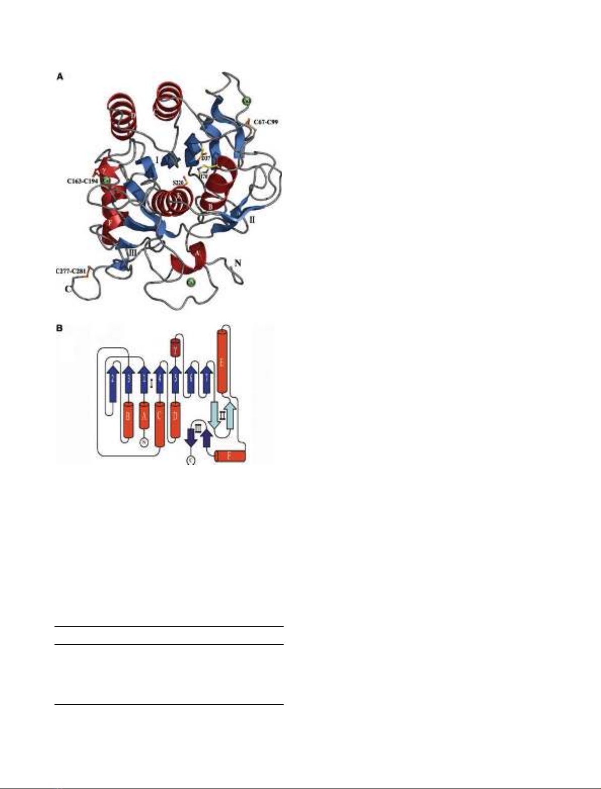
Vibrio proteinase has three calcium ions associated with its structure, one
of which is in a calcium-binding site not described in other subtilases.
832 FEBS Journal 272 (2005) 832–845 ª2005 FEBS
stress imposed by extreme thermal motion. As a
response, they have evolved enzymes that are highly
stable against heat and other denaturants. The
increased stability of enzymes from thermo- and hyper-
thermophiles is considered to reflect structural rigidity,
which in turn would account for their poor catalytic
efficiency at low temperatures. The properties of ther-
mophilic enzymes have aroused great interest as they
have potential in biotechnology and diverse industrial
processes [3,4]. In addition, the production of thermo-
philic recombinant enzymes is facilitated by their relat-
ively straightforward overexpression and purification,
which makes them feasible candidates for various bio-
chemical experiments as well as for crystal structure
determination. These factors have enhanced research
on thermostability, which has been studied extensively
in the past, mainly by comparing the structural proper-
ties of thermo- and mesophilic enzymes, as well as
by using mutagenic experiments [5]. In contrast to
enzymes from thermophiles, cold-adapted enzymes are
relatively poorly examined, in particular considering
their extensive distribution and occurrence in our bio-
sphere. Organisms occupying permanently cold areas
that dominate the earth’s surface, collectively called
psychrophiles, have to rely on enzymes that can com-
pensate for low reaction rates at their physiological
temperatures. The properties that characterize and dis-
tinguish cold-adapted enzymes from enzymes origin-
ating at higher temperatures are their increased
turnover rate (k
cat
) and inherent higher catalytic effi-
ciency (k
cat
K
m
) at low temperatures [6]. It is assumed
that optimization of the catalytic parameters in cold-
adapted enzymes is accomplished by developing
increased structural flexibility, allowing the conforma-
tional changes required for catalysis at low tempera-
tures [7]. In recent years, a few crystal structures of
cold-adapted enzymes have been determined [8–16].
These structures have served as a basis in comparative
studies on structural aspects of cold adaptation. Also,
information from site-directed mutagenesis experi-
ments, homology modelling and directed evolution has
been used in an effort to shed light on the molecular
principles underlying the adaptation of enzymes to low
temperatures [17–24]. In general, regardless of whether
research is directed at thermo- or psychrophilic adap-
tation, the results show that each protein family adopts
its own strategies for coping at extreme temperatures.
Although no general rules have been found to apply in
temperature adaptation in enzymes, some structural
tendencies have emerged. The most frequently reported
features related to temperature adaptation, going from
higher to lower temperatures, are a reduced number of
noncovalent intra- and intermolecular interactions, less
compact packing of the hydrophobic core, an
increased apolar surface area, decreased metal ion
affinity, longer surface loops and a reduced number of
prolines in loops [5,6,8,25–28]. In general, in naturally
occurring enzymes, a correlation is seen between cata-
lytic efficiency at low temperatures and susceptibility
to heat and other denaturants [29]. However, using
directed evolution methods, mutants have been
obtained with changes in one of the properties, stabil-
ity or catalytic efficiency, indicating that these pro-
perties are not essentially interlinked [22,23]. The
observed instability of cold-adapted enzymes is regar-
ded not as a selected for property, but rather as a
consequence of the reduction in stabilizing features
arising from the need for increased flexibility to main-
tain catalytic efficiency at low temperatures [30].
Structural flexibility in cold-adapted enzymes is, as
yet, a poorly defined term for which little direct experi-
mental evidence is available. Attempts to assess and
compare the structural flexibility of a psychrophilic
a-amylase and more thermostable homologues using
dynamic fluorescence quenching supported the idea of
an inverse correlation between protein stability and
structural flexibility [31]. Comparisons of hydrogen–
deuterium exchange rates as a way of estimating flexi-
bility in enzymes originating at different temperatures
[32] have supported the idea of ‘corresponding states’
[33], which assumes that, at their physiological temper-
atures, enzymes possess comparable flexibility and a
structural stability adequate to maintain their active
conformation.
In order to improve the understanding of the struc-
tural principles of temperature adaptation we studied a
subtilisin-like serine proteinase from the psychrotrophic
marine bacterium, Vibrio sp. PA-44. The Vibrio prote-
inase belongs to the proteinase K family and has a high
sequence identity of 60–87% with several meso- and
thermophilic family members [34]. Furthermore, it has
41% sequence identity and 57% similarity with protein-
ase K, the best characterized representative of this pro-
tein family, the three-dimensional structure of which
has been determined to atomic resolution [35]. The
Vibrio proteinase has been identified as showing clear
cold-adaptive traits in comparison with its meso- and
thermophilic homologues [36]. Thorough sequence and
computer model comparisons performed on the Vibrio
proteinase and its most closely related meso- and
thermophilic enzymes have revealed some differences,
possibly relevant to temperature adaptation [34]. The
results have given rise to ongoing mutagenic research in
which single and combined amino acid substitutions
aimed at increasing the stability of the Vibrio protein-
ase are being tested. Elucidation of the Vibrio protein-
J. Arno
´rsdo
´ttir et al. Structural aspects of cold adaptation
FEBS Journal 272 (2005) 832–845 ª2005 FEBS 833
ase structure, the first structure of a cold-adapted subti-
lase to be determined, enables a more focused examina-
tion of plausible determinants of different temperature
adaptation among subtilases.
We crystallized the cold-adapted Vibrio proteinase in
the presence of bound inhibitor, phenyl-methyl-sulfo-
nate, and the structure was refined at 1.84 A
˚resolu-
tion. In order to identify parameters that might be
important with respect to cold adaptation we analysed
and compared structural features in Vibrio proteinase
and the two most closely related enzymes of known
three-dimensional structure, proteinase K from the
mesophilic fungi Tritirachium album Limber and thermi-
tase from the thermophilic eubacterium Thermoactino-
mycetes vulgaris.
Results
The crystal structure of the Vibrio proteinase
The obtained Vibrio proteinase crystals formed clusters
of needles, which transformed into thin platelets within
a few days. The crystals belong to space group P2
1
with
unit cell dimensions of a ¼43.2 A
˚,b¼36.9 A
˚,c¼
140.5 A
˚and b¼97.8. The Matthews coefficient [37]
(V
m
¼1.9 A
˚
3
Da) suggested two molecules in the
asymmetric unit with a solvent content of 36.3%. The
structure was determined by molecular replacement
using a homology model based on the known structure
of proteinase K (PDB accession number, 1IC6) as a
search model. The crystallized 30 kDa catalytic domain
of Vibrio proteinase encompasses amino acids 140–420
of the 530 amino acid prepro-enzyme [34]. The model
was refined at a resolution of 1.84 A
˚with an R-factor
of 14.1% and an R
free
value of 19.6% (Table 1).
Figure 1 shows the three-dimensional structure of
Vibrio proteinase, hereafter referred to as 1SH7
according to its PDB accession number. The structure
shows the abscaffold characteristic of subtilisin-like
serine proteinases. It consists of six ahelices, one
310 helix, a bsheet made of seven parallel strands
and two bsheets made of two antiparallel strands
(Fig. 1B). Determination of the structure confirms the
presence of three previously predicted disulfide bonds,
Cys67–Cys99, Cys163–Cys194 and Cys277–Cys281
[34]. Three calcium-binding sites are found in 1SH7,
two of which were predicted based on sequence align-
ments and one as yet not described in other subtilases.
The active site of 1SH7 consists of the catalytic triad
Asp37, His70 and Ser220, and substrate recognition
and binding sites that are well conserved among subti-
lases [38]. The substrate-binding site in 1SH7 appears
on the surface as a relatively distinct cleft (see below,
‘Surface properties and packing’) in which the sub-
strate is accommodated by forming a triple-stranded
antiparallel bsheet with residues of the S4- and S3-
binding sites (nomenclature of subsites, S4–S2¢,is
according to Schechter and Berger [39]). The bottom
of the S1 substrate-binding pocket is made up of resi-
dues A154–A155–G156 and the oxyanion hole residue
N157. The substrate-binding cleft appears to be relat-
ively open with T105 at the rim of S4; in many subti-
lases this site is occupied by a larger residue, typically
a tyrosine (e.g. subtilisin and proteinase K), which is
assumed to form a flexible lid on the S4 pocket [40].
Overall structure comparison with related
enzymes from meso- and thermophiles
A 0.98 A
˚resolution structure of proteinase K (PDB
accession number 1IC6) and a 1.37 A
˚resolution struc-
ture of thermitase (PDB accession number 1THM),
were used for structural comparison with 1SH7. The
high resolution of all three structures allows reasonable
comparison with respect to the quality of the models.
Pairwise least square superposition of the three
Table 1. Data collection and refinement statistics for 1SH7. Num-
bers in parenthesis refer to the highest resolution shell.
Data collection
Resolution range (A
˚) 40–1.81 (1.87–1.81)
Space group P2
1
Unit cell parameters
a¼43.2 A
˚
b¼36.9 A
˚
c¼140.5 A
˚
?¼97.80
Number of reflections 135,690
Unique reflections 37,893
Completeness (%) 93.2 (50.4)
R
syma
(%) 9.3 (50.3)
Average I r13.0 (2.7)
Refinement statistics
Resolution range (A
˚) 30–1.84 (1.88–1.84)
R
cryst
R
freeb
(%) 14.1(22.6) 19.6(29.8)
Rms deviation from ideality
Bonds (A
˚)angles () 0.014 1.521
Average B-values (A
˚
2
)
Protein water PMSF Ca
2+
13.3 25.4 34.1 11.9
Ramachandran plot
c
Most favoured, additional,
generously allowed (%)
89.9 9.9 0.2
a
R
sym
¼100ÆS
h
S
i
|I
i
(h) < I(h) > | ⁄S
h
I(h), where I
i
(h) is the ith meas-
urement of the h reflection and < I(h) > is the average value of the
reflection intensity.
b
R
cryst
¼S|F
o
F
c
|⁄S |F
o
|, where F
o
and F
c
are
the observed and calculated structure factors, respectively. R
free
is
R
cryst
with 10% of test set structure factors.
c
Calculated with PRO-
CHECK [82].
Structural aspects of cold adaptation J. Arno
´rsdo
´ttir et al.
834 FEBS Journal 272 (2005) 832–845 ª2005 FEBS
structures, with a cut-off distance of 3.5 A
˚showed that
85–93% of the Ca-atoms lie at common positions and
gave a root mean square deviation of 0.84–1.21 A
˚
(Table 2, Fig. 2). The structural resemblance with
regard to root mean square deviation, fraction of com-
mon Ca-atoms and the amino acid sequence identity,
is in the order 1SH7–1IC6 > 1SH7–1THM > 1IC6–
1THM. The distance deviations of the superposed
structures and the locations of insertions and or dele-
tions are restricted to a few parts of the structure. The
most distinct differences are seen in the N- and C-ter-
minal regions, where 1THM aligns poorly with both
1SH7 and 1IC6. The C-termini of 1IC6 and 1SH7 also
diverge; the last four residues of 1IC6 are not equival-
ent to residues in 1SH7. Furthermore, 1SH7 has an
extended C-terminus relative to 1IC6. The four regions
that deviate considerably owing to multiple residue
insertions and deletions are marked in Fig. 2 as des-
cribed below. First, a surface loop region, Phe57–
Asn68 in 1SH7 does not align with 1IC6. This loop is
identical in 1SH7 and 1THM and hosts a calcium-
binding site that has been described as a medium–
strong calcium-binding site in thermitase [41]. Second,
relative to both 1THM and 1SH7, 1IC6 has an inser-
tion in an extended surface loop, residues 119–125 in
1IC6. This surface loop in 1IC6 contains some plaus-
ible stabilizing features, a disulfide bridge, Cys34–
Cys123, and a salt bridge, Asp117–Arg121. Third, a
loop region connecting ahelices E, carrying the Ser of
the catalytic triad, and the succeeding ahelix F is not
well conserved among the enzymes and the structures
are accordingly variable. Fourth, 1SH7 contains a new
calcium-binding site. This part of the structure is
noticeably different from the corresponding regions in
proteinase K and thermitase. If the allowed distance
between equivalent Ca-atoms is defined as being within
2A
˚, the ratio of Ca-atoms common to 1SH7 and the
other two structures remains > 80%. The high struc-
tural homology of these enzymes which originate at
different temperatures gives an opportunity to examine
structural features that might contribute to their differ-
ent temperature adaptation.
Charged residues and ion pairs
Thermitase contains 30 charged side chains, whereas
proteinase K and the Vibrio proteinase each contain
38. The Vibrio proteinase differs from the enzymes
with which it is compared in that it has a higher pro-
portion of negatively charged side chains (Table 3).
Charged residues reside on the protein surface in
regions that are the least conserved. Superposition of
1SH7, 1IC6 and 1THM revealed that at seven sites
there are identically charged side chains in all three
proteins. Also, each pair of enzymes, 1SH7–1IC6,
1SH7–1THM and 1IC6–1THM, has 4–6 side chains
with the same charge in equivalent positions. Thus,
Fig. 1. (A) Model of the crystal structure of the Vibrio proteinase.
The residues of the catalytic triad, S220, H70 and D37 are shown
in yellow, the calcium ions as green spheres and the disulfide brid-
ges in orange. (B) A topology diagram of the Vibrio proteinase
structure.
Table 2. Pairwise superposition of Ca-atoms in 1SH7, 1IC6 and
1THM with a cut-off of 3.5 A
˚.
1SH7–1IC6 1IC6–1THM 1SH7–1THM
Number of residues 281–279 279–279 281–279
Aligned residues 261 (93%) 238 (85%) 246 (88%)
Identities 120 (43%) 86 (31%) 93 (33%)
Root mean square
deviation (A
˚)
0.84 1.21 1.11
J. Arno
´rsdo
´ttir et al. Structural aspects of cold adaptation
FEBS Journal 272 (2005) 832–845 ª2005 FEBS 835
conservation of charged residues is comparable with
the overall homology of these structures, being in the
range of 30–40%.
The tendency for more salt-bridges with increasing
temperature of origin, which is frequently observed
when comparing related structures, cannot be con-
firmed for the enzymes in this study. Ionic interactions,
as defined here, are restricted to two oppositely
charged residues (Asp, Glu, Arg and Lys) within a dis-
tance of 4 A
˚. The meso- and psychrophilic structures
have the same number of salt-bridges and only two
fewer than the thermophilic structure (Table 3). An
important aspect of the proposed contribution of salt-
bridges to protein stability resides in their location and
distribution. Bae and Phillips [13] recently defined as
‘critical ion pairs’ for temperature adaptation, those
ion pairs that are not conserved between the structures
compared and bridging residues of distant regions
(> 10 residues) of the polypeptide chain. Four non-
conserved ion pairs in 1IC6 link residues that are four
or fewer residues apart in the polypeptide chain. In
contrast, all the salt-bridges in 1SH7 and all but one
in 1THM, involve residues more than 10 residues apart
(Table 4). The higher number of critical ion pairs in
1SH7 and 1THM, which contain seven such inter-
actions each, compared with three in 1IC6, supports
the possible significance of salt-bridges in the adapta-
tion of enzymes to hot as well as to cold environments
Fig. 2. Stereoview of the superposition of
the cold-adapted Vibrio proteinase (1SH7,
blue) with (A) proteinase K (1IC6, green) and
(B) thermitase (1THM, red). Calcium ions
(same colour as the protein they belong to)
and a sodium ion (beige) bound to
thermitase are shown as spheres. The
numbering relates to the four regions that
deviate due to multiple insertion and
deletions as described in the text.
Table 3. Comparison of structural features of 1SH7, 1IC6 and
1THM.
1SH7 1IC6 1THM
Number of charged residues 38 38 30
(D + E) (R + K) 24 14 18 20 15 15
Number of noncompensated
charged residues
23 23 15
(D + E) (R + K) (16 7) (10 13) (7 8)
Number of ion pairs
a
88 10
Number of hydrogen bonds
Main chain–main chain 152 157 161
Main chain–side chain 87 68 76
Side chain–side chain 23 10 30
Total 262 235 267
Exposed surface area
b
(A
˚
2
) 10 115 10 079 9822
Apolar
c
(A
˚
2
) 4989 5024 4732
Buried surface area
b
(A
˚
2
) 31 695 32 013 31 714
Apolar
c
(A
˚
2
) 18 601 19 288 19 234
a
An interaction is assigned to a salt bridge where distance
between atoms of opposite charge is within 4 A
˚. Interactions invol-
ving histidine are not included.
b
Solvent accessible surface area for
residues 1–275 of each enzyme.
c
Carbon and sulphur atoms.
Structural aspects of cold adaptation J. Arno
´rsdo
´ttir et al.
836 FEBS Journal 272 (2005) 832–845 ª2005 FEBS