Crystal structures of isomaltase from
Saccharomyces cerevisiae and in complex with its
competitive inhibitor maltose
Keizo Yamamoto
1
, Hideo Miyake
2
, Masami Kusunoki
3
and Shigeyoshi Osaki
1
1 School of Medicine, Nara Medical University, Japan
2 Graduate School of Bioresources, Mie University, Japan
3 Faculty of Engineering, University of Yamanashi, Japan
Introduction
Oligo-1,6-glucosidase (EC 3.2.1.10; oligosaccharide
oligo-1,6-glucosidase) hydrolyzes the a-1,6-glucosidic
linkage of isomalto-oligosaccharides and dextran [1,2].
However, unlike a-1,4-glucosidases (EC 3.2.1.20), oligo-
1,6-glucosidase fails to hydrolyze the a-1,4-glucosidic
bonds of maltosaccharides [2–8].
On the basis of amino acid sequence similarities, oligo-1,
6-glucosidase has been classified as a member of the
retaining glycoside hydrolase (GH) family 13, also
called the a-amylase family. GH family 13 includes
a-amylases, a-glucosidases, cyclodextrin glucantransfe-
rases, pullulanases, isoamylases, branching enzymes,
and neopullulanases [9,10]. The members of GH
family 13 share little amino acid sequence similarity.
However, they contain four highly conserved regions
(regions I–IV), and three catalytic acidic residues
Keywords
crystal structure; glycoside hydrolase
family 13; isomaltase; substrate binding;
substrate specificity
Correspondence
K. Yamamoto, School of Medicine, Nara
Medical University, 840 Shijo, Kashihara,
Nara 634-8521, Japan
Fax: +81 744 29 8810
Tel: +81 744 29 8810
E-mail: kama@naramed-u.ac.jp
Database
Structural data are available in the Protein
Data Bank under the accession numbers
3AJ7 and 3A4A
(Received 2 June 2010, revised 27 July
2010, accepted 5 August 2010)
doi:10.1111/j.1742-4658.2010.07810.x
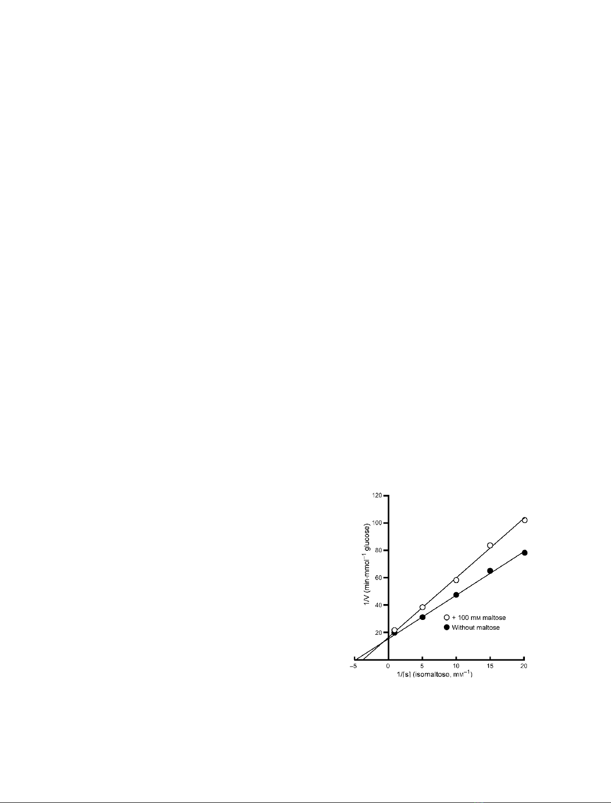
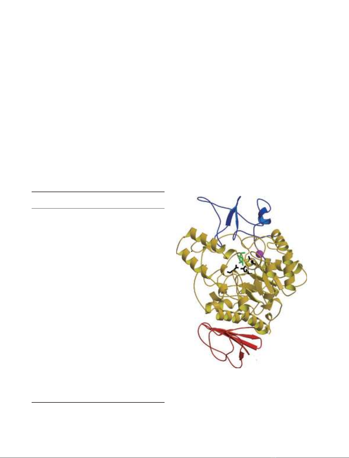
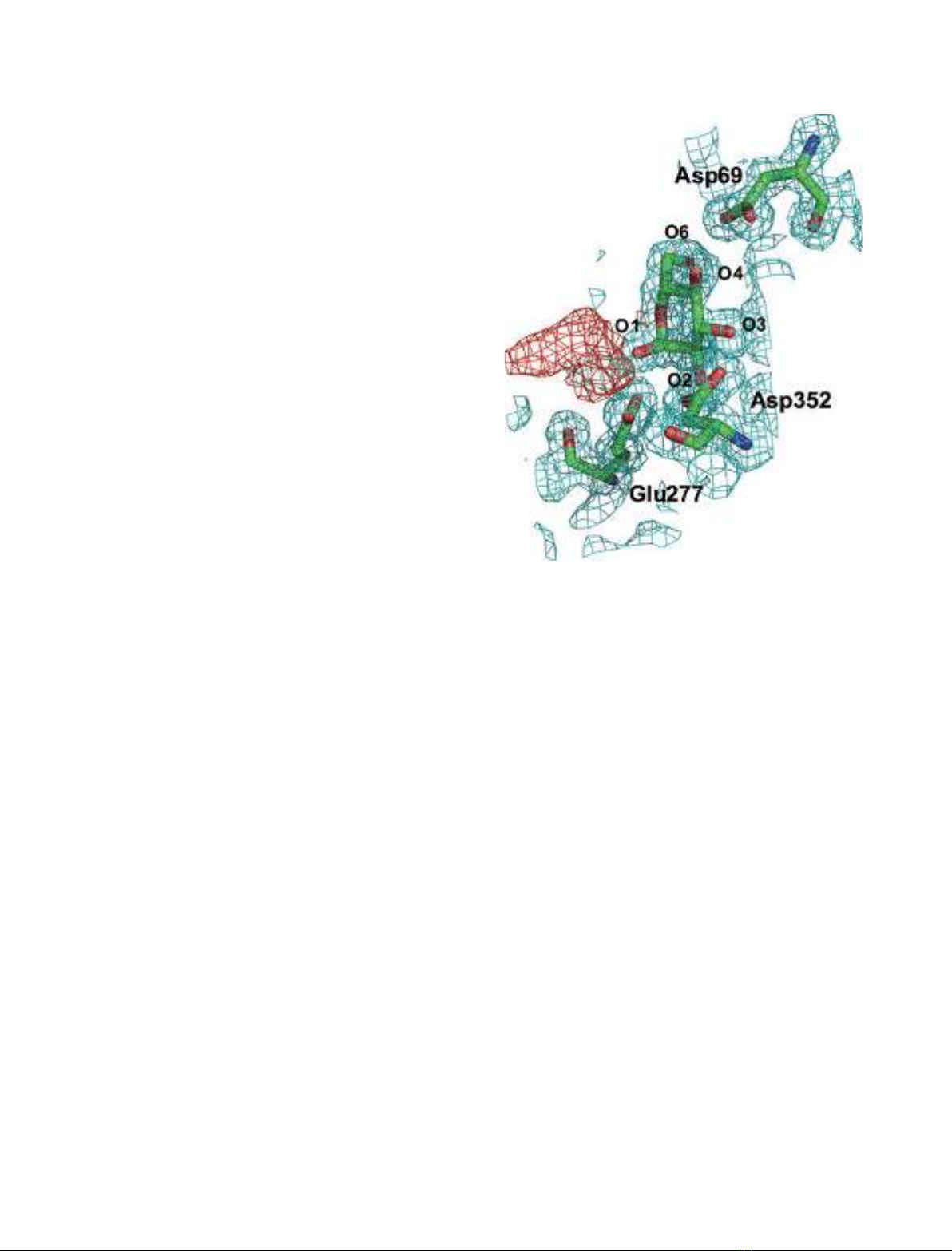
The structures of isomaltase from Saccharomyces cerevisiae and in complex
with maltose were determined at resolutions of 1.30 and 1.60 A
˚, respec-
tively. Isomaltase contains three domains, namely, A, B, and C. Domain A
consists of the (ba)
8
-barrel common to glycoside hydrolase family 13.
However, the folding of domain C is rarely seen in other glycoside hydro-
lase family 13 enzymes. An electron density corresponding to a nonreduc-
ing end glucose residue was observed in the active site of isomaltase in
complex with maltose; however, only incomplete density was observed for
the reducing end. The active site pocket contains two water chains. One
water chain is a water path from the bottom of the pocket to the surface
of the protein, and may act as a water drain during substrate binding. The
other water chain, which consists of six water molecules, is located near
the catalytic residues Glu277 and Asp352. These water molecules may act
as a reservoir that provides water for subsequent hydrolytic events. The
best substrate for oligo-1,6-glucosidase is isomaltotriose; other, longer-
chain, oligosaccharides are also good substrates. However, isomaltase
shows the highest activity towards isomaltose and very little activity
towards longer oligosaccharides. This is because the entrance to the active
site pocket of isomaltose is severely narrowed by Tyr158, His280, and loop
310–315, and because the isomaltase pocket is shallower than that of other
oligo-1,6-glucosidases. These features of the isomaltase active site pocket
prevent isomalto-oligosaccharides from binding to the active site
effectively.
Abbreviations
DGase, dextran glucosidase; GH, glycoside hydrolase; O16G, oligo-1,6-glucosidase from Bacillus cereus;a-MG, methyl a-D-glucopyroside.
FEBS Journal 277 (2010) 4205–4214 ª2010 The Authors Journal compilation ª2010 FEBS 4205
located in these regions [11–15]. Furthermore, all mem-
bers of this enzyme family are composed of three
domains, namely, A, B, and C [16]. Domain A is a cat-
alytic domain containing a (ba)
8
-barrel. Domain B is
inserted between the third b-sheet and the helix of the
(ba)
8
-barrel of domain A. Domain C comprises eight
antiparallel b-strands in a double Greek-key motif.
The conserved Asp (region II) and Glu (region III)
located at the C-termini of b-strand 4 and b-strand 5
act as the catalytic nucleophile and general acid–base
catalyst, respectively. The third conserved residue, Asp
(region IV), located between b-strand 7 and helix 7,
may stabilize the substrate during catalysis to help
accelerate the acid–base hydrolysis reaction [17]. The
three-dimensional arrangement of the three catalytic
residues is also highly conserved among GH family 13
enzymes [18]. Thus, the difference in substrate specific-
ity among GH family 13 enzymes must depend on the
structure of the active site.
Saccharomyces cerevisiae contains two a-glucosidases,
namely, a-1,4-glucosidase (maltase) and oligo-1,6-gluco-
sidase (isomaltase). The expression levels of these
enzymes are independently regulated by the MAL and
MGL polymeric genes [19–21]. Maltase preferentially
hydrolyzes maltose, but not isomaltose or methyl
a-d-glucopyroside (a-MG), whereas isomaltase hydro-
lyzes isomaltose and a-MG, but not maltose [22,23].
S. cerevisiae isomaltase shows notable substrate
specificity for chain length. In general, oligo-1,6-gluco-
sidase enzymes preferentially hydrolyze isomaltotriose,
and are highly active towards isomalto-oligosaccha-
rides and dextran [1,2,24]. On the other hand, dextran
glucosidase (DGase) prefers dextran, and hydrolyzes
panose and isomalto-oligosaccharides [25]. However,
S. cerevisiae isomaltase shows high activity towards
isomaltose and a-MG, but does not readily hydrolyze
isomaltotriose and isomaltotetraose [8]. The crystal
structure of oligo-1,6-glucosidase from Bacillus cereus
(O16G) was solved at a resolution of 2.0 A
˚[26].
Recently, the structures of the uncomplexed DGase
and its mutant in complex with isomaltotriose have
been determined at a resolution of 2.2 A
˚[27]. The
amino acid identities between isomaltase and O16G
and DGase are 41.0% and 35.7%, respectively. The
overall structures of O16G and DGase are very similar
to each other [26,27]. However, the substrate specificity
for chain length is different among the three enzymes.
Thus, determination of the precise three-dimensional
structure of isomaltase will contribute to a better
understanding of the structure–function relationships
of oligo-1,6-glucosidases with their substrates.
In the present article, we report the crystal structures
of isomaltase with no ligand bound at the active site
(free form) and in complex with its competitive inhibi-
tor maltose at resolutions of 1.30 and 1.60 A
˚, respec-
tively. A comparison of the structures of isomaltase
with those of O16G and DGase may reveal the differ-
ences in the active site structures that determine the
substrate specificity of oligo-1,6-glucosidases.
Results and Discussion
Isomaltase substrate specificity and inhibition
constant
Measurement of isomaltase activity was performed
according to a previously described method [8]. One
unit of isomaltase activity was defined as the amount
that released 1 lmol glucoseÆmin
)1
under the condi-
tions described in Experimental procedures. When the
activity of isomaltase with isomaltose was 100%, its
activities with isomaltotriose and isomaltotetraose were
3.5% and 0.1%, respectively, demonstrating that iso-
maltase cannot hydrolyze isomaltopentaose or isomal-
tohexaose.
The Lineweaver–Burk plot (Fig. 1) of isomaltase
revealed that maltose acts as a competitive inhibitor of
isomaltase. The inhibition constant (K
i
) was 289 mm.
Quality of the model
The crystal structures of free isomaltase and the iso-
maltase–maltose complex were determined by the
molecular replacement method, with O16G as the
search model. For free isomaltase, the model was
Fig. 1. Lineweaver–Burk plot for isomaltase with or without
100 mMmaltose. Isomaltose (50, 66.7, 100, 200 and 500 mM) was
used as the substrate.
Crystal structure of isomaltase K. Yamamoto et al.
4206 FEBS Journal 277 (2010) 4205–4214 ª2010 The Authors Journal compilation ª2010 FEBS
refined to a crystallographic R-factor of 17.2%
(R
free
= 18.6%) for 15 5721 unique reflections in the
resolution range 18.70–1.30 A
˚, and for the isomaltase–
maltose complex, to a crystallographic R-factor of
15.9% (R
free
= 17.4%) for 87 353 unique reflections in
the resolution range 23.22–1.60 A
˚. Table 1 summarizes
the details of data collection and the results of the
crystallographic refinement. The electron densities of
the Met1–Ile3 in both structures, and the reducing end
of maltose in the isomaltase–maltose complex struc-
ture, were very low; thus, their coordinates were not
determined. These residues may have been disordered.
Analysis of the main chain torsion angles of both
structures showed that 88.9% of the nonglycine
residues are located in the most favored regions of the
Ramachandran plot, and the remaining 10.9% are in
the additionally allowed regions. Only Gln279 is in the
disallowed region, located in the tight turn between
b-strand 5 and helix 5 of domain A, but this residue
was well identified in the electron density.
Overall structure
The overall structural features of isomaltase are similar
to those of O16G [26], DGase [27], and a-amylases
[11,14,15]. Isomaltase consists of three domains, A, B,
and C, as shown in Fig. 2. Domain A (residues 1–113
and 190–512) comprises eight alternating parallel
b-strands and a-helices, which make up the (ba)
8
-bar-
rel common to GH family 13 enzymes. Domain A also
contains the catalytic residues Asp215, Glu277 and
Asp352 at the C-terminal side of the barrel. Domain B
(residues 114–189) has a loop-rich structure containing
one short helix and an antiparallel b-sheet. The active
site cleft created by domains A and B forms a pocket-
shaped structure similar to that seen in the structures
Table 1. Data collection and refinement statistics. R
merge
=R
hkl
R
i
|I
(hkl)I)<I(hkl)>| R
hkl
I(hkl). R
work
=R(F
obs
)F
calc
)R(F
obs
). R
free
: cryst-
allographic R-factor based on 5% of the data withheld from the
refinement for cross-validation.
Free
isomaltase
Maltose
complex
Data collection
Beamline BL5A, PF BL44XU,
SPring-8
Wavelength (A
˚) 1.0000 0.9000
Space group C2C2
a(A
˚) 95.52 95.43
b(A
˚) 115.54 115.40
c(A
˚) 61.76 61.61
b() 91.05 91.19
Resolution range (A
˚) 50.00–1.30
(1.32–1.30)
25.00–1.60
(1.66–1.60)
Total reflections 646 080 370 160
Unique reflections 155 721 87 353
Completeness
a
(%) 95.1 (93.1) 99.7 (97.4)
Average Ir(I)
a
34.0 (8.4) 25.8 (13.8)
R
merge
(%)
a
3.8 (14.4) 4.2 (9.0)
Refinement
Resolution limit (A
˚) 18.70–1.30 23.22–1.60
R
work
R
free
0.172 0.186 0.159 0.174
No. of protein atoms 4835 4835
No. of water molecules 608 582
No. of calcium ions 1 1
No. of glucose atoms 12
rmsd
Bond length (A
˚) 0.007 0.009
Bond angle () 1.397 1.175
Average B-factors (A
˚
2
)
Main chain 10.15 10.16
Side chain 11.75 11.83
Water molecules 19.16 20.37
Glucose 11.07
a
The values for the highest-resolution shells are given in parentheses.
Fig. 2. Stereoview of the overall structure of isomaltase in complex
with maltose. Domain A (residues 1–113 and 190–512) is shown in
yellow, domain B (residues 114–189) in blue, and domain C (resi-
dues 513–589) in red. A calcium ion is shown as a magenta sphere.
The reducing end of the glucose residue is displayed in green. The
three catalytic acidic residues are shown as a stick model.
K. Yamamoto et al. Crystal structure of isomaltase
FEBS Journal 277 (2010) 4205–4214 ª2010 The Authors Journal compilation ª2010 FEBS 4207
of O16G [26], DGase [27], amylosucrase [28], and
sucrose phosphorylase [29]. It is notable that the fold
of domain C is different from those of other GH
family 13 enzymes. Usually, domain C of the GH
family 13 hydrolases consists of an antiparallel eight-
stranded b-barrel motif (composed of five b-strands
and three b-strands). However, domain C (residues
513–589) of S. cerevisiae isomaltase consists of an anti-
parallel five-stranded b-sheet; thus, only half of the
eight-stranded b-barrel motif close to domain A is
present in isomaltase. A similar fold has been found
only in the structure of sucrose phosphorylase [29].
The rmsd values between the corresponding main
chain atoms and all atoms of the free isomaltase and
isomaltase–maltose complex structures were found to
be 0.13 and 0.45 A
˚, respectively. Furthermore, all con-
formations of the active site amino acids were similar
between free isomaltase and the isomaltase–maltose
complex. Thus, inhibitor binding does not affect the
overall structure of isomaltase.
One calcium ion is bound at the loop located just
before helix 1 of domain A (Fig. 2). This calcium ion
has an octahedral geometry coordinated by the side
chains of Asp30 OD1, Asn32 OD1, Asp34 OD1, and
Asp38 OD2, the main chain oxygen of Trp36, and a
water molecule. A similar structure is found in native
DGase [27]. During purification and crystallization,
calcium salt was not added to the buffer solutions.
Thus, binding of this calcium ion must be very tight.
All known a-amylases contain a conserved calcium
ion that is located at the interface between domains A
and B [11,14,30–32]. This calcium ion is essential for
the stability and activity of the enzyme [33]. However,
the calcium-binding site of isomaltase is not identical
to that of a-amylases.
Active site structure
Figure 3 shows the electron density map around the
bound maltose in the isomaltase–maltose complex
structure. In this structure, the bound nonreducing end
of maltose was identified as electron densities in the
active site pocket. However, the reducing end of malt-
ose was not observed, probably because of the fluctua-
tion in the glucose residue. The glucose residue was
found to be in the a-anomer state. Moreover, the
incomplete electron density continued from O1 of the
nonreducing end of the glucose residues to the left side
of the figure. Because of the fluctuation in the reducing
end of maltose, only incomplete electron density was
observed. Isomaltase showed no activity towards malt-
ose, even at higher enzyme concentrations (Fig. 4).
Thus, the 1,4-glucosidic linkage of maltose is not
hydrolyzed, and maltose remains bound to the enzyme,
acting as a competitive inhibitor of isomaltase.
Prior to substrate or inhibitor binding, the active site
of isomaltase is occupied by five water molecules
(Fig. 5A). The positions of three of these five water
molecules are nearly identical to those of O2, O3 and
O4 of the glucose residue, and the remaining two
water molecules are near O1 and O6 of the glucose
residue. The hydrogen bond interactions are listed in
Table 2. These water molecules must be displaced by
incoming substrate. After substrate or inhibitor bind-
ing, the nonreducing end of the glucose residue is
bound at the bottom of the active site pocket (subsite
)1) by nine hydrogen bonds (Fig. 5B), and stacked
against Tyr72. The three catalytic acidic residues are
also involved in the hydrogen bond network. Asp215
OD2 forms a hydrogen bond with O6 of the glucose
residue in subsite )1. The distance between C1 of the
glucose residue and Asp215 OD1 is 2.6 A
˚. Glu277,
which acts as a general acid–base catalyst, forms a
hydrogen bond with O1 of the glucose residue, with a
bond distance of 2.8 A
˚. The lengths of hydrogen bonds
between Asp352 OD1 and O3 of the glucose residue
Fig. 3. Magnified view of the nonreducing end of maltose in the
active site of isomaltase. The final 2F
o
)F
c
electron density con-
toured at 1ris shown in cyan, and the F
o
)F
c
electron density
contoured at 3ris shown in red.
Crystal structure of isomaltase K. Yamamoto et al.
4208 FEBS Journal 277 (2010) 4205–4214 ª2010 The Authors Journal compilation ª2010 FEBS
and between OD2 and O2 are 2.7 and 2.5 A
˚, respec-
tively. The hydrogen bond interactions between the
protein and glucose residue are also listed in Table 2.
Seven of the nine hydrogen bonds are conserved
among the a-amylase and other GH family 13 enzymes
[34]. The remaining two hydrogen bonds are specific to
oligo-1,6-glucosidases. Asp69 OD2 and Arg442 NH1
form hydrogen bonds with O4 of the glucose residue.
Recognition of the nonreducing end of glucose is pre-
dominantly accomplished by these two interactions.
Here, isomaltase differs from other GH family 13
enzymes, although a similar structure is observed in
DGase [27]. In the case of DGase, the side chains of
the corresponding Asp and Arg residues (Asp60 and
Arg398 of DGase) together form a salt bridge, and
OD1 of Asp also forms a hydrogen bond with O4 of
the glucose residue [27]. These amino acids play an
important role in the interaction between the substrate
and the water path. However, in isomaltase, Asp69
OD1 also forms a hydrogen bond with Arg442 NH2,
but the conformations of the side chains of Asp69 and
Arg442 are slightly different from those of DGase.
The role of the water chains in the active site
pocket
Two chains of water molecules were identified in the
active site pocket. One water chain is a water path
located in the bottom of the active site pocket. In this
water path, three water molecules are arranged in a
nearly straight line from the bottom of the active site
pocket to the surface of the other side of the entrance
of the active site (Fig. 6). These water molecules are
labeled Wat1011, Wat1033 and Wat1099 in the free
isomaltase, and Wat713, Wat734 and Wat779 in the
isomaltase–maltose complex. These water molecules
form direct hydrogen bond contacts with each other.
Wat1011 forms hydrogen bonds with Asp69 OD1 and
Arg446 NH2. Wat1033 is bound to Asp69 OD1,
Asp409 OD1, and Arg446 NH1. Wat1099 forms
hydrogen bonds with the main chain carbonyl oxygen
atom of Glu408 and the main chain nitrogen atom of
Val410 and Glu408 OE1. An identical row of water
molecules is also found in the structures of DGase
[27], O16G [26], amylosucrase [28], and sucrose phos-
phorylase [29], all of which contain a pocket-shaped
active site. In DGase, for example, Wat1 forms hydro-
gen bonds with Asp60 and Arg 398 NH2. Wat2 forms
hydrogen bonds with Asp60, Asp369, and Arg398.
Wat3 is bound to the carbonyl oxygen atom of
Asp368, Asp368OD1, and the main chain nitrogen
atom of Ile370 [27].
The active site of free isomaltase is occupied by five
water molecules prior to substrate binding (Fig. 5A).
The positions of these water molecules are similar to
those of O1, O2, O3, O4 and O6 of the glucose residue
(Fig. 5B). When a substrate enters the active site
pocket, these water molecules must be displaced from
their binding sites. However, the entrance into the
pocket is very narrow, as shown in Fig. 7A; hence,
there is no available space to accommodate both the
incoming substrate and the outgoing water molecules.
Therefore, the water path may function as a water
drain. The binding water molecules are pushed out
from the pocket through the drain by the incoming
substrate.
The existence of such a water drain was first sug-
gested by Hondou et al. [27], on the basis of the
DGase structure. However, in this structure, the
DGase active site was occupied by Tris and glycerol.
Amylosucrase and sucrose phosphorylase were also
bound by glucose and Tris, respectively. Thus, it is
uncertain whether the water molecules exist in the
Fig. 4. TLC detection of hydrolytic products of isomaltose and
maltose. Standards: lane 1, isomaltose; lane 2, maltose; lane 3, glu-
cose. Products of the reaction of isomaltase with isomaltose are
shown in lane 4, and products of the reaction of isomaltase with
maltose are shown in lane 5.
K. Yamamoto et al. Crystal structure of isomaltase
FEBS Journal 277 (2010) 4205–4214 ª2010 The Authors Journal compilation ª2010 FEBS 4209