doi:10.1046/j.1432-1033.2003.03545.x
Eur. J. Biochem. 270, 2353–2362 (2003) (cid:1) FEBS 2003
Detection and characterization of a novel extracellular fungal enzyme that catalyzes the specific and hydrolytic cleavage of lignin guaiacylglycerol b-aryl ether linkages
Yuichiro Otsuka1, Tomonori Sonoki1, Seiichiro Ikeda1, Shinya Kajita1, Masaya Nakamura2 and Yoshihiro Katayama1 1Graduate School of Bio-Applications and Systems Engineering, Tokyo University of Agriculture and Technology, Koganei, Tokyo, Japan; 2Forestry and Forest Products Research Institute, Microbial Technology Laboratory, Tsukuba, Norin Kenkyu Danchi-nai, Ibaraki, Japan
into the extracellular fraction. The b-aryl ether cleavage enzyme converts the guaiacylglycerol b-O-guaiacyl ether (GOG) to guaiacylglycerol and guaiacol. It requires the Ca alcohol structure and p-hydroxyl group and specifically attacks the b-aryl ether linkage of high-molecular mass lignins with addition of two water molecules at the Ca and Cb positions.
Keywords: lignin biodegradaion; b-aryl ether linkage; fungi; guaiacylglycerol b-O-guaiacyl ether; extracellular enzyme. Cleavage of the arylglycerol b-aryl ether linkage is the most important process in the biological degradation of lignin. The bacterial b-etherase was described previously and shown to be tightly associated with the cellular membrane. In this study, we aimed to detect and isolate a new extracellular function that catalyses the b-aryl ether linkage cleavage of high-molecular lignin in the soil fungi. We screened and isolated 2BW-1 cells by using a highly sensitive fluorescence assay system. The b-aryl ether cleavage enzyme was pro- duced by a newly isolated fungus, 2BW-1, and is secreted
lignins, b-O-4-linkages are the most abundant and b-b-, b-5-, 5-5- and 5-O-4-linkages are also found. Therefore, lignins have very complicated structures with C-C and C-O-C linkages, and it is difficult for living organisms to degrade them. However, many soil microorganisms can easily digest lignins to fulfill important roles in the earth’s carbon cycle.
Lignins are the most abundant high-molecular mass aromatic compounds in plants. In trees, high levels of lignin are synthesized in wood and account for 15–36% of the dry weight of wood. Lignins are complex phenolic polymers that reinforce the walls of certain cells in the vascular tissues of higher plants. Lignin plays an import- ant role in mechanical support, water transport and pathogen resistance. The lignification process encompasses the biosynthesis of monolignols such as p-coumaryl, coniferyl and synapyl alcohols, and polymerization into the final molecule. Polymerization is thought to result from oxidative (radical-mediated) coupling between a monolignol and the growing oligomer/polymer. The oxidative coupling between monolignols can result in the formation of several different interunit linkages. In native
Correspondence to Y. Otsuka, Graduate School of Bio-Applications and Systems Engineering, Tokyo University of Agriculture and Technology, Koganei, Tokyo, Japan. Fax: + 81 42 388 7364, Tel.: + 81 42 388 7364, E-mail: y-otuka@cc.tuat.ac.jp Abbreviations: GOU, guaiacylglycerol-b-O-4-methylumbelliferone; GOG, guaiacylglycerol b-O-guaiacyl; GOU aO, a-O-methylumbel- liferyl-b-hydroxyl-propiovanillone; GOG aO, a-O-guaiacyl- b-hydroxyl-propiovanillone; GOUbz, O-benzyl-guaiacylglycerol- b-O-4-methylumbelliferone; GOGbz, O-benzyl-guaiacylglycerol- b-O-guaiacyl; DHP-GOU, reduced and polymerized form of GOU; DHP-GOU, fluorescent-labeled synthetic lignin; 4MU, 4-methyl- umbelliferone; MnP, manganese peroxidase; LiP, peroxidase; SYK-6, Sphingomonas paucimobilis SYK-6. (Received 1 October 2002, revised 18 December 2002, accepted 27 February 2003)
Lignin-biodegradation systems in nature can be sum- marized as follows. Initially, basidiomycetes secrete peroxi- dases and/or laccases and degrade the aromatic polymer lignin [1–7]. The role of each enzyme in this complicated process is an active area of research and debate. Thus far, mainly white rot fungi, Phanerochaete chrysosporium and Trametes (Coriolus) versicolor, have been studied regarding these peroxidases. P. chrysosporium produces two types of peroxidases, manganese peroxidase (MnP) and lignin per- oxidase (LiP) and T. versicolor generally produces laccase. Laccase reacts with polyphenols including lignin, and other lignin-derived aromatic compounds, that, in turn, can be both polymerized and depolymerized. MnP can oxidize Mn2+ to Mn3+; Mn3+, in turn, is able to oxidize a wide range of phenolic substrates including phenolic lignin. LiP can directly oxidize a variety of phenolic and nonphenolic aromatic compounds. These peroxidases remove an elec- tron and a proton from phenolic hydroxyl, aromatic amino groups or other aromatic side chains to form free radicals. Although this acts to cleave Ca-Cb linkages and b-O-4 linkages in the lignin structure, the free radicals cause random depolymerization of lignin. The various low- molecular mass lignins produced by these peroxidases and/or laccase are decomposed to carbon dioxide and water by specific bacterial enzymes, such as ring-fission enzymes [8,9], demethylases [10,11] and b-etherases [12–14].
2354 Y. Otsuka et al. (Eur. J. Biochem. 270)
(cid:1) FEBS 2003
type) were analysed using a JEOL-GX270 (solvent dimeth- ylsulfoxide-d6) (Fig. 2). For the Ca position reduction of GOU aO in DHP-GOU (Ca carbonyl type), 1 g each of DHP-GOU and sodium borohydride were dissolved in dioxane/methanol (4 : 1) and stirred for 12 h at 4 (cid:2)C. A large excess of water was added and the resultant precipitate was collected as DHP-GOU.
Isolation of the fungi and enzyme
In our current studies, we have focused on b-aryl ether linkages in lignin. Such linkages account for more than 50% of the intermolecular linkages in lignin. In Shingomonas paucimobilis SYK-6 (SYK-6), we have already identified the enzyme that specifically cleaves lignin dimmers with b-aryl ether linkages [12]. This enzyme belongs to the family of glutathione S-transferases (GSTs) and reductively cleaves the b-aryl ether bond [14]. However, this enzyme does not attack b-aryl ether linkages in high-molecular mass mate- rials in vivo because it acts at the intramolecular level and cannot gain access to the b-aryl ether linkages in high- molecular mass lignins [12].
If we could identify and characterize a secretory enzyme that cleaves b-aryl ether bonds in high-molecular mass lignins, we would uncover a new field of lignin degradation in nature. In addition, such an enzyme would convert high- molecular mass lignins into low-molecular mass lignins that retain benzene rings. Such lignins would have enormous biomass and be useful as an industrial material. Therefore in this study, we tried to detect and characterize such an enzyme in various fungi.
As described below, we succeeded in characterizing the production and reaction mechanism of a novel secretory enzyme that cleaves b-aryl ether bonds. Activity of the b-aryl ether cleavage enzyme was assayed as described in a previous report [12]. Soil samples were collected from several sites in Futyunomori Park in Tokyo. Each soil sample was suspended in 2 mL of Vogel’s medium (VM) [16]. After 7 days of stationary culture at 28 (cid:2)C, 10 lg of GOU were added and incubation was continued for 12 h. The fluorescence of each sample was examined under a UV illuminator (model TDS-15, Upland, Japan). Fluorescence- emitting samples were streaked onto VM plates for the isolation of single colonies. After incubation at 28 (cid:2)C for 3 days, 1400 colonies were selected randomly from the plates and suspended separately in liquid VM. After stationary culture for 7 days at 28 (cid:2)C, 10 lg of GOU were added to each sample. After further incubation for 12 h at 28 (cid:2)C, the fluorescence of cultures was examined.
Materials and methods
Chemicals
Activity of the b-aryl ether cleavage enzyme was assayed as described previously [12]. Cultures of fluorescing cells were centrifuged at 15 000 g for 10 min. Supernatants (1 mL) were added to 1.0 mL of 200 mM glycine/NaOH buffer (pH 10.0). Fluorescence of 4-methylumbelliferone released from GOU was measured with excitation at 360 nm and emission at 450 nm with a fluorophotometer (Shimadzu, Japan RF-1200).
The production of b-aryl ether cleavage enzyme by 2BW-1 cells
2BW-1 cells were suspended in VM and cultured without agitation at 28 (cid:2)C. Pieces of hyphae were collected with 1.0 mL of culture solution at 24 h intervals, and 10 lg of GOU were added to each collected sample. After incubation for 12 h at 28 (cid:2)C, b-aryl ether cleavage activity was measured as described above.
Localization of b-aryl ether cleavage activity
A 14-day-old culture (4 mL) of 2BW-1 cells was separated into supernatant and residue by centrifugation at 4000 g for 15 min at 20 (cid:2)C. Additional supernatant was removed from the cell debris by ultra centrifugation at 60 000 g for 60 min at 4 (cid:2)C. The resulting extracellular fraction (EC) was used for the assay of b-aryl ether cleavage activity. One gram of residue was washed twice with 100 mL of 0.8% (w/v) Nail solution and centrifuged at 4000 g for 15 min. Half of the residue was designated the hyphae fraction (HP) and the remainder was homogenized with mortar and pestle for 5 min in liquid nitrogen and suspended in 15 mL of 10 mM of Tris buffer (pH 7.5) at 4 (cid:2)C for 60 min. The resulting suspension was separated into supernatant and residue by centrifugation at 60 000 g for 60 min at 4 (cid:2)C. The resultant supernatant and residue were designated the cytoplasm fraction (CY) and the membrane fraction (M), respectively. Guaiacylglycerol-b-O-4-methylumbelliferone (GOU), guaiacylglycerol b-O-guaiacyl (GOG), a-O-methylumbel- liferyl-b-hydroxyl-propiovanillone (GOU aO) and a-O-gua- iacyl-b-hydroxyl-propiovanillone (GOG aO) were prepared as described previously [12]. O-Benzyl-guaiacylglycerol-b- O-4-methylumbelliferone (GOUbz) and O-benzyl-guaiacyl- glycerol-b-O-guaiacyl (GOGbz) were synthesized by as described in a previous report [15]. A reduced and polymerized form of GOU (DHP-GOU) was synthesized as follows. Acetone (50 mL) containing 0.2% GOU aO and 0.2% conyferyl alcohol, and 20 mL of 3% H2O2 were dropped into 430 mL of 100 mM potassium phosphate [pH 6.0, containing 30% acetone and 6 mg horseradish peroxidase (44 UÆmg)1, Sigma)] and stirred for 14 h at 20 (cid:2)C. After the additions, 4 mg of peroxidase was added and the reaction mixture was stirred for additional 12 h. The resultant precipitate was collected and washed three times with 10 mL water and centrifuged at 4000 g for 10 min. The precipitate was dried completely on the phosphorus (V) oxide. Dioxane/water (9 : 1) solution (used to dissolve the crude DHPs) was poured into 300 mL of diethyl ether for recrystallization. The precipitate was washed four times with diethyl ether and dispersed in distilled water. After lyophilization, the fluorescent-labeled synthetic lignin (DHP-GOU Ca carbonyl type) was pro- duced. DHP-GOU (Ca carbonyl type) was fractionated by gel permeation chromatography on an Asahipack GS310 column (500 mm in length · 7.6 mm diameter). N,N- Dimethylformamide containing 0.1 M lithium chloride was used as the eluant at a flow rate 0.5 mLÆmin)1. Relative molecular mass was estimated by calibration with polysty- lene standard (Mr ¼ 175 000, 9000, 4000, 2000, 800) (Fig. 1). The 1H-NMR spectra of DHP-GOU (Ca carbonyl
The fungal b-aryl ether cleavage enzyme (Eur. J. Biochem. 270) 2355
(cid:1) FEBS 2003
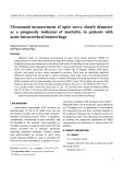
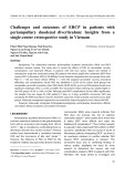
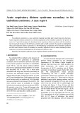
Fig. 1. A highly sensitive assay system for b-aryl ether cleavage function. (A) Scheme of 4-methylumbelliferone (4MU) released from guaiacylglycerol b-O-4-methylumbelliferone (GOU) and DHP-GOU. Upon cleavage of the b-aryl ether linkage, 4-methylumbelliferone is released and emits powerful fluorescence. Fluorescence of 4MU was measured with excitation at 360 nm and emission at 450 nm. (B) Gel filtration chromatogram of DHP-GOU (Ca alcohol type). Fractions used in this study are shaded and substrate molecular mass is more than 1000. Relative molecular mass was calibrated using polystylene standard series (175 000, 9000, 4000, 2000, 800). (C) 1H-NMR spectrum of acetylated DHP-GOU (Ca alcohol type).
For EC and CY, reactions were initiated by addition of 0.1 mL of EC or CY to 1.0 mL of VM containing 10 lg of GOU. For HP and M, reactions were initiated by addition of 0.1 g of HP or M to 1.0 mL of VM containing 10 lg of GOU. After incubation for 12 h at 28 (cid:2)C, the reaction mixture was centrifuged at 15 000 g for 10 min. Reactions were terminated by addition of 1.0 mL of 200 mM glycine/ NaOH buffer (pH 10.0). Fluorescence was measured with excitation at 360 nm and emission at 450 nm.
Purification of the b-aryl ether cleavage enzyme
enzyme activity was defined as the amount of b-aryl ether cleavage enzyme that released 1 ng of 4-methylumbellifer- one (4MU) per hour. Two hundred millilitres of EC were concentrated with an Ultrafree 30k system (Millipore, Tokyo, Japan) to 5 mL. The concentrated solution was purified by gel filtration through a Sephadex G-75 column (0.5 cm · 30 cm, Pharmacia, Tokyo, Japan) with 10 mM of Tris/HCl buffer as the mobile phase. The most active fractions were further purified by ion-exchange chromato- graphy on a Mono Q column (0.5 cm · 0.5 cm, Pharmacia) and eluted with a gradient of 0 mM to 1 M (NH4)2SO4 in water. Proteins eluted from the column were detected by monitoring the absorbance at 280 nm and examined by SDS/PAGE. b-aryl ether cleavage activity was measured by using a GOU fluorometric assay of b-aryl ether cleavage. One unit of
2356 Y. Otsuka et al. (Eur. J. Biochem. 270)
(cid:1) FEBS 2003
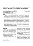
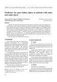
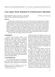
Fig. 2. Phylogenetic tree of 2BW-1 based on 18S rDNA sequence comparisons of sequences of 18S rDNA and drawn using GENETYX version 10.1 software. The numbers on some branches refer to confidence levels estimated by bootstrap analysis (100 replications).
SDS/PAGE
was measured under oxygen-saturated (100%; 7.5 mgÆO)1) conditions or low-oxygen (17%; 1.3 mgÆO)1) conditions with surrounding argon gas. The method for measurement of activity is described above. To examine the enzymatic incorporation of 18O2 and 18O-labeled water into GG, reactions were initiated by the addition of 20 lL of a
SDS/PAGE was performed with a stacking gel of 7.5% (w/v) acrylamide and a separating gel of 12.5% (w/v) acryl amide, as described by Laemmli [17]. The molecular mass and subunit composition of b-aryl ether cleavage enzyme were determined by electrophoresis under reducing condi- tions. An MW-Marker (SDS) kit (Oriental Yeast Industry Co., Japan) was used as the source of standard proteins. Protein bands were visualized with a Coomassie/Brilliant Blue-staining procedure.
Enzymic reaction and analysis of metabolites
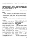
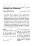
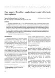
Fig. 3. Localization of b-aryl ether cleavage function and assay for high- molecular mass lignin structure. (A) Localization of b-aryl ether clea- vage enzyme in 2BW-1. HP, hyphae fraction; EC, extracellular frac- tion; CY, cytoplasmic fraction. Control, GOU added to 1 mL of VM. (B) Assay of b-aryl ether cleavage by the extracellular fraction with a model compound that resembles high-molecular mass lignin.
Reactions were initiated by the addition of 100 ll of a solution of enzyme (200 lgÆmL)1) to 0.9 mL of a solution of 50 lg of substrate in 10% (v/v) dimethyl sulfoxide and incubated for 12 h at 28 (cid:2)C. Then, reaction mixtures were acidified to pH 2 by the addition of 12 M HCl and extracted three times with 300 lL of ethyl acetate. The extract was then dried on a rotary evaporator (REN-1, Iwaki Glass. Co. Ltd, Iwaki, Japan) with a vacuum controller (FTP-10; Asahi Techno Glass, Japan). The residue was dissolved in 20 lg of pyridine and treated with N,O-bis(trimethyl- silyl)trifluoroacetamide (BSTFA; Tokyo Kasei Co., Tokyo, Japan) to prepare trimethylsilyl derivatives. Then 1 lg of the solution of these derivatives was subjected to gas chromatography (model 390, GL Science, Tokyo, Japan) and gas chromatography-mass spectrometry (GC-MS; Auto Mass System II; JEOL, Tokyo, Japan). A fused silica capillary column (CP-Sil 5CB; 25 m · 0.32 mm; i.d., 0.25 lm; Chrompack, the Netherlands) was used as the stationary phase. The temperature of the eluant was raised at 5 (cid:2)CÆmin)1 from 100–300 (cid:2)C. The eluant was detected by a flame ionization detector.
To clarify whether the enzymatic reaction was a hydro- lytic reaction or an oxidative reaction, the enzymic activity
The fungal b-aryl ether cleavage enzyme (Eur. J. Biochem. 270) 2357
(cid:1) FEBS 2003
Table 1. Purification of b-aryl ether cleavage enzyme in 2BW-1.
Purification (fold)
Recovery
Step
Vol (mL)
Protein (mg)
Activity (UÆmg)1)
Culture medium Ultrafiltration (30 kDa) Sephadex G-75 Mono Q
200 5 8 2
5520 3254 591 426
3 3.9 31.3 49.4
1 1.3 10.27 16.2
100 115 38 34
solution of enzyme (1 mgÆmL)1) and 20 lL of a solution of GOG in dimethyl sulfoxide (50 lgÆmL)1) to 160 lL Tris/ HCl buffer (pH 7.5) with bubbling 18O2 gas for 5 min (Nippon Sanso Co., Kawasaki, Japan) or 18O-labeled water (94% 18O atom; Nippon Sanso Co., Kawasaki, Japan) and incubated for 12 h at 28 (cid:2)C. After the incubation, the mixture was analysed as described above.
Results and discussion
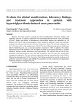
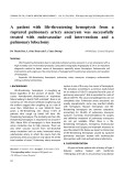
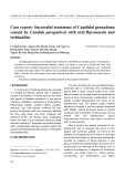
Fig. 4. Analysis by SDS/PAGE of the purified b-aryl ether cleavage enzyme from 2BW-1.
In our previous study, we characterized the b-etherase and the nucleotide sequences of the ligE and ligF genes of S. paucimobilis SYK-6 [13]. b-Etherase is a member of the glutathione-S-transferase superfamily [14]. It catalyzes the reductive cleavage of the b-aryl ether linkage of GOU aO (Fig. 7, structure V) to produce b-hydroxypropiovanillone and 4MU [12,14]. However, the b-etherase is associated tightly with cell membranes and was not secreted into the extracellular fraction [12]. Therefore, this b-etherase cannot cleave the b-aryl ether linkages of high-molecule-mass lignins.
Isolation of microorganisms that can cleave b-aryl ether linkages
Fig. 5. The time course of cleavage the b-aryl ether linkage by purified b-aryl ether cleavage enzyme in various condition. GOU was used as substrate and measured of fluorescence intensity.
used guaiacylglycerol
Using GOU, we isolated six fungi from soil samples with enzymes able to cleave b-aryl ether linkages. One isolate, 2BW-1, generated the strongest fluorescence and therefore, it appeared that 2BW-1 cleaved the b-aryl ether linkage efficiently. 2BW-1 also cleaved the b-aryl ether linkages of DHP-GOU (data not shown), a result that suggested that 2BW-1 might cleave b-aryl ether linkages in high-molecular- mass materials. A very sensitive assay was necessary for isolation of microorganisms that can cleave b-aryl ether linkages, so b-O-4-methylumbelliferone we (GOU) for our screening tests (Fig. 1A). In addition, we synthesized DHP-GOU as a fluorescent-labeled synthetic lignin to assay activity for high-molecular mass lignin (see Materials and methods). The synthesized DHP-GOU (Ca carbonyl type) was fractionated by gel permeation chromatography (Fig. 1B). We used the mixture that was contained from 9000–17 000 Mr as DHP-GOU (Ca car- bonyl type) in this study. The 1H-NMR spectrum of acetylated DHP-GOU (Ca carbonyl type) was analysed (Fig. 1C). A signal at 3.8 p.p.m. was assigned to the methoxyl group (OCH3) of the guaiacyl structure in conyferyl alcohol and GOU aO. The signal at 2.2 p.p.m. was assigned to the methyl group (CH3) originating from the CH3 of 4MU in GOU aO (Ca carbonyl type). The area ratio between the signals at 3.8 p.p.m and 2.2 p.p.m. was calculated as 10 : 1. It was considered that DHP-GOU (Ca carbonyl type) contained conyferyl alcohol and GOU aO at the ratio of 9 : 1 by chemical structure. To prepare DHP-GOU we used the reduction of the Ca position of GOU aO in DHP-GOU (Ca carbonyl type; see Materials and methods). When the b-aryl ether linkage of GOU structure is cleaved, the 4-methylumbelliferone (4MU) generated can be detected with high sensitive because of its strong fluorescence.
2358 Y. Otsuka et al. (Eur. J. Biochem. 270)
(cid:1) FEBS 2003
Fig. 6. GC and GC-MS analysis of TMS-derivatives of metabolites produced from GOG by the purified enzyme. See text for details.
Taxonomic position of 2BW-1 Cell growth and the production of the b-aryl ether cleavage enzyme
these results
We cultured 2BW-1 in stationary test tubes at 28 (cid:2)C. To observe the expression of b-aryl ether cleavage activity, we collected the culture solution that contained hyphae at 24 h intervals during a 3 week incubation. The enzymatic activity in the culture was determined as emitted fluorescence generated by cleavage of a b-aryl ether linkage. The enzymatic activity was not detected until cultures were 6-days-old. In 7-day-old cultures, we detected weak activity. The activity increased for 7 more days and then decreased (data not shown). This result suggested that production of the b-aryl ether cleavage enzyme might be induced under specific conditions. Analysis of the products of the reaction revealed the presence of guaiacylglycerol (GG) and 4MU (data not shown).
Localization of enzymatic activity
To confirm the localization of the b-aryl ether cleavage activity, we prepared a hyphae fraction (HP), an extracel- lular fraction (EC), a cytoplasmic fraction (CY) and a membrane fraction (M) from cultures of 2BW-1 (see Materials and methods). Enzymatic activity was determined 2BW-1 did not produce spores under any tested condi- tions. Therefore, to determine the taxonomic position of this novel fungus, we determined the nearly complete sequence of its 18S rDNA. The sequence of the 18S rDNA of 2BW-1 was strongly homologous to that of the ascomycetes, Chaetomium elatum, Podospora anserina, Sordaria fimicola and Neurospora crassa (Fig. 2). We had considered that 2BW-1 would be a member of indicate that basidiomycetes. However, 2BW-1 belongs rather to ascomycetes. The sequence from 2BW-1 was very similar to that from C. elatum and C. globosum (more than 99% homology). These results suggest that 2BW-1 is a member of the genus Chaeto- mium. C. globosum and C. elatum have been studied as wood-rotting fungi. They are able to grow on wood chips and decompose wood via the degradation of cellulose [22]. However, in Chaetomium sp., the degradation system of lignin or lignin related compounds have not been studied. Similarly Chaetomium sp. 2BW-1 was able to grow on wood chips as the sole carbon source. In addition, 2BW-1 also grew in the lignin-related compounds, p-hydroxy- benzoic acid, gallic acid and vanillic acid, as a sole source of carbon.
The fungal b-aryl ether cleavage enzyme (Eur. J. Biochem. 270) 2359
(cid:1) FEBS 2003
Fig. 7. Substrate specify of the b-aryl ether cleavage activity of 2BW-1. Structure I, gua- iacylglycerol b-O-4-methylumbelliferone; II, guaiacylglycerol; III, O-benzyl-guaiacylgly- cerol b-O-4-methylumbelliferone; IV, O-benzyl-guaiacylglycerol b-O-guaiacyl; V, a-O-(b-methylumbelliferyl)-b-(hydroxy) propriovanillone; VI, a-O-(b-guaiacyl)- b-(hydroxy)propriovanillone.
as the emitted fluorescence from GOU incubated with each fraction. We detected strong fluorescence only with EC (Fig. 3A). These results indicated that the b-aryl ether cleavage enzyme accumulated and was stable in the extracellular fraction. The extracellular fraction of 2BW-1 generated abundant GG and 4MU from GOU by cleaving the ether linkage. In addition, this extracellular enzyme cleaved the b-aryl ether linkages of DHP-GOU, a high- molecular mass compound (Fig. 3B). purification factor was about 16.2-fold, and the final yield was 34%. The final product had a specific activity of about 49.4 UÆmg)1 (Table 1). The molecular mass of the purified enzyme was estimated to be about 65 kDa. The time course of cleavage of the b-aryl ether linkage by the purified enzyme (20 lgÆmL)1) was followed with GOU as the substrate and by measurement of fluorescence intensity (Fig. 5). From this result, we used an enzymatic reaction time of 12 h in further experiments.
Reaction mechanism and substrate specificity Purification and characterization of the b-aryl ether cleavage enzyme
To identify the reaction mechanism of the enzyme, we analysed the reaction products by GC and GC-MS. 4-Methylumbelliferone (m/z 248) and guaiacylglycerol (m/z 502) were detected as major reaction products by GC-MS analysis, indicating that the enzyme cleaved the b-aryl ether linkage in GOU specifically to produce GG and 4MU. In addition, the enzyme also cleaved the b-aryl ether bond in GOG to produce GG and guaiacol (Fig. 6).
To characterize in further detail the b-aryl ether cleavage enzyme, we tried to purify it from the extracellular fraction, as summarized in Table 1. The EC was concentrated by ultrafiltration and applied to a gel-filtration column of Sephadex G-75. The fractions with the highest activity were collected and subjected to anion-exchange chromatography. The active fraction yielded only a single band after SDS/ PAGE (Fig. 4), indicating that the b-aryl ether cleavage enzyme had been purified to homogeneity. The overall To clarify the substrate specificity of the b-aryl ether cleavage enzyme, we synthesized the substrates GOUbz
2360 Y. Otsuka et al. (Eur. J. Biochem. 270)
(cid:1) FEBS 2003
Fig. 8. Mass spectra of guaiacylglycerol and guaiacol, products of the b-aryl ether cleavage reaction. The reaction products of GOG generated by the b-aryl ether cleavage enzyme in 18O-labeled water, in an atmosphere of 18O2 and in a control reaction (no radiolabel).
Fig. 9. The proposed mechanism of catalysis of the b-aryl ether cleavage enzyme.
(structure III) and GOGbz (structure IV) by replacing the p-hydroxyl group of GOU (structure I) and GOG (structure II) by a benzyl group (Fig. 7). The b-aryl ether cleavage enzyme could not cleave the b-aryl ether linkages of GOUbz (III) and GOGbz (IV). In addition, the b-aryl ether cleavage enzyme failed to cleave the b-aryl ether linkage of GOU aO (Fig. 7 structure V). Thus, the b-aryl ether cleavage enzyme required a p-hydroxyl group and a Ca alcohol structure for activity. Despite the high speci- ficity of the Ca structure and p-hydroxyl group, the b-aryl ether cleavage enzyme could react with DHP-GOU. This result indicates reactivity for the structure that retained the Ca alcohol and p-hydroxyl group that exists on the surface of DHP, as shown in Fig. 1. Therefore, the enzyme activity for DHP-GOU was lower than GOU. However, this enzyme did not act on substrates such as guaiacol and a- and b-naphthol (data not shown). The enzyme produced GG and 4MU from GOU, suggesting that the cleavage of the b-aryl ether bond might be a hydrolytic reaction. We examined whether this enzyme catalyzed an oxidative or a hydrolytic reaction. If the enzyme catalysed an oxidative reaction, its activity should reflect the level of oxygen in the atmosphere. Therefore, we measured the activity in low-oxygen (1.3 mg O) atmosphere. In the absence of oxygen, the enzyme reaction was very slow or none-existent (Fig. 5). Therefore, the reaction seemed to resemble the mono- oxygenase reaction of P450. We examined the incorpor- ation of oxygen using GOG in an atmosphere of 18O2 and analysed the reaction mixture by GC-MS. Figure 8 shows the mass spectrum of GG; no 18O were found in the reaction products. Therefore, we examined the incorpor- ation of the water molecule using GOG in 18O-labeled water. The mass spectrum revealed that two molecules
The fungal b-aryl ether cleavage enzyme (Eur. J. Biochem. 270) 2361
(cid:1) FEBS 2003
4. Bogan, B.W. & Lamar, R.T. (1996) Polycyclic aromatic hydro- carbon-degrading capabilities of Phanerochaete laevis HHB-1625 and its extracellular lignolytic enzymes. App. Env. Microbiol. 62, 1597–1603.
the products of
5. Eggert, C., Temp, U., Dean, J.F. & Eriksson, K.E. (1996) A fungal metabolites degradation of non-phenolic lignin structures and synthetic lignin by laccase. FEBS Lett. 391, 144–148.
6. Camarero, S., Sarkar, S., Ruiz-Duenas, F.J., Martinez, M.J. & Martinez, A.T. (1999) Description of a versatile peroxidase involved in the natural degradation of lignin that has both man- ganase peroxidase and lignin peroxidase substrate interaction sites. J. Biol. Chem. 274, 10324–10330.
7. Roy, B.P., Paice, M.G., Archibald, F.S., Misra, S.K. & Misiak, L.E. (1994) Creation of metal-complexing agents, reduction of man- ganase dioxide, and promotion of manganese peroxidase-medi- ated Mn (III) production by cellobiose: quinone oxidoreductase from Trametes versicolor. J. Biol. Chem. 269, 19745–19750.
of 18O-labeled water were incorporated into GG. In addition, we found the radiolabeled oxygen at the Ca and Cb positions in a comparison of the mass spectrum with that of the reaction with GG and unlabeled water. The incorporation of 18O from radio- labeled water was not observed with guaiacol.
8. Noda, Y., Nishikawa, S., Shinozuka, K., Kadokura, H., Nakaj- ima, H., Yoda, K., Katayama, Y., Morohoshi, N., Haraguchi, T. & Yamasaki, M. (1990) Molecular cloning of the protocatechuate 45-dioxygenase genes of Pseudomonas paucimobilis. J. Bacteriol. 172, 2704–2709.
9. Peng, X., Egashira, T., Hanashiro, K., Masai, E., Nishikawa, S., Katayama, Y., Kimbara, K. & Fukuda, M. (1998) Cloning of a Sphingomonas paucimobilis SYK-6 gene encoding a novel oxyge- nase that cleaves lignin-related biphenyl and characterization of the enzyme. Appl. Environ. Microbiol. 64, 2520–2527.
10. Kaufmann, F., Wholfarth, G. & Diekert, G. (1997) Isolation of O-demethylase, an ether-cleaving enzyme system of the homo- acetogenic strain MC. Arch. Microbiol. 168, 136–142.
11. Sonoki, T., Obi, T., Kubota, S., Higashi, M., Masai, E. & Katayama, Y. (2000) Coexistence of two different O demethyla- tion systems in lignin metabolism by Sphingomonas paucimobilis SYK-6: cloning and sequencing of the lignin biphenyl-specific O-demethylase (LigX) gene. Appl., Environ. Microbiol. 66, 2125– 2132.
12. Masai, E., Katayama, Y., Nishikawa, S., Yamasaki, M., Morohoshi, N. & Haraguchi, T. (1989) Detection and localization of a new enzyme catalyzing the beta-aryl ether cleavage in the soil bacterium (Pseudomonas paucimobilis SYK-6). FEBS Lett. 249, 348–352.
13. Masai, E., Katayama, Y., Kawai, S., Nishikawa, S., Yamasaki, M. & Morohoshi, N. (1991) Cloning and sequencing of the gene for a Pseudomonas paucimobilis enzyme that cleaves beta-aryl ether. J. Bacteriol. 173, 7950–7955.
the reaction mechanism of
14. Masai, E., Katayama, Y., Kubota, S., Kawai, S., Yamasaki, M. & Morohoshi, N. (1993) A bacterial enzyme degrading the model lignin compound beta-etherase is a member of the glutathione- S-transferase superfamily. FEBS Lett. 323, 135–140.
to the Ascomycetes
15. Weinstein, A.D. & Gold, H.M. (1979) Synthesis of guaiacyl- glycerol and glycerol-b-O-(b-methylumbelliferyl) ethers: Lignin model substrates for the possible fluorometric assay of b-etherases. Holzforschung 33, 134–135.
16. Peizer, L.R. & Vogel, H. (1969) An autoclavable medium for the
isolation of mycobacteria. Am. Rev. Respir. 100, 582–583.
history fungal of It was clear that the b-aryl ether cleavage enzyme catalyzed the addition of two molecules of H2O (at Ca and Cb positions) and cleavage the b-aryl ether bond. In addition, under differing enzyme conditions, although the fluorescent segregation quantity was proportional to the enzyme amount, the reaction rate remained mainly constant (Fig. 5). If this enzyme reaction was only a onestep reaction, the reaction rate must be faster where more enzyme exists. Therefore, we considered that the enzyme reaction was a twostep reaction where 4MU was released at the second step. Figure 9 shows a model of this novel hydrolytic enzyme [18–21,23–25]. Enzymatic dehydration generates the quinonemethide from GOG (structure II). The reaction mixture turned yellow as a result of formation of the quinonemethide. The scheme is consistent with the fact that the b-aryl ether cleavage enzyme requires a hydroxyl group and a Ca alcohol structure. Probably, at this time, molecular oxygen affects the formation of the quinonemethide. Then, water attacks the Ca position in the quinonemethide and the b-aryl ether linkage is cleaved. Another water molecule then attacks the Ca position to is generate GG. As the result, GOG (structure II) converted to GG and guaiacol. In addition, this reaction model is consistent with the fact that the initial reaction rate of this enzyme was very slow (Fig. 5). There are no reports of similar enzymatic reactions, to our knowledge. In this report, we have described a new secretory enzyme that specifically cleaves the b-aryl ether linkage of the major intramolecular bond in lignins. The b-aryl ether cleavage enzyme was produced by a newly isolated fungus, 2BW-1 and is secreted into the extracellular fraction. It attacks the b-aryl ether linkage of high- molecular mass lignins with the addition of two water molecules at positions, Ca and Cb. In addition, 2BW-1 did not belong to the Basidiomycetes (known as lignin (known degradation fungi) but mainly as cellulolytic fungi. Therefore, further charac- terization of this enzyme and isolation of its gene should contribute to improved utilization of high-molecular mass lignins and provide a new perspective on the lignin-degradation evolutionary systems.
References
1. Andrawis, A., Johnson, K.A. & Tien, M. (1988) Studies on compound I formation of the lignin peroxidase from Phaner- ochaete chrysosporium. J. Biol. Chem. 263, 1195–1198.
17. Cleveland, D.W., Fischer, S.G., Kirschner, M.W. & Laemmli UK. (1977) Peptide mapping by limited proteolysis in sodium dodecyl sulfate and analysis by gel electrophoresis. J. Biol. Chem. 252, 1102–1106.
18. Kido, T. & Soda, K. (1976) A new oxigenase, 2-nitropropane dioxygenase of Hansenula mrakii. J. Biol. Chem. 251, 6994–7000. 19. Umezawa, T. (1988) Mechanisms of fungal degradation of lignin.
Wood research, no. 75, 26–78.
2. Gold, M.H., Kuwahara, M., Chiu, A.A. & Glenn, J.K. (1984) Purification and characterization of an extracellular H2O2- requiring diarylpropane oxygenase from the white rot basidio- mycete, Phanerochaete chrysosporium. Arch. Biochem. Biophys. 234, 353–362.
3. Cai, D. & Tien, M. (1993) Lignin-degrading peroxidases of Pha-
20. Sharp, R.F. (1975) The microbial colonization of some woods of small dimensions buried in soil. Can. J. Micrlbiol. 21, 784–793.
nerochaete chrysosporium. J. Biotechnol. 30, 79–90.
2362 Y. Otsuka et al. (Eur. J. Biochem. 270)
(cid:1) FEBS 2003
21. Greaves, H. (1975) Microbiological aspects of wood chip storage
in tropical environments. Aust. J. Biol. Sci. 28, 323–330.
24. Gellerstedt, G. & Gierer, J. (1968) The reactions of lignin during neutral sulphite cooking, Part I. The behaviour of b-arylether structures. Acta. Chem. Scand. 22, 2510.
22. Mohtashamipur, E. & Norpoth, K. (1990) Release of mutagens after chemical degradation of beech wood lignin. Toxicol. Lett. 51, 277–285.
25. Freudenberg, K., Chen, L.C. & Cardinale, G. (1962) Die Oxy- dation des methylierten nauturlichen und kunstlichen Lignins Chem. Ber. 95, 2814.
23. Sarkanen, V.K. & Ludwig, H.C. (1971) Lignins: occurrence, for- mation, structure and reactions. Wiley-Interscience, John Wiley & Sons, Inc. NY, USA.