
Chiang et al. Journal of Biomedical Science 2010, 17:46
http://www.jbiomedsci.com/content/17/1/46
Open Access
RESEARCH
© 2010 Chiang et al; licensee BioMed Central Ltd. This is an Open Access article distributed under the terms of the Creative Commons
Attribution License (http://creativecommons.org/licenses/by/2.0), which permits unrestricted use, distribution, and reproduction in
any medium, provided the original work is properly cited.
Research
Enhancement of tolerance development to
morphine in rats prenatally exposed to morphine,
methadone, and buprenorphine
Yao-Chang Chiang, Tsai-Wei Hung, Cynthia Wei-Sheng Lee, Jia-Ying Yan and Ing-Kang Ho*
Abstract
Background: Abuse of addictive substances is a serious problem that has a significant impact on areas such as health,
the economy, and public safety. Heroin use among young women of reproductive age has drawn much attention
around the world. However, there is a lack of information on effects of prenatal exposure to opioids on their offspring.
In this study, an animal model was established to study effects of prenatal exposure to opioids on offspring.
Methods: Female pregnant Sprague-Dawley rats were sub-grouped to receive (1) vehicle, (2) 2-4 mg/kg morphine (1
mg/kg increment per week), (3) 7 mg/kg methadone, and (4) 3 mg/kg buprenorphine, subcutaneously, once or twice
a day from E3 to E20. The experiments were conducted on animals 8-12 weeks old and with body weight between 250
and 350 g.
Results: Results showed that prenatal exposure to buprenorphine caused higher mortality than other tested
substance groups. Although we observed a significantly lower increase in body weight in all of the opioid-
administered dams, the birth weight of the offspring was not altered in all treated groups. Moreover, no obvious
behavioral abnormality or body-weight difference was noted during the growing period (8-12 weeks) in all offspring.
When the male offspring received morphine injection twice a day for 4 days, the prenatally opioid-exposed rats more
quickly developed a tolerance to morphine (as shown by the tail-flick tests), most notably the prenatally
buprenorphine-exposed offspring. However, the tolerance development to methadone or buprenorphine was not
different in offspring exposed prenatally to methadone or buprenorphine, respectively, when compared with that of
the vehicle controlled group. Similar results were also obtained in the female animals.
Conclusions: Animals prenatally exposed to morphine, methadone, or buprenorphine developed tolerance to
morphine faster than their controlled mates. In our animal model, prenatal exposure to buprenorphine also resulted in
higher mortality and much less sensitivity to morphine-induced antinociception than prenatal exposure to morphine
or methadone. This indicates that buprenorphine in higher doses may not be an ideal maintenance drug for treating
pregnant women. This study provides a reference in selecting doses for clinical usage in treating pregnant heroin
addicts.
Background
Opioid drugs are the most effective therapeutic analgesic
for chronic pain and cancer pain. Continual use of opi-
oids, however, results in the development of tolerance
and dependence. Moreover, widespread abuse of opioids
(heroin and/or morphine) causes serious social and eco-
nomic problems around the world. According to the U.S.
National Survey on Drug Use and Health, 5.2% of preg-
nant women ages 15 to 44 used illicit drugs in 2006-2007
[1]. In the United States, the average rate of illicit drug
use increased slightly from 3.9% in 2004-2005 to 5.2% in
2006-2007. The U.S. study indicates that illicit drug use
during pregnancy is a growing problem. In opioid addic-
tion, children born to heroin- or morphine-addicted
mothers have been known to suffer from higher mortality
and deficiency in the central nerve system [2,3]. Those
* Correspondence: iho@nhri.org.tw
1 Division of Mental Health & Addiction Medicine, Institute of Population
Health Sciences, National Health Research Institutes, 35 Keyan Road, Zhunan,
Miaoli County 35053, Taiwan ROC
Full list of author information is available at the end of the article

Chiang et al. Journal of Biomedical Science 2010, 17:46
http://www.jbiomedsci.com/content/17/1/46
Page 2 of 10
children may present long-term neuropsychological
sequel caused by dysfunction in intellectual ability and in
emotional control during their school years [4-6]. These
findings underscore the importance of investigating the
effects of prenatal opioid exposure in offspring.
Methadone is a synthetic μ-opioid receptor agonist; it is
also an antagonist for N-methyl-D-aspartate (NMDA)
receptor, which is based on its racemic structure [7].
Methadone is commonly utilized in detoxification and
maintenance programs for heroin-addicted patients,
including pregnant women [8-10]. Methadone mainte-
nance treatment for heroin addicted mothers had been
reported to result in lower maternal morbidity/mortality
rates and to promote fetal stability and growth, as com-
pared with pregnant women not under methadone main-
tenance treatment [8,10]. However, high doses of
methadone have been found to cause higher neonatal
abstinence syndrome (NAS) in offspring [11], suggesting
that methadone is not ideal to treat pregnant opioid
addicts.
Buprenorphine is a well-established opioid analgesic
that recently has been used to treat heroin addiction.
Buprenorphine shows complex interactions with various
opioid receptor subtypes. It has high affinity to μ- and κ-
opioid receptors and also binds to ORL-1 (opioid recep-
tor-like 1) receptor [12,13]. Mu- and κ-opioid and ORL-1
receptor are all expressed in the central nerve system dur-
ing early prenatal development, hence the use of opioids
may affect these receptors during the prenatal period.
Recent studies show that buprenorphine maintenance, a
new approach to treat heroin dependence, has a lower
risk of neonatal abstinence syndrome than methadone
[14,15], suggesting that buprenorphine is safer than
methadone to treat opioid-addicted women during preg-
nancy. However, animal studies showed that prenatal
exposure to higher dose (1 mg/kg) of buprenorphine
affected the myelination in the developing brain [16],
indicating that opioid signals played an important role in
regulating the brain development of innervations, espe-
cially in neuronal axons. The long-term effects of
buprenorphine treatment during pregnancy in offspring
await further investigation.
Tolerance, the progressive diminution of the suscepti-
bility to the effects of a drug, is an important phenome-
non that occurs after chronic opioid administration.
Tolerance to morphine-induced analgesia has been found
in prenatally morphine-exposed offspring [17-19]. Yet,
more studies are needed to investigate tolerance or cross-
tolerance development after prenatal exposure to mainte-
nance drugs such as methadone and buprenorphine.
Therefore, we aimed to investigate if the prenatal
administration of opioids altered antinociceptive effects
of supraspinal analgesia induced by postnatal systemic
morphine, methadone, or buprenorphine. The results
demonstrate that prenatal administration of morphine,
methadone, and buprenorphine brought about the devel-
opment of a cross tolerance to morphine in the offspring
of rats.
Methods
Animals
Pregnant Sprague-Dawley rats (BioLASCO Taiwan Co.,
Ltd) and their offspring were used in the experiments.
After arrival, the dams were acclimatized to a room with
controlled temperature (25°C), humidity (50 ± 10%) and a
12-h day-night cycle (light on 07:00-19:00 h) for 24 hours
before experimentation. Pregnant rats were kept individ-
ually in separate cages, and their offspring were housed 2-
3 per cage after weaning. All animals were provided with
food (Western Lab 7001, Orange, CA, USA) and water ad
libitum. The ethical guidelines provided by Laboratory
Animal Center of the National Health Research Institutes
were followed throughout the study.
Drugs
Morphine (NBCD, Taiwan), methadone (USP, USA), and
buprenorphine (Sigma Aldrich, USA) were dissolved in
distilled water and were administrated subcutaneously
(s.c.) in a volume of 1.0 ml/kg of body weight.
Heroin is a major drug of abuse by addicts, however, it
is rapidly converted to morphine after crossing the blood
brain barrier into the central nervous system. Accord-
ingly, we used morphine directly as a test agent in this
study.
Prenatal treatments
Pregnant Sprague-Dawley female rats, 10-12 weeks old
and weighing 200-250 g, were randomly assigned to dif-
ferent groups and were s.c. injected with opioids or vehi-
cle during the gestational period (E3 to E20). The dose of
opioids used in pregnant rats was selected based on the
studies reported previously [17,20]. The treatment proto-
cols for these groups are as follows. Group 1 (vehicle con-
trol) rats received 1X phosphate buffer saline 1 ml/kg, s.c.,
twice a day from E3 to E20. Group 2 (morphine) rats
received morphine, 2 mg/kg (initial dose), s.c., twice a day
in the first week; the dose was increased by 1 mg/kg every
week until the final dose reached 4 mg/kg. Group 3
(methadone) rats received methadone, 7 mg/kg, s.c.,
twice a day from E3 to E20. Group 4 (buprenorphine) rats
received buprenorphine, 3 mg/kg, s.c., once a day from E3
to E20. The offspring were weaned at postnatal day 28
and were maintained until use. The animals at the time of
the experiments were 8-12 weeks old with body weight
between 250 and 350 g.
Drug injection protocols
To measure antinociceptive effects of morphine on off-
spring prenatally exposed to morphine, methadone, and

Chiang et al. Journal of Biomedical Science 2010, 17:46
http://www.jbiomedsci.com/content/17/1/46
Page 3 of 10
buprenorphine, rats were administrated morphine, 10
mg/kg, s.c., and subjected to the tail-flick test. Rats were
treated with morphine twice a day (9:00 and 17:00), and
the morphine-induced antinociception was measured
after the first injection of morphine every day. To investi-
gate antinociceptive effects of methadone on prenatally
methadone-exposed offspring, the testing dose of metha-
done was 5 mg/kg. Although methadone has a longer
duration of action than morphine in humans, its half-life
is similar to morphine (70-90 minutes) in rats [21]. In this
test, the methadone injection protocol was similar to the
morphine protocol as described above. To measure anti-
nociceptive effects of buprenorphine on the prenatally
buprenorphine-exposed offspring, rats were injected
with buprenorphine, 1.5 mg/kg, s.c., and underwent the
analgesic test [22]. Buprenorphine has longer duration of
action than morphine and methadone in rats; hence, they
were injected with buprenorphine only once a day.
Analgesia Test
The tail flick test was carried out on rats using a modified
method of Dai et al. [23]. The tail flick latency was
defined by the time (seconds) the animal withdrew the
tail from a heat source (bulb, 8 V/50 W, OSRAM, Ger-
many), and was measured using a semiautomated
machine (Model 7369, Ugo Basile, Italy). The infrared
intensity of the tail-flick machine was set at 45, which
produced a baseline tail flick latency of 2-3 seconds and
the cut-off time was set as 10 sec to prevent tissue dam-
age. The rat was put in a restrainer for 5 min for adaption
before the tail-flick test was performed. To measure the
analgesic effect of opioid agonists, animals were sub-
jected to the tail-flick procedure once a day to minimize
the learning effects. All experimental animals were ran-
domly selected from different litters to ensure a general
effect in the population. The antinociceptive effects were
presented as the area under the time-response curve
(AUC = latency × time).
Data analyses and statistics
All data were analyzed using GraphPad Prism software.
Results were expressed as mean ± SEM. Behavioral data
were analyzed by an unpaired Student's t-test, linear
regression, and one-way or two-way ANOVA followed by
post-hoc Tukey's multiple comparison. A P value < 0.05
was considered significant.
Results
Prenatal effects of opioids on the offspring
Results showed that administration of all three opioids
(full μ-receptor agonist-morphine/methadone and partial
agonist-buprenorphine) decreased the total body weight
gain from E3 to E20 in dams. Though the body weight
significantly decreased in dams after chronic opioid
administration, the average number of pups per litter and
the average body weight of the offspring on the first day
of birth did not differ significantly from the saline con-
trols. One week after birth, the body weight of the off-
spring showed a lower increase in prenatally
buprenorphine-exposed rats. This phenomenon, how-
ever, did not occur in adulthood (8-12 weeks) (data not
shown). There was no difference in the fatality of neona-
tal rats between the saline and morphine/methadone
groups; fatality, however, was significantly higher in the
prenatally buprenorphine-exposed group than in the
saline controls. Fatality among the offspring at P2-P10 of
the prenatally buprenorphine-exposed group was also
significantly higher than the morphine or methadone
prenatally exposed group and saline controls. The results
reveal that opioid administration caused changes in
weight and neonatal mortality, especially for prenatal
exposure to buprenorphine. Effects of prenatal opioid
administration on the gross observations of the offspring
are summarized in Table 1.
Effects of prenatal morphine administration on morphine-
induced supraspinal antinociception
There was a significant decrease of the antinociceptive
activity in prenatally morphine-exposed rats in compari-
son with the prenatal saline controls after the first injec-
tion of morphine (Figure 1A). Daily administration of
morphine resulted in tolerance development in rats. At
the 7th systemic injection of morphine, antinociceptive
activity was significantly different between the prenatally
saline- and morphine-exposed offspring, with the latter
group showing remarkably fewer antinociceptive effects
than the saline controls (Figure 1B). The daily recording
of the antinociceptive response to morphine revealed that
the prenatally morphine-exposed offspring developed a
tolerance to morphine more quickly than the saline group
(F(1, 114) = 4.333, p < 0.05) (Figure 1C). Female offspring
exhibited results similar to those of the male offspring in
morphine-induced antinociceptive effects (data not
shown). These results indicate that prenatally morphine-
exposed animals developed a tolerance to morphine
more quickly after multiple systemic morphine injec-
tions.
Effects of prenatal methadone administration on
methadone-induced supraspinal antinociception
Postnatal acute treatment with methadone did not result
in different antinociceptive response between the prena-
tally methadone-exposed offspring and the saline con-
trols (Figure 2A). Rats in both groups also developed
tolerance to methadone after repeated injection of the
drug. At the 7th methadone injection, animals exhibited a
decreased analgesic effect of methadone; but there was
no difference between the prenatally methadone-exposed

Chiang et al. Journal of Biomedical Science 2010, 17:46
http://www.jbiomedsci.com/content/17/1/46
Page 4 of 10
group and the saline controls (Figure 2B). The analysis of
the daily changes in methadone-induced tolerance on the
prenatal methadone-exposed offspring showed no differ-
ence from the saline controls (F(1, 31) = 0.535, p = 0.471)
(Figure 2C). In the female offspring, similar results were
obtained (data not shown). These results indicate that
acute methadone administration produced the same anti-
nociceptive activity in both prenatally methadone-
exposed and saline groups. It also shows that the toler-
ance development to methadone was not altered in pre-
natally methadone-exposed offspring.
Effects of prenatal buprenorphine administration on
buprenorphine-induced supraspinal antinociception
Results showed that postnatal acute injection with
buprenorphine did not result in a different antinocicep-
tive response between the prenatally buprenorphine-
exposed offspring and the saline controls (Figure 3A).
The animals showed a limited antinociceptive response
of buprenorphine at the 4th injection of buprenorphine
(Figure 3B). In addition, the daily recoding of the data
presented a similar development of tolerance between the
two groups (F(1, 31) = 0.073, p = 0.789) (Figure 3C). The
female offspring exhibited similar results (data not
shown). The antinociceptive response of buprenorphine
showed no difference in the offspring of prenatally
exposed buprenorphine and saline controlled group.
Duration of antinociception in prenatally saline-exposed
animals to morphine, methadone, and buprenorphine
Analyses of the data from the above mentioned experi-
ments on the antinociception in prenatally exposed saline
animals are presented in Figure 4. In animals receiving
the first injection of buprenorphine, the duration of anti-
nociception was longer than the ones received morphine
or methadone (Figure 4A). However, there was no differ-
ence in antinociceptive activity between the morphine-
and methadone-injected groups (Figure 4A). Further-
more, there was a notable decrease in antinociceptive
response after the 2nd administration of buprenorphine,
compared with that of the animals receiving the 3rd
administration of morphine or methadone (Figure 4B);
moreover, the slope of tolerance development was
steeper than that of the morphine or methadone group
(Figure 4C). These results suggest that acute buprenor-
phine administration produced better antinociceptive
ability than that of the morphine or methadone treated
group. In contrast, chronic buprenorphine exposure
developed faster tolerance than the other two opioids in
rats.
Effects of prenatal morphine, methadone and
buprenorphine administration on morphine-induced
supraspinal antinociception
The offspring of all three opioids prenatally exposed rats
developed a faster tolerance to morphine. As shown in
Figure 5A, the antinociceptive effect was decreased in all
prenatally opioid-exposed offspring after acute morphine
treatment. Similar analgesic response curves were found
in both morphine and methadone prenatally exposed
rats. However, buprenorphine prenatally exposed rats
were less responsive to morphine-induced antinocicep-
tion (Figure 5A). All prenatally opioid-exposed groups
developed tolerance to morphine after repeated adminis-
tration of morphine (Figure 5B). The prenatally
buprenorphine-exposed group, however, exhibited much
less sensitivity to morphine-induced analgesic effects, as
compared to morphine or methadone prenatally treated
groups (Figure 5B). Comparing the AUC of 1st and 7th
morphine administration in different prenatally opioid-
exposed rats, these results revealed that the rates (slopes)
of tolerance development to morphine in all opioid-
Table 1: Effects of prenatal exposure to opioids on offspring
Saline Morphine Methadone Buprenorphine
Mean ± SEM
Number of offspring per litter 10.9 ± 0.2 10.5 ± 0.3 9.8 ± 0.3 10.2 ± 0.3
Fatality (%) 0.69 ± 0.33 0 0 7.1 ± 2.38**
Fatality occurred in the offspring (%) (P2-P10) 0.21 ± 0.14 0 0.56 ± 0.56 12.14 ± 7.02*
Body weight increase in the dams (g) (E3-E20) 148.1 ± 2.7 132.3 ± 4.2** 121.3 ± 3.3*** 136.4 ± 3.7*
Body weight of the offspring at birth (g) 6.8 ± 0.1 7 ± 0.1 6.6 ± 0.1 6.9 ± 0.1
Body weight of the offspring on day 7 (g) 14.7 ± 0.3 16.3 ± 0.5* 14.5 ± 0.4 13.5 ± 0.4*
*Significantly different compared to saline group, p < 0.05
**Significantly different compared to saline group, p < 0.01
***Significantly different compared to saline group, p < 0.001
Chiang et al. Journal of Biomedical Science 2010, 17:46
http://www.jbiomedsci.com/content/17/1/46
Page 5 of 10
exposed groups were faster than the saline control (mor-
phine, F(1, 71) = 4.411, p < 0.05; methadone F(1, 55) = 14.771,
p < 0.001; buprenorphine, F(1, 55) = 72.624, p < 0.001).
However, the development of tolerance to morphine did
not differ between the morphine and methadone prena-
tally treated groups (F(1, 54) = 0.684, p = 0.412). Similar
results were also obtained in the female offspring (data
not showed). These results indicate a cross-tolerance
occurred in the prenatally opioid-exposed offspring after
postnatal morphine administration. The prenatally
buprenorphine-exposed offspring showed a significantly
higher cross-tolerance to morphine than the prenatally
morphine- or methadone-exposed offspring.
Discussion
The goal of this study was to compare effects of prenatal
exposure to morphine, methadone, and buprenorphine
on offspring when they were re-exposed to opioids at
adulthood. Treatments with all these opioids caused
weight loss in dams but did not directly affect the birth
weight of the offspring. Though the weight of the off-
spring did not differ on the first postnatal day, the pups in
the buprenorphine group showed a significant loss in
body weight after one week, that may reflect the potential
existence of neonatal abstinence syndrome. During the
tail-flick testing period at age 8-12 weeks, there was no
difference in the average of body weight in all of the opi-
oid treated groups. Prenatal exposure to morphine
enhanced the rate of tolerance development to morphine
in the offspring. However, development of tolerance to
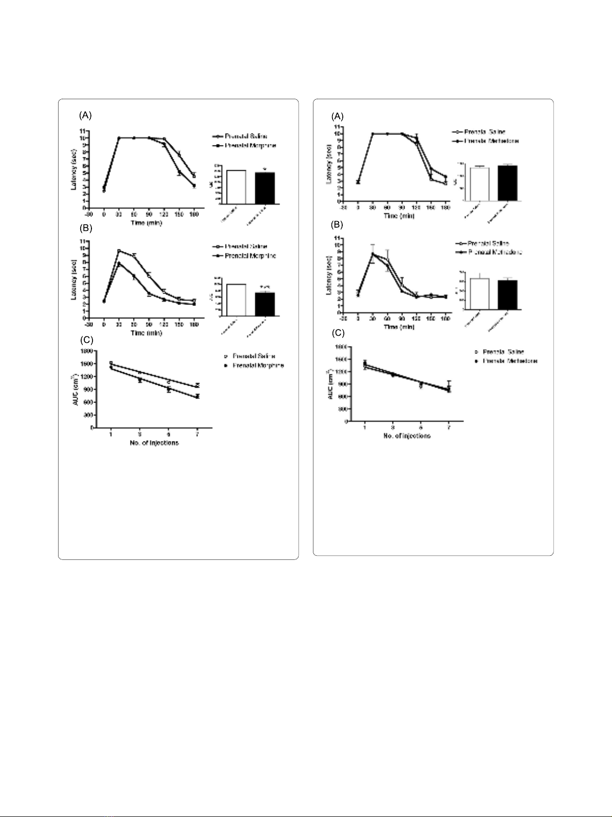
Figure 1 Tolerance development to morphine in prenatally mor-
phine-exposed male rats. (A) The latency and AUC of rats after receiv-
ing first injection of morphine. (B) The latency and AUC of rats after
receiving 7th injection of morphine. (C) Rate of tolerance development
to morphine in morphine or saline prenatally exposed rats. The ani-
mals more quickly developed a tolerance to morphine than the prena-
tally saline-exposed controls (F(1, 114) = 4.333, p < 0.05). All data are
expressed as mean ± S.E.M, (N = 19 per group), *p < 0.05, ***p < 0.001
compared to saline control.
Figure 2 Tolerance development to methadone in prenatally
methadone-exposed male rats. (A) The latency and AUC of rats after
receiving first injection of methadone. (B) The latency and AUC of rats
after receiving 7th injection of methadone. (C) Rate of tolerance devel-
opment to methadone in methadone or saline prenatally exposed rats.
There was no difference in tolerance development to methadone (F(1,
31) = 0.535, p = 0.471) between the methadone and saline prenatally
exposed groups. All data are expressed as mean ± S.E.M, (N = 4 per
group).


%20--%3e%3cdefs%3e%3cstyle%3e%20.st0%20{%20fill:%20%23fff;%20}%20.st1%20{%20fill:%20%237800fa;%20}%20%3c/style%3e%3c/defs%3e%3cpath%20class='st1'%20d='M117.78,12.18H43.11c2.9,3.47,4.65,7.94,4.65,12.82,0,5.6-2.3,10.66-6.01,14.29h76.02l7.22-13.56-7.22-13.56Z'/%3e%3cg%3e%3cpath%20class='st0'%20d='M53.58,26.17h-.59v-1.46h.59v-4.96h2.83c1.78,0,2.67.94,2.67,2.82v5.76c0,1.87-.89,2.81-2.67,2.81h-2.83v-4.96ZM55.36,21.37v3.34h1.1v1.46h-1.1v3.34h1.01c.61,0,.91-.37.91-1.1v-5.93c0-.74-.3-1.1-.91-1.1h-1.01Z'/%3e%3cpath%20class='st0'%20d='M65.99,31.14h-1.8l-.31-2.07h-2.19l-.31,2.07h-1.64l1.82-11.39h2.62l1.82,11.39ZM65.28,18.04c-.25.46-.51.77-.75.94-.21.15-.47.22-.79.22-.26,0-.57-.07-.92-.22l-.38-.15c-.14-.05-.26-.07-.37-.07-.3,0-.53.18-.71.54l-.91-.68c.25-.46.51-.77.75-.94.21-.14.48-.21.79-.21.26,0,.57.07.92.21l.38.15c.14.05.26.07.37.07.3,0,.53-.18.71-.54l.91.68ZM61.91,27.52h1.73l-.87-5.76-.87,5.76Z'/%3e%3cpath%20class='st0'%20d='M74.53,26.89v1.52c0,1.91-.89,2.86-2.67,2.86s-2.67-.95-2.67-2.86v-5.93c0-1.91.89-2.86,2.67-2.86s2.67.95,2.67,2.86v1.11h-1.69v-1.22c0-.75-.31-1.12-.93-1.12s-.93.37-.93,1.12v6.15c0,.74.31,1.11.93,1.11s.93-.37.93-1.11v-1.63h1.69Z'/%3e%3cpath%20class='st0'%20d='M81.4,31.14h-1.8l-.31-2.07h-2.19l-.31,2.07h-1.64l1.82-11.39h2.62l1.82,11.39ZM75.9,19.2l1.52-1.91h1.71l1.51,1.91h-1.61l-.76-.95-.75.95h-1.61ZM77.32,27.52h1.73l-.87-5.76-.87,5.76ZM83.1,15.99l-1.76,1.91h-1.26l1.17-1.91h1.86Z'/%3e%3cpath%20class='st0'%20d='M84.86,19.75c1.78,0,2.67.94,2.67,2.82v1.48c0,1.87-.89,2.81-2.67,2.81h-.85v4.28h-1.79v-11.39h2.64ZM84.01,21.37v3.86h.85c.58,0,.87-.36.87-1.08v-1.71c0-.71-.29-1.07-.87-1.07h-.85Z'/%3e%3cpath%20class='st0'%20d='M93.51,19.75c1.78,0,2.67.94,2.67,2.82v1.48c0,1.87-.89,2.81-2.67,2.81h-.85v4.28h-1.79v-11.39h2.64ZM92.66,21.37v3.86h.85c.58,0,.87-.36.87-1.08v-1.71c0-.71-.29-1.07-.87-1.07h-.85Z'/%3e%3cpath%20class='st0'%20d='M98.8,31.14h-1.79v-11.39h1.79v4.88h2.03v-4.88h1.83v11.39h-1.83v-4.88h-2.03v4.88Z'/%3e%3cpath%20class='st0'%20d='M105.36,24.55h2.46v1.62h-2.46v3.34h3.09v1.63h-4.88v-11.39h4.88v1.63h-3.09v3.18ZM108.17,17.29l-1.76,1.91h-1.26l1.17-1.91h1.86Z'/%3e%3cpath%20class='st0'%20d='M112.2,19.75c1.78,0,2.67.94,2.67,2.82v1.48c0,1.87-.89,2.81-2.67,2.81h-.85v4.28h-1.79v-11.39h2.64ZM111.35,21.37v3.86h.85c.58,0,.87-.36.87-1.08v-1.71c0-.71-.29-1.07-.87-1.07h-.85Z'/%3e%3c/g%3e%3ccircle%20class='st1'%20cx='25'%20cy='25'%20r='20'/%3e%3cpath%20class='st0'%20d='M32.78,19.27c2.92,0,4.43,2.55,5.28,5.33l.71,2.17c.14.38-.33.75-.71.75h-5.61c.19-.33.24-.71.09-1.08l-.75-2.45c-.43-1.32-.99-2.64-1.79-3.77.75-.57,1.65-.94,2.78-.94h0ZM25,18.38c3.25,0,4.9,2.78,5.89,5.89l.76,2.45c.14.42-.33.8-.8.8h-11.69c-.42,0-.94-.38-.8-.8l.75-2.45c.99-3.11,2.64-5.89,5.89-5.89h0ZM25,11.35c1.74,0,3.11,1.37,3.11,3.11s-1.37,3.11-3.11,3.11-3.11-1.41-3.11-3.11,1.41-3.11,3.11-3.11h0ZM17.27,19.27c1.08,0,1.98.38,2.73.94-.8,1.13-1.37,2.45-1.74,3.77l-.8,2.45c-.14.38-.05.75.09,1.08h-5.56c-.42,0-.9-.38-.75-.75l.71-2.17c.9-2.78,2.41-5.33,5.33-5.33h0ZM17.27,12.91c1.51,0,2.78,1.27,2.78,2.83s-1.27,2.83-2.78,2.83-2.83-1.27-2.83-2.83,1.27-2.83,2.83-2.83h0ZM32.78,12.91c1.56,0,2.78,1.27,2.78,2.83s-1.23,2.83-2.78,2.83-2.83-1.27-2.83-2.83,1.27-2.83,2.83-2.83h0ZM27.07,28.56v.09c0,.57-.24,1.08-.61,1.46h0v.05c-.38.33-.9.57-1.46.57s-1.08-.24-1.46-.61h0c-.38-.38-.61-.9-.61-1.46v-.09h1.41v.09c0,.19.05.38.19.47v.05c.09.09.28.19.47.19s.38-.09.47-.19v-.05c.14-.09.24-.28.24-.47t-.05-.09h1.41ZM30.99,28.56v.09c0,1.65-.66,3.16-1.74,4.24-1.08,1.08-2.59,1.79-4.24,1.79s-3.16-.71-4.24-1.79l-.05-.05c-1.04-1.08-1.7-2.55-1.7-4.2v-.09h1.41v.09c0,1.27.47,2.4,1.27,3.25h.05c.85.85,1.98,1.37,3.25,1.37s2.4-.52,3.25-1.37c.85-.8,1.37-1.98,1.37-3.25v-.09h1.37ZM34.99,28.56v.09c0,2.78-1.13,5.28-2.92,7.07-1.79,1.79-4.29,2.92-7.07,2.92s-5.23-1.13-7.07-2.92c-1.79-1.79-2.92-4.29-2.92-7.07v-.09h1.41v.09c0,2.4.94,4.53,2.5,6.08,1.56,1.56,3.72,2.5,6.08,2.5s4.52-.94,6.08-2.5c1.56-1.56,2.5-3.68,2.5-6.08v-.09h1.41Z'/%3e%3c/svg%3e)