7
Journal of Medicine and Pharmacy, Volume 11, No.07/2021
The correlation between femoral intima-media thickness (F.IMT) and
the severity of coronary artery damage in patients with coronary
artery disease
Nguyen Quoc Viet1, Ho Anh Binh2*, Nguyen Phuoc Bao Quan2
(1) Da Nang General Hospital, Vietnam
(2) Hue Central Hospital, Vietnam
Abstracts
A pre-clinical sign of atherosclerisis is hypertrophy of arterial wall. Femoral intima-media thickness is non-
invasive marker of arterial wall alteration, which can easily be assessed by high resolusion B mode ultrasound.
Aims: To investigate the correlation between femoral intima-media thickness and the severity of coronary
artery diseases. Methods: 111 consecutive patients with coronary artery diseases were enrolled. Femoral
intima-media thickness was assessed by B mode ultrasound with 7.5 - 10 MHz probe about 10 - 15 mm
before bifurcation to profond and superfacial femoral arteries. The femoral intima-media thickness < 1.0 mm
is named as “normal”, 1.0 mm is “thick” and 1.5 mm is defined as “atherosclerosic femoral plaque”. The
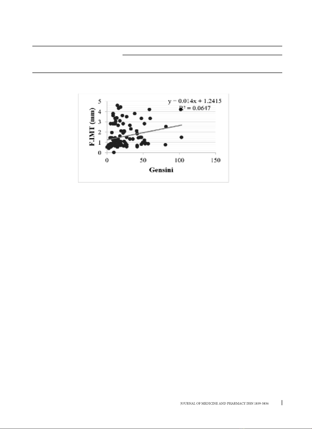
severity of coronary artery diseases was calculated by Gensini Score. Results: Mean femoral intima-media
thickness was 1.57 ± 1.23 mm, 55% patients with abnormal femoral intima-media thickness (male 57.0%
female 50.0%), 36.9% of patients with coronary artery diseases had atherosclerosic femoral plaque. There
was a good correlation between femoral intima-media thickness and severity of coronary artery diseases
by Gensini score and its risk factors (age, plasma glucose, smoking, hypertension…). Conclusion: Patients
with coronary artery diseases are likely to have concomittant peripheral artery disease with high frequency
of femoral artery wall changes. Femoral intima-media thickness could be a helpful diagnostic marker and
therapeutic points.
Keywords: atherosclerisis, Femoral intima-media thickness, coronary artery diseases, femoral intima-
media thickness (F.IMT).
1. INTRODUCTION
Atherosclerosis has been discovered in Egypt
since the 50s BC. The pathogenesis of atherosclerosis
is not entirely clear. Peripheral vascular disease is
an important complication of atherosclerosis. The
risk factors for atherosclerosis such as smoking,
diabetes, dyslipidemia, hypertension and elevated
homocysteine… are also considered major risk
factors for lower limb artery disease [1], [2], [11].
Lower extremity atherosclerosis, which early sign
in the preclinical stage as thickening of the intima-
media layer, can be detected early and accurately
by Doppler ultrasound. The femoral intima-media
thickness (F.IMT) is considered to be an overall
cardiovascular risk factor, was strongly correlation
with coronary artery damage and cardiovascular
events [16], [17], [18].
From the clinical practice, the lower limb artery
disease is often not properly focused, leading to
a missed diagnosis, which can lead to dangerous
complications for the patients because treatment is
too late. Therefore, we implement this study for two
purposes:
1. To assess the Femoral intima-medina thickness
by Doppler ultrasound in patients with coronary
artery diseases.
2. To evaluate the relationship between lower
extremity artery lesions with several cardiovascular
risk factors and severity of lesions to coronary artery
diseases.
2. MATERIALS AND METHODS
A cross-sectional study was conducted on
111 patients with coronary artery disease in Hue
Central Hospital from March 2013 to June 2014. All
participants were provided with written informed
consent and agreed to join our study; and the
protocol was approved by the Ethical Review
Committee of Hue University of Medicine and
Pharmacy, Vietnam
Assessment of severity of coronary artery
disease
All patients were diagnosed with coronary
artery disease based on coronary angiography
Corresponding author: Ho Anh Binh, email: drhoanhbinh@gmail.com
Recieved: 5/1/2021; Accepted: 8/10/2021; Published: 30/12/2021
DOI: 10.34071/jmp.2021.7.1
8
Journal of Medicine and Pharmacy, Volume 11, No.07/2021
3. RESULTS
3.1. General characteristics of the study population
Table 1. General characteristics of study subjects
General features Male (n=79) Female (n=32) Total P
n % n % n %
n 79 71.2 32 28.8 111 100 < 0.05
Mean age 64.48 ± 11.10 68.84 ± 9.65 65.74 ± 10.84
> 0.05
Hypertension 44 55.7 21 65.6 67 58.6
History of coronary artery disease 32 40.51 16 50 48 43.2
Smoking 57 72.2 0 0.0 57 51.4
Diabetes 20 25.32 8 28.6 28 25.2
p > 0.05
Hypertotalcholesterolemia 29 36.7 18 56.3 47 42.3
Hypertriglyceridemia 35 44.9 15 46.9 50 45.5
Hyper-LDLCholesterolemia 23 29.1 9 28.1 32 28.8
Hypo-HDLCholesterolemia 12 15.2 412.5 16 14.4
Study subjects include 79 male patients (71.2%) and 32 female patients (28.8%). The mean age was
65.74 ± 10.84 years. There were a 58.6% patients with hypertension (55.7% male and 65.6% female). The
proportion of patients who smoke was 51.4%, of which 72.2% was male and there was no female patients
smoke. There were 25.2% patients with type 2 diabetes (25.32% male and 28.6% female).
3.2. Coronary artery lesions on DSA:
Table 2. Rate of lesions to the main branches of coronary arteries
Gender
Left Main
(1)
Right Coronary
Artery (2)
Left Anterior
Descending
Artery (3)
Left Circumflex
Artery (4) P
n % n % n % n %
Male (1) 1 1.3 56 71.8 64 82.1 43 55.1 P 3,4 < 0.05
Female (2) 26.3 27 79.4 27 79.4 15 44.1 P 2,4; 3,4 < 0.05
Total 3 2.7 83 74.1 91 81.3 58 51.8 P2,4; 3,4 < 0.001
p (1),(2) > 0.05
LAD lesion is the highest at 81.3%, followed by RCA with 74.1% and LCX with 51.8%. Only 2.7% had a slight
stenosis of the left main coronary artery.
with significant lesion which was > 50% diameter
of stenosis and assess its severity according to the
Gensini score [3].
Bilateral Femoral Arteries Findings by
Ultrasonography
Patients were guided to lay on the supine position
with flexible lower extremities. According to the
standardized protocol for ultrasound in Vietnam,
experienced ultrasound practitioners investigated
femoral arteries from the common femoral arteries
to the bifurcation of the femoral artery into the
superficial artery and the profunda femoral artery.
Colored Doppler and continuous Doppler modes
were employed to investigate the morphology and
functions of arteries. The IMT was measured from
the boundary of the vascular intima and lumen to the
boundary of tunica media and tunica adventitia at end-
diastole B-mode. IMT measurements were performed
at both left and right femoral artery alternatively and
the highest IMT was reported as an IMT variable for
each patient, which classified into 3 categories: (i)
normal IMT (less than 1mm); (ii) thick IMT (1 IMT <
1.5mm); (iii) atherosclerosis (IMT 1.5mm) based on
the classification for carotid artery [9], [10], [11].
9
Journal of Medicine and Pharmacy, Volume 11, No.07/2021
Table 3. Rate of the number of lesion to the main branches of coronary arteries
1-vessel (1) 2-vessel (2) 3-vessel (3) P
n % n % n %
Male (1) 23 29.1 27 34.2 29 36.7 p>0.05
Female (2) 721.9 13 40.6 10 31.3
Total 30 27.03 40 36.04 39 35.13
P (1) (2) p>0.05 p>0.05 p>0.05
The rate of 1-vessel of coronary artery was 27.03%, (male and female were 29.1% and 21.9%, respectively),
2-vessel accounted for 36.04% (male and female were 34.2% and 40.6%, respectively). There was 35.13% of
patients (36.7% male and 31.3% female) have 3-vessel coronaries. Thus, the proportion of patients who have
multiple vessel diseases were 72.97% (the rate of lesion to 2,3 and 4 main vessel coronaries were 36.04%,
35.13% and 1.80%, respectively).
Table 4. The severity of coronary artery lesions by the Gensini score
Diagnosis Male (1) Female (2) Total (3) P
(1),(2)
n Gensini n Gensini nGensini
Stable angina 29 14.41 ± 16.10 13 8.92 ± 6.76 42 12.71 ± 14.04
< 0.01
Unstable
angina 27 24.82 ± 24.66 16 20.25 ± 17.09 43 23.12 ± 22.04
NSTEMI 734.67 ± 11.50 230.00 ± 22.63 9 33.50 ± 13.13
STEMI 16 37.37 ± 22.88 110.00 ± 0.00 17 36.71 ± 23.21
Total 79 24.48 ± 22.2 32 15.94 ± 14.82 111 22.00 ± 20.70
The severity of coronary artery lesions calculated on the Gensini score of study subjects was 22.00 ± 20.70
points, of which 24.48 ± 22.2 points for male and 15.94 ± 14.82 points for female.
3.3. Lesions of the lower limb arteries on B-mode and Doppler ultrasound
Table 5. Average femoral intima-media thickness by gender
Male (1) Female (2) Total P
(1),(2)
M ± SD (mm) M ± SD (mm) M ± SD (mm)
Right side (1) 1.47 ± 1.06 1.54 ± 1.18 1.49 ± 1.09
> 0.05
Left side (2) 1.40 ± 1.01 1.40 ± 1.04 1.40 ± 1.02
F.IMT (3) 1.56 ± 1.10 1.59 ± 1.19 1.57 ± 1.23
P (1) (2) > 0.05 > 0.05 > 0.05
The mean thickness in male was 1.56 ± 1.10 (mm), in female it was 1.59 ± 1, 19 (mm) and for both gender
was 1.57 ± 1.23 (mm).
Table 6. Mean F.IMT by number of damaged coronary vessels
Age group 1-vessel (1) 2- vessel (2) 3-vessel (3) P (1), (2), (3)
n X ± SD (mm) nX ± SD (mm) n X ± SD (mm)
Male 23 1.10 ± 0.86 27 1.43 ± 0.90 29 2.07 ± 1.28
< 0.05Female 71.43 ± 1.15 13 1.22 ± 1.01 10 2.06 ± 1.26
Total 30 1.18 ± 0.93 40 1.36 ± 0.92 39 2.06 ± 1.25
The mean of the femoral intima-media thickness in patients with 1-vessel coronary lesion was 1.18 ± 0.93
(mm), 2-vessel lesion was 1.36 ± 0.92 (mm) and 3-vessel lesion was 2.06 ± 1.25 (mm). The thickness of the
femoral intima-media in patients with 1, 2 and 3 main artery disease tends to increase.
10
Journal of Medicine and Pharmacy, Volume 11, No.07/2021
Table 7. Ratio of femoral intima-media thickness and atheroma
Male (1) Female (2) Total (3) P (1),(2)
n%n%n%
Thick IMT
(IMT ≥ 1.0 mm) 45 57.0 16 50.0 61 55.0 < 0.05
Atheroma/femoral
(IMT ≥ 1.5 mm) 29 36.7 12 37.5 41 36.9 > 0.05
The rate of patients with thick of the intima-media layer femoral artery on ultrasound was 55.0%, of
which 57.0% for male and 50.0% for female. The detection rate of femoral atheroma (with femoral IMT ≥ 1.5
mm) was 36.9%, of which 36.7% for male and 37.5% for female.
Table 8. F.IMT according to several risk factors for coronary artery disease
Risk factor of CAD Yes (1) No (2) P
(1) and (2)
n M ± SD (mm) n M ± SD (mm)
Hypertension 65 1.71 ± 1.26 46 1.38 ± 0.89 p=0.132
History of CAD 48 1.64 ± 1.14 63 1.49 ± 1.11 p=0.015
Hyperglycemia 28 2.02 ± 1.18 83 1.42 ± 1.08 p=0.019
Hyper-totalcholesterolemia 47 1.57 ± 1.17 64 1.58 ± 1.10 p=0.532
Hypertriglyceridemia 50 1.51 ± 1.11 60 1.60 ± 1.14 p=0.66
Hyper-LDLCholesterolemia 32 1.77 ± 1.24 79 1.49 ± 1.07 p=0.25
Hypo-HDLCholesterolemia 48 1.49 ± 1.11 63 1.64 ± 1.14 p=0.511
Smoking 57 1.65 ± 1.14 54 1.49 ± 1.11 p=0.228
For a group of patients with a history of coronary artery disease and diabetes, mean femoral intima-
media thickness was statistically significant compared with the group without.
3.4. The correlation between lower extremity artery damage on B-mode and Doppler ultrasound and
coronary artery diasease:
Table 9. Correlation between F.IMT with age, blood pressure, glucose and blood lipids
Age Blood
pressure Glucose Total –C LDL_C TG HDL_C
F.IMT r=0.319
p<0.01
r=0.351
p<0.05
r=0.404
p<0.001
r=0.205
p<0.05
r=0.170
p>0.05
r=0.035
p>0.05
r=-0.001
p>0.05
Figure 1. Correlation between F.IMT with age and plasma glucose.
There was a statistically significant and positive correlation (0.3 r < 0.5 and p < 0.01) between the
thickness of the femoral intima-media with age and plasma glucose level (r = 0.404 and p < 0.001)
11
Journal of Medicine and Pharmacy, Volume 11, No.07/2021
Table 10. Correlation between the thickness of the femoral intima-media with the number of main
coronary vessel damage:
Number of main coronary vessel
rp
F.IMT r=0,282 p < 0.001
Correlation between the thickness of the femoral intima-media with the number of main coronary vessel
damage was a weak positive correlation and statistically significant with r = 0.282 and p < 0.001.
Figure 2. Correlation between F.IMT and Gensini score.
There was a weak correlation and statistically significant between the thickness of the femoral intima-
media with the severity of coronary artery lesions according to the Gensini score with correlation coefficient
r = 0.247 and p < 0.05, and the linear regression equation y = 0.014x + 1.2415.
4. DISCUSSION
4.1. Femoral intima-media thickness on ultrasound:
According to Depairon et al. (2000) [8], F.IMT
study in 98 healthy patients (53 women and 45 men)
aged 20 to 60, with no risk factor of cardiovascular
diseases. F.IMT was 0.543 ± 0.0063 (mm) in women
and 0.562 ± 0.074 (mm) in men, annually increase
in F.IMT in women was 0.0012 (mm) and 0.0031
(mm) in men. According to Junyent M. et al. (2008)
[10], studied in the intima-medina thickness of the
femoral artery on 192 healthy subjects (85 men,
107 women, mean age 49 years) by ultrasound.
F.IMT values were ranged from 0.50 - 1.04 (mm) in
men aged 35 - 65 years and 0.40 - 0.53 (mm), F.IMT
correlated strongly with age and increased annually
about 0.016 (mm) in men and 0.008 (mm) in women.
F.IMT in our study was statistically significantly
higher than the results of the two above authors
with p < 0.001.
Compared to the study of Grozdinski (2009) on
87 patients with coronary artery diseases, the mean
F.IMT was 1.46 ± 0.41 (mm) compared with the group
of patients without stenosis was 0.85 ± 0.16 (mm) as
well as the control group of 32 healthy subjects was
0.81 ± 0.14 (mm). This difference compared to our
study is no statistically significant with p > 0.05 [9].
Table 6 showed: mean F.IMT in patients with
1-vessel coronary lesion was 1.18 ± 0.93 (mm),
2-vessel was 1.36 ± 0.92 (mm) and 3–vessel was
2.06 ± 1.25 (mm). F.IMT in patients with 1, 2 and 3 of
the main vessels tended to increase and differ from
statistical significance.
Lagroodi R. M. et al (2010), studied on 100
patients with coronary artery diseases divided into
4 groups: group with 1,2,3 vessel diseases and group
with left main coronary lesions. Results: 1-vessel
lesion group: mean F.IMT was 0.64 ± 0.11mm, 2
vessels were 0.73 ± 0.10mm; 3-vessel was 0.84 ±
0.15 and the left main lesion group was 0.85 ± 0.08
(mm). F.IMT increased gradually with the number of
vessel lesions, (p <0.001) [14].
Regarding the F.IMT value, currently there is
no value- approved universally on F.IMT value for
each age group and gender. Many authors agree
to choose the reference value (cut-off) F.IMT is 1
(mm) as Khoury Z. et al [11], Simon A. et al [19].
In this study, we defined femoral intima-media
thickness when F.IMT ≥ 1 (mm) and called femoral