Characterization of SCP-2 from Euphorbia lagascae reveals that a single Leu/Met exchange enhances sterol transfer activity Lenita Viitanen1*, Matts Nylund1*, D. Magnus Eklund2, Christina Alm2, Ann-Katrin Eriksson2, Jessica Tuuf1, Tiina A. Salminen1, Peter Mattjus1 and Johan Edqvist2,3
1 Department of Biochemistry and Pharmacy, A˚ bo Akademi University, Turku, Finland 2 Department of Plant Biology and Forest Genetics, Swedish University of Agricultural Sciences, Uppsala, Sweden 3 IFM-Biology, Linko¨ ping University, Sweden
Keywords b-oxidation; lipid transfer protein; peroxisome; sterol; sterol carrier protein-2
Correspondence J. Edqvist, Department of Plant Biology and Forest Genetics, Swedish University of Agricultural Sciences, Box 7080, 750 07 Uppsala, Sweden Fax: +46 18 673389 Tel: +46 18 673242 E-mail: Johan.Edqvist@vbsg.slu.se
*These authors contributed equally to this work
(Received 25 August 2006, revised 3 October 2006, accepted 23 October 2006)
doi:10.1111/j.1742-4658.2006.05553.x
is a small
Sterol carrier protein-2 (SCP-2) intracellular basic protein domain implicated in peroxisomal b-oxidation. We extend our knowledge of plant SCP-2 by characterizing SCP-2 from Euphorbia lagascae. This pro- tein consists of 122 amino acids including a PTS1 peroxisomal targeting signal. It has a molecular mass of 13.6 kDa and a pI of 9.5. It shares 67% identity and 84% similarity with SCP-2 from Arabidopsis thaliana. Proteo- mic analysis revealed that E. lagascae SCP-2 accumulates in the endosperm during seed germination. It showed in vitro transfer activity of BODIPY- phosphatidylcholine (BODIPY-PC). The transfer of BODIPY-PC was almost completely inhibited after addition of phosphatidylinositol, palmitic acid, stearoyl-CoA and vernolic acid, whereas sterols only had a very marginal inhibitory effect. We used protein modelling and site-directed mutagenesis to investigate why the BODIPY-PC transfer mediated by E. lagascae SCP-2 is not sensitive to sterols, whereas the transfer mediated by A. thaliana SCP-2 shows sterol sensitivity. Protein modelling suggested that the ligand-binding cavity of A. thaliana SCP-2 has four methionines (Met12, 14, 15 and 100), which are replaced by leucines (Leu11, 13, 14 and 99) in E. lagascae SCP-2. Changing Leu99 to Met99 was sufficient to con- vert E. lagascae SCP-2 into a sterol-sensitive BODIPY-PC-transfer protein, and correspondingly, changing Met100 to Leu100 abolished the sterol sensitivity of A. thaliana SCP-2.
new crops [2]. The large size of E. lagascae seeds also makes them attractive for proteomic, biochemical and physiological studies of seed germination. We have previously applied proteomics to E. lagascae endo- sperm to identify novel components involved in endo- sperm degradation, nutrient recycling, lipid catabolism and b-oxidation [3].
Abbreviations BODIPY-PC, BODIPY-phosphatidylcholine; DBP, D-bifunctional protein; DMPA, dimyristoylphosphatidic acid; EF1-a, elongation factor 1-a; GST, gluthathione S-transferase; PDB, Protein Data Bank; RET, resonance energy transfer; SCP-2, sterol carrier protein-2; SCP-X, sterol carrier protein-X.
FEBS Journal 273 (2006) 5641–5655 ª 2006 The Authors Journal compilation ª 2006 FEBS
5641
In Euphorbia lagascae, the storage triacylglycerol in the seed endosperm contains high amounts of the epoxidated fatty acid vernolic acid [(12S,13R)-epoxy- 12-octadecenoic acid]. Vernolic acid has potential industrial applications in the production of paints, coatings and lubricants as an alternative to petroleum- derived oils [1]. Our research interest was initially to increase our knowledge about the enzymatic reactions involved in mobilization and oxidation of vernolic acid in order to improve our potential to develop valuable In this report, we show that sterol carrier protein-2 (SCP-2) accumulates in the E. lagascae endosperm dur- ing germination. SCP-2 is an intracellular, small, basic
L. Viitanen et al.
SCP-2 from Euphorbia lagascae
sensitivity abolished sterol the of amino-acid substitutions that may explain the differ- ences in ligand-transfer activity, we used protein mod- elling and site-directed mutagenesis. Replacement of Leu99 with Met99 was sufficient to convert E. lagascae SCP-2 into a sterol-sensitive BODIPY-PC-transfer protein, and, correspondingly, changing Met100 to Leu100 the BODIPY-PC transfer mediated by A. thaliana SCP-2.
Results
E. lagascae SCP-2 accumulates in the endosperm during germination
protein domain that in vitro enhances the transfer of lipids between membranes [4,5]. In mammals, SCP-2 is implicated in peroxisomal b-oxidation, which is the repeated cleavage of 3-oxoacyl-CoAs into acyl-CoAs this process provides and acetyl-CoAs. In oilseeds, metabolic energy and carbon skeletons to fuel germi- nation and early postgerminative growth [6]. The exact function of the SCP-2 domain in b-oxidation is not clear, but it might facilitate the presentation and ⁄ or solubilization of the substrates and ⁄ or stabilization of the enzymes involved in catalysing the reaction cycles [7,8]. These hypotheses are mainly based on studies of the mammalian peroxisomal proteins, sterol carrier protein-X (SCP-X) and D-bifunctional protein (DBP), which both contain C-terminal SCP-2 domains. SCP-X consists of a 3-oxoacyl-CoA thiolase domain connected to a C-terminal SCP-2 domain [9], whereas DBP has domains for D-3 (equivalent to 3R)-hydroxyacyl-CoA dehydrogenase, 2-enoyl-CoA hydratase and SCP-2 [10,11]. Thus, the enzymatic domain of SCP-X cata- lyses the last step of the peroxisomal b-oxidation path- way, and the enzymatic domains of DBP catalyse the second and third steps. Although genes for DBP and SCP-X have not been identified in plant genomes, we recently showed that plants encode and express SCP-2. PSCP (At5g42890) encodes the Arabidopsis thaliana SCP-2, which is a 13.6-kDa protein with pI of 9.2 pre- dominantly localized in peroxisomes [12]. Curiously, A. thaliana SCP-2 and also all other plant SCP-2s that we have identified are single-domain polypeptides, whereas, as indicated above, SCP-2 domains in ani- mals and many other eukaryotes are often present at the terminus of polypeptides, which carry multiple pro- tein domains [13].
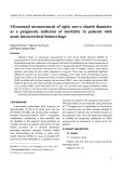
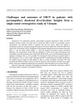
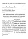
We recently reported the initial characterization of the endosperm proteome of E. lagascae and its changes dur- ing seed germination [3]. In the previous 2D gel electro- phoresis experiments, we used immobilized pH gradients of 3–10, and consequently did not obtain a perfect separation of proteins with high pI. In this set of experiments, our intention was to complement the previ- ous report by focusing on the identification of small and basic proteins that accumulate in the E. lagascae endo- sperm during germination. The aim was to identify novel components involved in b-oxidation, endosperm degradation, or nutrient recycling. Protein extracts from E. lagascae endosperm, collected 2, 4 and 6 days after the seeds had been sown, were loaded on to dry poly- acrylamide gel strips with immobilized pH gradients of 6–11. Electrophoresis in the second dimension was per- formed on 15% polyacrylamide gels. In the 2D gels, we identified spots which increased in size and density dur- ing germination. One such protein spot is indicated in Fig. 1. We cut this spot from the gels, digested the pro- tein with trypsin, extracted the peptides, and finally sequenced the peptides using a mass spectrometer equipped with an electrospray ion source. The peptide sequence analysis revealed that this spot corresponded to E. lagascae SCP-2. The spot corresponding to SCP-2 is barely detectable in samples collected 2 days after sowing, whereas a distinct spot is seen in samples collec- ted after 4 and 6 days (Fig. 1). Thus, there is an evident accumulation of SCP-2 in the endosperm during germi- nation.
Cloning and sequence analysis of E. lagascae SCP-2
cDNA sequences
FEBS Journal 273 (2006) 5641–5655 ª 2006 The Authors Journal compilation ª 2006 FEBS
5642
The peptide sequences, obtained from tandem MS ana- lysis of the E. lagascae SCP-2 2D gel spot, were used in an expressed to search the sequence tag library constructed from mRNA isolated from germinating E. lagascae seeds [14]. One of the The physiological function of plant SCP-2 has not been determined, although its peroxisomal location and lipid-binding capabilities may suggest a role in peroxisomal b-oxidation. To extend our knowledge of the function and activity of plant SCP-2, we have com- pared the lipid-transfer activity of SCP-2 from E. la- gascae and A. thaliana. There are similarities but also quite a few interesting differences. We showed previ- ously that the A. thaliana SCP-2-stimulated transfer of BODIPY-phosphatidylcholine (BODIPY-PC) was unaffected by palmitic acid, indicating that this single- chain lipid is a poor transfer substrate for plant SCP-2 [12]. Therefore, we were surprised to discover that pal- mitic acid very efficiently inhibits BODIPY-PC transfer by E. lagascae SCP-2. Furthermore, in contrast with A. thaliana SCP-2, the E. lagascae SCP-2-mediated BODIPY-PC transfer was not affected by any of the sterols tested. To better understand the lipid–ligand binding mode of plant SCP-2 and to identify
L. Viitanen et al.
SCP-2 from Euphorbia lagascae
Fig. 1. Silver-stained 2D gels showing the differences in the endosperm proteome during different stages of seed germination. Proteins were extracted from E. lagascae endosperm 2, 4 and 6 days after sowing. Gels containing 15% polyacrylamide were used for the second- dimension electrophoresis. The numbers to the left indicate the position and size of the molecular mass protein standards in kDa. pI values indicated ranging from 6 to 11 apply to all gels. The arrows indicate the spots corresponding to SCP-2.
observations allowed us to conclude that E. lagascae SCP-2 is not encoded as a domain of a larger multi- functional protein.
Expression pattern of SCP-2 in E. lagascae
sequenced expressed sequence tag clones, BF12, was identified to encode the sequenced peptides derived from E. lagascae SCP-2. The clone BF12 has accession number BG507194 in GenBank. Complete sequence analysis of BF12 revealed that it lacked the 5¢-end of the coding region. RACE-PCR was carried out on mRNA isolated from the endosperm of germinat- ing seeds to obtain the full coding sequence of the E. lagascae SCP-2 cDNA.
Euphorbia lagascae SCP-2 cDNA sequence encodes a protein of 122 amino acids with a molecular mass of 13.6 kDa and pI of 9.5. It contains a PTS1 peroxisomal targeting signal at the C-terminus (SKL), suggesting that the protein is predominantly localized to peroxi- somes. The amino-acid sequence of E. lagascae SCP-2 shares 67% identity and 84% similarity with A. thaliana SCP-2, 66% identity and 80% similarity with a putative SCP-2 from the monocotyledon Oryza sativa, and 58% identity and 75% similarity with a putative SCP-2 from the moss Physcomitrella patens. Thus, the amino-acid sequence of SCP-2 is well conserved among land plants. The amino-acid identity shared between SCP-2 from land plants and the green algae Chlamydomonas rein- hardtii is less than 40%, which is about the same level of identity shared between land plant and mammalian SCP-2.
during flower also and and
FEBS Journal 273 (2006) 5641–5655 ª 2006 The Authors Journal compilation ª 2006 FEBS
5643
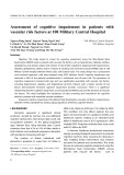
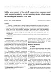
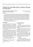
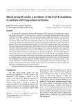
When confirmed and putative plant SCP-2 protein sequences ferns, from angiosperms, gymnosperms, mosses and green algae are aligned, it becomes evident that the C-terminal parts of the proteins are the most conserved regions from Gly86 in (Fig. 2). Thus, E. lagascae SCP-2, angiosperm SCP-2 domains share 100% similarity. As shown in Fig. 2, the start codons are well aligned in the plant SCP-2 sequences. More- in E. lagascae SCP-2 cDNA, an in-frame stop over, codon was detected upstream of the start codon. These Total RNA was isolated from various tissues, such as leaves, roots, stems, flowers and siliques of E. lagascae plants. We also isolated RNA from endosperm and hypocotyls of 4-day-old seedlings. The expression pat- tern of SCP-2 RNA was analysed by RT-PCR using gene-specific primers SCPElNE and SCPElCN. As a control for our RNA preparations and RT-PCR con- ditions, we also used primers ELEFF and ELEFR to analyse the expression of E. lagascae elongation factor 1-a (EF1-a), which is expected to show a stable expres- sion pattern. SCPElNE and SCPElCN amplify a 384- bp fragment from E. lagascae SCP-2, and a 290-bp fragment is amplified from EF1-a, using ELEFF and ELEFR. Analysis of the PCR products revealed that a PCR product from EF1-a was obtained from all sam- ples (Fig. 3). The E. lagascae SCP-2 primers SCPElNE and SCPElCN amplified a PCR product of the expec- ted size from samples from hypocotyls, endosperm, flowers, siliques, leaves and stems. A particularly large accumulation of the SCP-2 amplification product relat- ive to EF1-a was obtained from the endosperm sam- ple, suggesting that SCP-2 mRNA is most abundant in the endosperm of germinating seeds. Moreover, our results indicate that expression is higher in hypocotyls, flowers and siliques than leaves and stems. Thus, E. la- gascae SCP-2 seems to be mainly expressed during ger- mination, seed development. We did not detect any amplification product from the root sample, indicating that SCP-2 is expressed at very low levels in roots.
L. Viitanen et al.
SCP-2 from Euphorbia lagascae
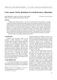
Fig. 2. Multiple sequence alignment of plant SCP-2 from angiosperms (dicotyledons: E. lagascae and A. thaliana; monocotyledon: O. sativa), gymnosperm (Pinus pinaster), fern (Ceratopteris richardii), moss (P. patens) and green algae (Chlamydomonas reinhardtii). Black boxes indi- cate that identical amino acids are present in at least 80% of the sequences, and shaded boxes indicate that amino acids with similar physico- chemical properties are present in at least 80% of the sequences. The sequences included in the analysis have the following GenBank accession numbers: E. lagascae, AAY42079; A. thaliana, NP_199103; P. patens, BJ200729.1; C. richardii, BE642073; O. sativa, AU030065.1; P. pinaster, BX249578.1; Chl. reinhardtii, BI729324.1.
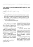
Fig. 3. RT-PCR analysis of SCP-2 in E. lagascae tissues. Total RNA isolated from hypocotyls (HY), endosperm (EN), flower (FL), silique (SI), stem (ST), root (RO) and leaf (LE) was analysed for the expres- sion of SCP-2 and EF1-a. The DNA size marker is shown to the left (MM), with numbers referring to sizes in bp of the corresponding bands.
Lipid-transfer capability of E. lagascae SCP-2
FEBS Journal 273 (2006) 5641–5655 ª 2006 The Authors Journal compilation ª 2006 FEBS
5644
Using a resonance energy transfer (RET) assay, we show that purified recombinant E. lagascae SCP-2 is capable of transferring fluorescently labelled phos- phatidylcholine (BODIPY-PC) between bilayer vesi- cles (Fig. 4, CTRL trace). The matrix lipids of the bilayer vesicles need to be unfavourable substrates for the transfer protein to allow a good transfer signal. A mixture of bovine brain sphingomyelin with choles- terol was chosen as it gives a tight membrane matrix with fluid BODIPY-PC clusters. This tightly packed matrix membrane is as resistant to the SCP-2-medi- ated lipid transfer as possible, and therefore the transfer protein preferentially transfers the labelled BODIPY-PC. To examine the ability of E. lagascae SCP-2 to recognize lipids other than BODIPY-PC as potential substrates, we used a competition assay [12]. In this assay set-up, BODIPY-PC and the added unlabelled lipids compete as substrates for SCP-2. The unlabelled lipids were added as multilamellar aggregates. No additional increase in fluorescence intensity or light scattering was detected as a result of the addition of the unlabelled lipids. This indicates that they remain as a third distinct entity during the measurements. We analysed the inhibiting ability of a range of lipids (Table 1 and Fig. 4). The rate of transfer of BODIPY-PC after the addition of lipo- somes was almost completely inhibited by bovine liver phosphatidylinositol and palmitic acid, both lipids yielding a normalized decrease in BODIPY-PC trans- fer activity of 0.8. The decrease in transfer activity was 0.6 in the presence of stearoyl-CoA or vernolic acid and 0.3 in the presence of dimyristoylphosphatidic
L. Viitanen et al.
SCP-2 from Euphorbia lagascae
Fig. 4. SCP-2 competition assay. BODIPY- PC transfer mediated by E. lagascae SCP-2 before (arrow A) and after (arrow B) addition of competing unlabelled lipids. The lipids were added as multilamellar aggregates that remain as a third entity in the assay. Phos- phatidylinositol, palmitic acid, stearoyl-CoA and vernolic acid have a dramatic competing effect on BODIPY-PC transfer. DMPA has a weak effect, whereas the rest of the lipids analysed interfere marginally with E. lagas- cae SCP-2-mediated BODIPY-PC transfer. The control (CTRL) trace is E. lagascae SCP-2-mediated BODIPY-PC transfer with- out addition of any competitors. MGDG, monogalactosyldiacylglycerol; SPM, palmi- toyl-sphingomyelin; GalCer, galactosyl cera- mide; blPI, bovine liver phosphatidylinositol.
Structural model of E. lagascae SCP-2 in Triton-bound conformation
transfer at all. The the
FEBS Journal 273 (2006) 5641–5655 ª 2006 The Authors Journal compilation ª 2006 FEBS
5645
acid (DMPA). Ergosterol, sitosterol and cholesterol only showed a marginal competing effect, and steryl glucoside, trimyristin, monogalactosyldiacylglycerol, galactosylceramide and palmitoyl-sphingomyelin did not affect results are surprising bearing in mind that, with SCP-2 from A. thaliana, the transfer of BODIPY-PC was almost completely inhibited after the addition of ergosterol, whereas palmitic acid and stearoyl-CoA did not affect the transfer at all [12]. To study the basis for the ligand-binding preference of E. lagascae SCP-2, we constructed a structural model in the Triton-bound conformation (E. lagascae Tr- SCP-2) (Fig. 5A) based on the Triton X-100-bound structure of the SCP-2-like domain of human DBP [Protein Data Bank (PDB) code 1IKT] [8]. The hydro- phobic end of the Triton X-100 molecule is buried in
L. Viitanen et al.
SCP-2 from Euphorbia lagascae
Table 1. E. lagascae SCP-2 in vitro lipid-transfer activity. E. lagas- cae SCP-2-mediated (10 lg) lipid transfer was examined using a fluorescence competition assay. The values, given as decrease in BODIPY-PC transfer rate on introducing the lipid, are mean ± SD from at least four different analyses. blPI, bovine liver phosphatidyl- inositol.
Decrease in BODIPY-PC transfer rate
Lipid
Palmitoyl-sphingomyelin Galactosyl ceramide Trimyristin Steryl glucoside (soybean) Cholesterol Cholesterol oleate Sitosterol Ergosterol DMPA Vernolic acid Stearoyl-CoA Palmitic acid blPI
0.00 ± 0.00 0.00 ± 0.00 0.00 ± 0.00 0.00 ± 0.00 0.11 ± 0.01 0.12 ± 0.01 0.13 ± 0.02 0.15 ± 0.02 0.27 ± 0.03 0.61 ± 0.03 0.63 ± 0.09 0.79 ± 0.01 0.83 ± 0.10
Ser120 and Glu22 are located at the first opening of the cavity. At the second cavity opening, the E. lagas- cae Tr-SCP-2 model has Gln91, Gln109 and Thr112, corresponding to Gln90, Gln108 and Gln111 in the structure of the SCP-2-like domain of human DBP (supplementary Table S1).
tail
The hydrophobic cavity of the E. lagascae Tr-SCP-2 model is extremely similar to the cavity of the A. thali- ana Tr-SCP-2 model (Fig. 5B) [12]. More than half of the amino acids in the cavity are conserved, including the three polar amino acids at the second opening. Nevertheless, we could identify some interesting differ- ences in the two proteins based on their structural models. The A. thaliana SCP-2 cavity has four methio- nines (Met12, 14, 15 and 100), which are replaced by leucines (Leu11, 13, 14 and 99) in E. lagascae SCP-2. Furthermore, the polar residue His18 in A. thaliana SCP-2 is replaced by Phe17 in E. lagascae SCP-2, and residue Glu22 in E. lagascae SCP-2 is the polar replaced by Ala23 in A. thaliana SCP-2. E. lagascae SCP-2 also has a phenylalanine at position 36, whereas this residue is an isoleucine (Ile37) in A. thaliana SCP- 2. Vice versa, Phe76 in A. thaliana SCP-2 is replaced by Leu75 in E. lagascae SCP-2 (Fig. 5A,B; supple- mentary Table S1).
Structural models of E. lagascae and A. thaliana SCP-2 with a bound palmitic acid
the inner cavity and the polar stretches out through the opening between helices D and E and b-strand V (Fig. 5C) [8]. According to the alignment used for modelling, the sequence identity is 36.7% between E. lagascae SCP-2 and the SCP-2-like domain of human DBP. Most of the residues that interact with the Triton X-100 molecule in the crystal structure of the SCP-2-like domain of human DBP are located on the C-terminal half of the protein sequence. The sequence identity between the C-terminal halves of E. lagascae SCP-2 and A. thaliana SCP-2 (starting from Asp77 and Asp78, respectively) is considerably higher (82.6%) than the identity between the N-ter- minal halves (58.4%), suggesting that the C-termini of E. lagascae SCP-2 and A. thaliana SCP-2 are import- ant for ligand binding.
FEBS Journal 273 (2006) 5641–5655 ª 2006 The Authors Journal compilation ª 2006 FEBS
5646
The fold of the E. lagascae and A. thaliana Tr-SCP- 2 models is a five-stranded (I–V) b-sheet covered on one side by five a-helices (A–E). The E. lagascae Tr-SCP-2 model (Fig. 5A) has an inner cavity that is lined by hydrophobic residues Ile10, Leu11, Leu13, Leu14, Phe17, Leu18, Val26, Phe36, Phe73, Leu75, Phe80, Leu83, Ala84, Pro90, Phe94, Leu99, Ile101, Leu105, Ala108, Phe111, Phe116, Pro117 and Pro119. Of these residues, Leu18, Val26, Leu75, Phe80, Pro90, Phe94 and Leu99 are conserved in the SCP-2-like domain of human DBP (supplementary Table S1). The hydrophobic cavity has two openings, of which the first one is formed by residues on helices A, C and E, and the second one is formed by residues on helices D and E and b-strand V (Fig. 5A). The polar residues shown to interfere with the Palmitic acid was BODIPY-PC transfer mediated by SCP-2 and would hence be a potential substrate of E. lagascae SCP-2 (Fig. 4), whereas BODIPY-PC transfer mediated by A. thaliana SCP-2 was not affected by the presence of palmitic acid [12]. This clearly indicates that interac- tions between the negative charge of palmitic acid and the positively charged SCP-2 s (at neutral pH) are not the sole inhibiting effect of BODIPY-PC transfer. To discover a structural reason for the difference in ligand preference, we constructed palmitic acid-bound models of E. lagascae and A. thaliana SCP-2 (pa-SCP-2) based on the mosquito SCP-2 structure (Fig. 5D) [15]. The initial assumption was that the binding mode of pal- mitic acid in E. lagascae SCP-2 might be similar to that in the mosquito SCP-2 structure, where the carb- oxylate moiety of the palmitic acid is bound by amino acids on a loop stretching upwards between helix A and b-strand I (Fig. 5D) [15]. The palmitic acid mole- cule in the mosquito SCP-2 structure is bound in the opposite direction compared with the Triton X-100 molecule in the SCP-2-like domain of human DBP (Fig. 5C) [8]. On the basis of the sequence alignments used for modelling, A. thaliana SCP-2 and E. lagascae
L. Viitanen et al.
SCP-2 from Euphorbia lagascae
A
B
D
C
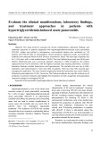
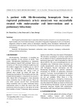
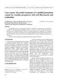
Fig. 5. Structural models of (A) E. lagascae SCP-2 and (B) A. thaliana SCP-2 in Triton- bound conformation (stereo view). The amino acids lining the inner cavity are shown. Residues studied by in vitro muta- genesis are in orange. (C) The crystal struc- ture of the SCP-2-like domain of human DBP [8], on which the models in (A) and (B) are based. (D) The crystal structure of yel- low fever mosquito SCP-2 [15]. The amino acids (grey) and the two water molecules that participate in the binding of the palmitic acid carboxylate are shown. The side chain of Arg24 (yellow) makes a direct bond to the carboxylate group. According to the pal- mitic acid-bound model of E. lagascae SCP-2, based on the mosquito SCP-2 structure, the residue corresponding to Arg24 (yellow) is Lys28 in E. lagascae SCP-2. Mosquito SCP-2 has four methionines (orange) posi- tioned in a row in the inner cavity. Mosquito SCP-2 Met90 corresponds to Met100 in A. thaliana SCP-2. The figures were pre- pared using MOLSCRIPT 2.1.2 [51], RASTER3D 2.7b [52], and GIMP 2.2 (http://www.gimp. org).
SCP-2 share 19.2% and 21.4% sequence identity, respectively, with mosquito SCP-2. Apart from the pal- mitic acid-binding loop, the E. lagascae and A. thaliana pa-SCP-2 models are very similar to the Tr-SCP-2 mod- els, and their inner cavities are lined by hydrophobic residues in the same way as in the Tr-SCP-2 models.
FEBS Journal 273 (2006) 5641–5655 ª 2006 The Authors Journal compilation ª 2006 FEBS
5647
In the mosquito SCP-2 template structure, the negat- ively charged carboxylate group of the palmitic acid molecule is bound by the positively charged Arg24 [15]. The corresponding residue in the (Fig. 5D) E. lagascae pa-SCP-2 model is also a positively charged residue, Lys28, and the corresponding residue in the A. thaliana pa-SCP-2 model is the negatively charged Glu29. To examine whether Lys28 in E. lagascae SCP-2 is in fact involved in palmitic acid binding, we chose this residue for in vitro mutagenesis.
L. Viitanen et al.
SCP-2 from Euphorbia lagascae
Table 2. Lipid-transfer activity of E. lagascae and A. thaliana SCP-2 mutants. Normalized decrease in BODIPY-PC transfer rate mediated by different A. thaliana and E. lagascae SCP-2 mutants (10 lg) upon introducing ergosterol or palmitic acid to the sample. The values are mean ± SD from at least four different analyses.
Plant
Type
Ergosterol
Palmitic Acid
E. lagascae E. lagascae E. lagascae E. lagascae A. thaliana A. thaliana A. thaliana
0.15 ± 0.02 0.14 ± 0.02 0.81 ± 0.10 0.11 ± 0.02 0.91 ± 0.08 0.11 ± 0.02 0.78 ± 0.09
0.79 ± 0.01 0.78 ± 0.02 0.76 ± 0.06 0.77 ± 0.02 0.00 ± 0.00 0.00 ± 0.00 0.09 ± 0.01
Wild-type Lys28Glu Leu99Met Leu13Met ⁄ Leu14Met Wild-type Met100Leu Met14Leu ⁄ Met15Leu ⁄ His18Phe
Docking of ergosterol into the models of A. thaliana SCP-2 and E. lagascae SCP-2 in Triton-bound conformation
converted His18 of A. thaliana SCP-2 into Phe18, and Lys28 of E. lagascae SCP-2 into Glu28. The BODIPY- PC-transfer activity for the different mutants differed only slightly from each other and from the wild-type E. lagascae or A. thaliana SCP-2. Replacing Leu99 with Met is sufficient to convert E. lagascae SCP-2 into a protein that is sensitive to sterols, as the rate of BODIPY-PC transfer was clearly diminished after the addition of ergosterol. The normalized decrease in BODIPY-PC-transfer activity after ergosterol addition was 0.15 for wild-type E. lagascae SCP-2 and 0.81 for the Leu99Met mutant (Table 2). In comparison with the wild-type, BODIPY-PC-transfer activity in the presence of ergosterol did not change for the other E. lagascae mutants, Lys28Glu and Leu13Met ⁄ Leu14Met. Changing Met100 to Leu abolished the sterol sensitivity of A. thaliana SCP-2 BODIPY-PC transfer. The normalized decrease in activity in the presence of ergosterol was 0.91 for wild-type A. thali- ana and 0.11 for the A. thaliana SCP-2 Met100Leu (Table 2). For the A. thaliana SCP-2 triple mutant mutant, Met14Leu ⁄ Met15Leu ⁄ His18Phe, the decrease in BODIPY-PC-transfer activity after the addition of ergosterol was large (0.78) and not significantly differ- ent from that of wild-type A. thaliana SCP-2. None of the mutations in E. lagascae SCP-2 or A. thaliana SCP-2 caused any changes in BODIPY-PC-transfer activity in the presence of palmitic acid (Table 2).
Discussion
Ergosterol was one of the preferred substrates of A. thaliana SCP-2 [12], whereas E. lagascae SCP-2 only showed a slight transfer of ergosterol (Fig. 4). To dis- cover a structural explanation for this substrate selec- tivity, we docked ergosterol into the E. lagascae and A. thaliana Tr-SCP-2 models (Fig. 5A,B) using the program gold 2.2 [17,18]. This found two alternative binding modes for ergosterol with the A. thaliana Tr-SCP-2. The docking result with the higher fitness positioned ergosterol with the hydrocarbon chain pointing towards the cavity opening located between helices D and E and b-strand V (supplementary Fig. S1A). The hydroxy group of ergosterol was posi- tioned close to His18 from helix A. The other docking result, which had a lower fitness, positioned ergosterol in the opposite way (supplementary Fig. S1B). The hydrocarbon chain of ergosterol pointed towards the opening located between helices A, C and E, and the hydroxy group was positioned in the centre of the cavity near Met100. The same two binding modes for ergosterol with the E. lagascae Tr-SCP-2 model were found with gold, but with a lower fitness than with the A. thaliana Tr-SCP-2 model. On the basis the docking results, A. thaliana SCP-2 residues of Met14, Met15, His18, Met100 and the corresponding E. lagascae SCP-2 residues were chosen for in vitro mutagenesis.
Mutant lipid-transfer activity
FEBS Journal 273 (2006) 5641–5655 ª 2006 The Authors Journal compilation ª 2006 FEBS
5648
The sequence identity between E. lagascae SCP-2 and A. thaliana SCP-2 is high, and the inner cavities of the two proteins are accordingly extremely similar, which would suggest that the proteins have similar ligands. Therefore, we were surprised to discover that palmitic acid very efficiently inhibits BODIPY-PC transfer mediated by E. lagascae SCP-2, whereas we showed previously that the A. thaliana SCP-2-stimulated trans- fer of BODIPY-PC was unaffected by palmitic acid. To test the importance of specific amino acids for lipid-transfer activity, we constructed genes encoding variants of A. thaliana and E. lagascae SCP-2. In par- ticular, we replaced some of the Met residues in the hydrophobic cavity of A. thaliana SCP-2 with Leu, and vice versa in E. lagascae SCP-2. Furthermore, we
L. Viitanen et al.
SCP-2 from Euphorbia lagascae
We first thought that the binding mode of palmitic acid to E. lagascae SCP-2 might be the same as in the mosquito SCP-2 structure (Fig. 5D) [15], where Arg24 binds the carboxylate group of palmitic acid. The structural model of palmitic acid-bound E. lagascae SCP-2 suggests that Lys28 is important in palmitic acid binding. To test this hypothesis, Lys28 in E. lagascae SCP-2 was mutated to Glu, which is the in A. thaliana SCP-2. This corresponding residue Lys28Glu mutant showed no decrease in BODIPY- PC-transfer activity compared with the wild-type when palmitic acid was present. Therefore, we now conclude that Lys28 is not crucial for palmitic acid binding to E. lagascae SCP-2.
in the binding of ergosterol (supplementary Fig. S1A). the A. thaliana The competition assay showed that SCP-2 Met100Leu mutant had lost its ergosterol sensi- tivity, and the E. lagascae SCP-2 Leu99Met mutant had acquired ergosterol sensitivity. Hence, Met100 is crucial for ergosterol binding in A. thaliana SCP-2, and, even more surprising, introducing this methionine to the E. lagascae SCP-2 cavity is enough to provide sterol-binding properties. However, the other methio- nines located on helix A in A. thaliana SCP-2 are apparently of little or no importance for ergosterol binding, as, in the presence of ergosterol, the A. thali- ana and SCP-2 Met14Leu ⁄ Met15Leu ⁄ His18Phe E. lagascae SCP-2 Leu13Met ⁄ Leu14Met mutants had similar BODIPY-PC-transfer activities to their corres- ponding wild-type proteins. The role of His18 in ligand binding to A. thaliana SCP-2 was also examined using the Met14Leu ⁄ Met15Leu ⁄ His18Phe mutant. His18 seems to have no effect on binding of ergosterol, as wild-type A. thaliana SCP-2 and the A. thaliana Met14Leu ⁄ Met15Leu ⁄ His18Phe mutant showed sim- ilar decreases in BODIPY-PC-transfer activity after addition of ergosterol.
is, thus, for residues are
How does E. lagascae SCP-2 then actually bind palmitic acid? One possibility is that another residue from the loop between helix A and b-strand I is responsible for the binding. Suitable candidates for binding would be Lys25 and Gln27, corresponding to Glu26 and Thr28 in A. thaliana SCP-2. Another pos- sibility is that E. lagascae SCP-2 binds palmitic acid in a similar way to the binding of Triton X-100 to the SCP-2-like domain of human DBP [8], i.e. with the charged moiety in the completely opposite direc- tion. In this binding mode, the polar residues (Gln91, Gln109 and Thr112) on helix E in E. lagascae SCP-2 (Fig. 5A) could provide a suitable surrounding for the carboxylate group of palmitic acid. These residues are, however, completely conserved in A. thaliana SCP-2 (Fig. 5B) and therefore cannot contribute to the different binding preferences of A. thaliana and E. lagascae SCP-2. A possible explanation is that the hydrophobic residues, Leu75, Leu83 and Leu99 in E. lagascae SCP-2 (Fig. 5A), corresponding to Phe76, Val84 and Met100 in A. thaliana SCP-2 (Fig. 5B), make the shape and size of the binding cavities in E. lagascae and A. thaliana SCP-2 somewhat different and, thus, affect ligand specificity. structures of crystal the the
FEBS Journal 273 (2006) 5641–5655 ª 2006 The Authors Journal compilation ª 2006 FEBS
5649
The X-ray crystallographic structures of SCP-2 domains also show methionines in their hydrophobic tempting to speculate that cavity, and it these sterol binding. important Mosquito SCP-2 has five methionines, four of which are positioned close to each other in a row (Fig. 5D) [15]. One of these methionines (Met90) corresponds to Met100 in A. thaliana SCP-2, and there are experi- mental results showing that yellow fever mosquito SCP-2 has high affinity for cholesterol [19]. Mamma- lian SCP-2 has also been shown to transfer choles- terol [20,21], and rabbit SCP-2 has two methionines in its hydrophobic cavity [16]. Furthermore, there are published structures of protein–sterol complexes in which methionines participate in binding of the sterol, e.g. fungal protein b-cryptogein in complex with ergosterol and choles- terol show that three methionines positioned close to each other in the binding cavity interact with the two the sterol molecules (PDB code methyl groups of 1BXM) [22] (PDB code 1LRI) [23]. In the crystal structure of human 17b-hydroxysteroid dehydrogenase in complex with estradiol, two methionines are in contact with the sterol (PDB code 1FDS) [24]. The O3-hydroxy group of estradiol is bound by a histi- dine, but one of the two methionines is close to the hydroxy group. Further studies are needed to eluci- date how A. thaliana SCP-2 binds to ergosterol and to determine the exact role of Met100 in the binding process. Another intriguing discovery was that ergosterol, which efficiently inhibited BODIPY-PC transfer by A. thaliana SCP-2, has only a marginal effect on E. lagascae SCP-2-mediated BODIPY-PC transfer. The docking of ergosterol in A. thaliana SCP-2 gave two solutions (supplementary Fig. S1A,B) which are quite similar to the respective binding modes of Triton X-100 in the SCP-2-like domain of human DBP [8] and palmitic acid in mosquito SCP-2 (Fig. 5C) (Fig. 5D) [15]. A comparison of the hydrophobic cavit- ies in A. thaliana and E. lagascae SCP-2 revealed that A. thaliana SCP-2 has four methionines, which are replaced by leucines in E. lagascae SCP-2 (Figs 2 and 5A,B). On the basis of the docking analysis, we suggest that the methionines in A. thaliana SCP-2 are involved
L. Viitanen et al.
SCP-2 from Euphorbia lagascae
the SCP-2-mediated transfer of
to the active sites of Thus, the role of SCP-2 would mainly be to facilitate b-oxidation of some substrates. Plant SCP-2 may also interact with other b-oxidation enzymes such as the 3-oxoacyl-CoA thiolases and acyl-CoA oxidases. Alter- natively or additionally, SCP-2 may improve the cata- the b-oxidation process by recruiting lytic rate of substrates the b-oxidation enzymes.
Being basic at neutral pH, SCP-2 is likely to inter- act with negatively charged interfaces. Thus, we can- not rule out that negatively charged lipids will inhibit to some extent its substrates and that SCP-2 might be sensitive to neg- atively charged membranes analogous to the glyco- lipid transfer protein [25]. However, it is important to note that whereas some negatively charged lipids, such as bovine liver phosphatidylinositol, efficiently inhibited the BODIPY-PC transfer mediated by E. lagascae SCP-2, other negatively charged lipids, such as DMPA, only had a rather marginal inhibi- tory effect. Furthermore, A. thaliana SCP-2 is also positively charged at neutral pH and seems able to transfer its substrate from negatively charged surfaces [12].
that interest vernolic
substrate the biological
It is possible that the distinctly different lipid-trans- fer properties of E. lagascae and A. thaliana SCP-2 reflect the lipid composition of the respective plant spe- cies. The differences may also indicate that E. lagascae and A. thaliana SCP-2 have adopted slightly different or overlapping physiological functions. We speculate that the sterol-binding property of A. thaliana SCP-2 may indicate an involvement of this protein in non- vesicular trafficking of sterols as well as its suggested role in peroxisomal b-oxidation. On the other hand, the lack of sterol-transfer activity shown for E. lagas- cae SCP-2 would suggest that it has a more specialized function in fatty acid b-oxidation. Our finding that changing one leucine residue to a methionine increased the affinity of SCP-2 for sterols will hopefully open the way for other experiments directed towards learning more about function of SCP-2. For instance, it will be of interest to express SCP-2 proteins with altered ligand-binding properties in plant or animal models in which phenotypes for deletion and overexpression of SCP-2 proteins have already been assessed.
Experimental procedures
Plants, bacteria and chemicals
Euphorbia lagascae Spreng. was germinated and grown as previously described [3]. Tissues were stored at )80 (cid:2)C, for shorter periods of time, until used. The E. lagascae seeds were a gift from S. Stymne, Department of Crop Science, SLU, Alnarp, Sweden. Cloning was performed in Escheri- chia coli DH5a, and E. coli BL21 cells were used for over-expression of E. lagascae and A. thaliana SCP-2. The fluorescent probes BODIPY FL C12 PC and DiI-C18 were from Invitrogen (Carlsbad, CA, USA). Egg sphingomyelin and bovine liver phosphatidylinositol were purchased from Avanti Polar Lipids (Alabaster, AL, USA). Galactosylcera- mide, trimyristin, cholesterol oleate, steryl glycoside (soy- bean), ergosterol, b-sitosterol, DMPA, palmitic acid, stearoyl-CoA and vernolic acid [(+)-(12S,13R)-epoxy-cis- 9-octadecenoic acid methyl ester] were from Larodan AB (Malmo¨ , Sweden). Cholesterol was from Sigma (St Louis, MO, USA). Palmitoyl-sphingomyelin was purified from egg sphingomyelin using RP-HPLC (Supelco, Bellefonte,
The expression pattern, the lipid binding and trans- fer capability, and the peroxisomal targeting signal allow us to suggest that E. lagascae SCP-2 is involved in the peroxisomal oxidation of lipids. It was of partic- ular acid interfered with BODIPY-PC transfer, as the storage triacylglycerols of E. lagascae largely consist of this fatty acid. These results from our indirect assay suggest that vernolic acid could be a favoured transfer for E. lagascae SCP-2 and supports our hypothesis that E. lagascae SCP-2 is involved in the catabolism of storage triacylglycerols in the endosperm. Our data from studies of A. thaliana suggest that A. thaliana SCP-2 is also involved in this process (B. S. Zheng and J. Edqvist, unpublished results). According to the cur- rent model of b-oxidation in A. thaliana [6], the ABC transporter CTS (also referred to as PED3 or PXA1) delivers fatty acids into the peroxisomes [26–28]. In the peroxisome, the acyl-CoA synthetases activate the fatty acids to acyl-CoA esters [29]. Finally, the b-oxidation enzymes [acyl-CoA oxidases ACX1–6, the multifunc- tional proteins MFP2 and AIM1, and the 3-oxoacyl- CoA thiolases (PED1, KAT1 and PKT2)] catalyse the repeated cleavage of the acyl-CoA esters to yield acetyl-CoA [30–34].
FEBS Journal 273 (2006) 5641–5655 ª 2006 The Authors Journal compilation ª 2006 FEBS
5650
How does SCP-2 fit into this scheme of peroxisomal b-oxidation in plants? In A. thaliana, SCP-2 is tightly coexpressed with MFP2 (Zheng and J. Edqvist, unpub- lished results) indicating a partnership between these two proteins. We suggest that SCP-2 interacts with the multifunctional protein to form a cavity for the hydro- phobic tails of some b-oxidation substrates, as has been suggested for the SCP-2-like domain of mamma- lian DBP [8]. The extended hydrophobic cavity will increase the accessibility and solubility for at least some of the b-oxidation substrates and consequently improve the catalytic rate of the b-oxidation process.
L. Viitanen et al.
SCP-2 from Euphorbia lagascae
PA, USA; Discovery C18; 5 lm particle size; 250 mm · 21.2 mm column dimensions at 9 mLÆmin)1 with methanol as the mobile phase).
TAGGCTAAAGTC-3¢). Synthetic oligonucleotides ELEFF and ELEFR were designed based on the E. lagascae cDNA-clone CE5 with accession number BG507239 [14].
to Boxshade
[12]
Total protein extracts were prepared from the endosperm of 2-, 4- and 6-day-old seedlings as described in [3]. The protein solutions were loaded on to dry polyacrylamide gel strips with immobilized pH gradients of 6–11 (GE Health- care, Chalfont St Giles, UK). First and second dimension gel electrophoresis, silver staining, and analysis of gels were carried out as previously described [3]. Peptide analysis by MS was performed by a slightly modified version [3,35] of a method described by Wilm et al. [36]. To acquire peptide sequence data, a quadropole time-of-flight mass spectrome- try instrument (Micromass Q-Tof; Waters Corp., Milford, MA, USA) with a nanospray ion source was used. The capillary voltage was set to 800–900 V and the cone volt- age to 40 V. Argon was used as collision gas, and the kin- etic energy was set to between 20 and 40 eV. Peptide sequence data were analysed using the biolynx program of the MassLynx NT software package (version 3.4; Waters Corp.). Peptide sequences obtained by MassLynx were subjected to blast using blastp [37] at the National Center for Biotechnology Information (NCBI: http:// www.ncbi.nlm.nih.gov/BLAST/). Multiple sequence align- ment was created using clustalx [38], and the resulting similarities were then visualized by subjecting the align- ment (http://www.ch.embnet.org/software/ BOX_form.html).
Proteomics and bioinformatics Structural modelling of E. lagascae SCP-2
A structural model of E. lagascae SCP-2 in the Triton- bound conformation was constructed based on the crystal structure of the SCP-2-like domain of human DBP (PDB code 1IKT) [8], which has bound a Triton X-100 mole- is referred to as E. lagascae cule (Fig. 5C). This model Tr-SCP-2. Structural models of E. lagascae SCP-2 and A. thaliana SCP-2 with a bound palmitic acid were also constructed based on the crystal structure of yellow fever mosquito SCP-2 (PDB code 1PZ4) [15], which has a pal- mitic acid molecule bound to its inner cavity (Fig. 5D). These models are referred to as E. lagascae pa-SCP-2 and A. thaliana pa-SCP-2, respectively. The sequences of E. lagascae SCP-2, A. thaliana SCP-2, rabbit SCP-2, mos- quito SCP-2 and the SCP-2-like domain of human DBP were submitted to the PredictProtein server (available at http://cubic.bioc.columbia.edu/predictprotein/), which pre- dicts the secondary structure of a protein sequence [40]. The known SCP-2 crystal structures were superimposed using the program vertaa in the Bodil modelling envi- [41]. The E. lagascae and A. thaliana SCP-2 ronment sequences were aligned with the template sequences using the program malign within Bodil [42,43]. The sequence alignments used for modelling were refined in the same way as described in Edqvist et al. in order to con- struct models comparable to the previously made models of A. thaliana SCP-2 in Triton-bound conformation (A. thaliana Tr-SCP-2). The models were constructed using the program modeller [44], and the representative model was selected as previously described [12]. The cavities within the models and the known structures were identified by using the program surfnet [45] and visualized and examined with Bodil [41].
to
added
site
(Hetero-compound
The docking program gold 2.2 [17,18] was used to into the E. lagascae and A. thaliana dock ergosterol Tr-SCP-2 models. Hydrogens were the models before the gold runs using the program reduce [46]. The ergosterol structure was downloaded from the Information Centre– HIC-Up Uppsala, http://xray.bmc.uu.se/hicup/) [47]. Ten independ- ent genetic algorithm runs were made in gold for the ligand, using the default docking parameters. The binding site in the models was restricted within a 15 A˚ radius of the side-chain hydrogen (HZ) of Phe112 and Phe111 in the A. thaliana and E. lagascae Tr-SCP-2 models, respect- ively. The docking was stopped if the three best scoring solutions were within 1.5 A˚ rmsd of each other. The docking results were visualized and examined in Bodil [41].
Euphorbia lagascae total RNA was isolated from various tissues using a guanidine hydrochloride method previously described by Logemann et al. [39], or with the RNeasy Plant Mini Kit (Qiagen, Hilden, Germany). RACE-PCR, to obtain the full-length cDNA sequence of E. lagascae SCP-2, was performed with the FirstChoice RLM-RACE kit (Ambion, Austin, TX, USA). Outer (STOP, 5¢-GAC AAATTCGATCCAAATCCA-3¢) and inner (STPI, 5¢-CAG TATGCCTCACAGCTTCG) E. lagascae SCP-2 specific primers were designed based on the sequence of cDNA clone BF12 [14]. For RT-PCR, 2 lg total RNA was used for cDNA synthesis together with 200 ng poly(dT) primer in a polymerization reaction catalysed by Superscript III (Invitrogen) in a total volume of 20 lL. A 1-lL portion of the cDNA obtained was used in PCRs with gene-specific primers for E. lagascae SCP-2 (SCPElNE, 5¢-ACTGGAA TTCAACTCAAGTCCCAAAATATTTTGGAT-3¢; SCPElCN, 5¢-TCATGGCGGCCGCTCACAGCTTCGACGGCTTGG GGAA-3¢) and E. lagascae EF1-a (ELEFF, 5¢-TATGGT GGTTGAGACTTTTGCAG-3¢; ELEFR, 5¢-GCATTCCAC
FEBS Journal 273 (2006) 5641–5655 ª 2006 The Authors Journal compilation ª 2006 FEBS
5651
RACE and RT-PCR
L. Viitanen et al.
SCP-2 from Euphorbia lagascae
SCP-2 inserts were confirmed by DNA sequencing. Plas- mids carrying SCP-2 variants were transformed into E. coli BL21 cells.
and
Expression and purification of plant SCP-2 in E. coli
The coding region was amplified from E. lagascae cDNA by the use of primers SCPElNE (5¢-ACTGGAATTCAACT CAAGTCCCAAAATATTTTGGAT-3¢) SCPElCN (5¢-TCATGGCGGCCGCTCACAGCTTCGACGGCTTGG GGAA-3¢). The PCR fragment obtained was digested with restriction enzymes EcoRI and NotI, and subcloned into the EcoRI–NotI site of the vector pGEX-5X-2 (GE Healthcare) to obtain a gene fusion between gluthatione S-transferase (GST) and E. lagascae SCP-2. The E. lagascae SCP-2 insert in the obtained plasmid pGEX-SCPEl was confirmed by DNA sequencing. Plasmid pGEX-SCPEl was transformed into E. coli BL21 cells. The bacteria were cultured at 27 (cid:2)C. When A600 reached 1.1, the expression of GST-ElSCP-2 was induced by the addition of 0.6 mm isopropyl b-d-thiogalacto- side. Expression of A. thaliana SCP-2 in E. coli was per- formed as described previously [12]. Purification of the fusion proteins (supplementary Fig. S2) and cleavage with Factor Xa were carried out according to the handbook for the GST Gene Fusion System (GE Healthcare) as described in [12].
The RET method used for measuring the transfer of lipids between bilayer vesicles, which is based on a method pub- lished previously [49], has been described briefly [12]. BODIPY-PC (1%) was used as the energy donor and DiI- C18 (3%) was the energy acceptor. The labelled lipids were mixed with bovine brain sphingomyelin and cholesterol (6 : 1), dried under nitrogen and then dissolved in sodium phosphate buffer containing 10 mm sodium dihydrogen phosphate, 1 mm dithiothreitol and 1 mm EDTA. The small unilamellar vesicles (both donors and acceptors) were prepared by sonication for 10 min on ice with a Branson (Danbury, CT, USA) 250 sonifier, and then centrifuged for 15 min at 15 000 g with a Sorvall Biofuge Pico centrifuge (Thermo Electron Corporation, Waltham, MA, USA) to remove titanium probe particles, multilamellar vesicles and undispered lipids (negligible amount). The acceptor vesicles were used in 10-fold excess over donor vesicles. The trans- fer was started by introducing 10 lg protein to the assay.
RET assay for monitoring SCP-2-mediated lipid intermembrane transfer
In vitro mutagenesis of A. thaliana and E. lagascae SCP-2
the injection of
To analyse the ability of plant SCP-2 to transfer unlabelled lipids, a competition assay [12,50] was used. Although indi- rect, this assay enables us to address if unlabelled lipids interfere with the transfer of BODIPY-PC in an experimen- tal set-up that does not require the large amount of mater- ial that is often the case in conventional binding assays. The RET assay with BODIPY-PC, which has been shown to be a substrate, was started by addition of SCP-2. Two minutes after the protein, when the BODIPY-PC transfer was still ongoing, we introduced an unlabelled lipid (sonicated multilamellar aggregates) to the sample. If the added lipid was a substrate for SCP-2, it would result transfer of in a decrease in the rate of BODIPY-PC because of the competition from the presence of a new distinct transferable lipid pool. When the added lipid was not a substrate for SCP-2, no deviation in the slope of the transfer rate would occur. All the unlabelled lipids were sonicated (Finnsonic Bath Sonifier; Finnsonic, Lahti, Finland) for 6 min at 25 (cid:2)C. The amount of the added lipid was 400 nmol. No additional increase in the fluorescence intensity or light scattering was detected as a result of the addition of the multilamellar aggregates. This would indicate that no fusion or aggregation of the assay component occurred during the time frame of the measure- ments. The values are derived from using a control assay,
The following oligonucleotides were used for in vitro muta- genesis: ELM13M14 (5¢-AAGTCCCAAAATATTATGGA TATGATGGCTCGATTTCTCGAG-3¢); ELE28 (5¢-ATG AAAGTTCAAGAGAAGATCAATCTC-3¢); ELM99 (5¢- ATGAGGGGTGCTATGAAGATCAAGG-3¢); ATL100 (5¢-ATTAGGGGTGCGCTGAAGATCAAGG-3¢); ATL14- L15F18 (5¢-AAATCCGATGCAATCCTGGACCTGCTG AAGGAATTTCTTTCCACCGACGCC-3¢). For construc- tion of A. thaliana SCP-2 variants Met100Leu and Met14Leu ⁄ Met15Leu ⁄ His18Phe and E. lagascae SCP-2 var- iants Leu13Met ⁄ Leu14Met, Lys28Glu and Leu99Met, we utilized the megaprimer method [48]. The first round of PCRs was performed with the mutagenic primer in combi- nation with the M13 reverse primer. In the second round, the PCR product of the first round was used as primer in combination with the M13 forward primer. Plasmids pGEX-SCPAt [12] and pGEX-SCPEl served as template for both rounds of PCR. In a third round of PCR, the PCR products from round two were, in the case of A. thali- ana SCP-2, amplified with primers SCPAtCN and SCPAtNE, or, in the case of E. lagascae SCP-2, amplified with primers SCPElNE and SCPElNE. PCRs were per- formed with Phusion DNA polymerase (Finnzymes, Espoo, Finland). The obtained PCR fragments carrying the SCP-2 gene variants were digested with restriction enzymes EcoRI and NotI, and subcloned into the EcoRI–NotI site of the vector pGEX-5X-2 (GE Healthcare) to obtain gene fusions between GST and the SCP-2 genes. The sequences of the
FEBS Journal 273 (2006) 5641–5655 ª 2006 The Authors Journal compilation ª 2006 FEBS
5652
Competition assay for monitoring SCP-2-mediated lipid intermembrane transfer
L. Viitanen et al.
SCP-2 from Euphorbia lagascae
resolution. J Mol Biol
tional enzyme type 2 at 1.75 A˚ 313, 1127–1138.
9 Ohba T, Rennert H, Pfeifer SM, He Z, Yamamoto R, Holt JA, Billheimer JT & Strauss JF3rd (1994) The structure of the human sterol carrier protein X ⁄ sterol carrier protein 2 gene (SCP2). Genomics 24, 370–374.
10 Adamski J, Normand T, Leenders F, Monte D,
where the BODIPY-PC transfer is allowed to continue without disturbance. The fluorescence intensity increase at 3 min for the BODIPY-PC transfer without a competing lipid is set to 0. A straight line, indicating no BODIPY-PC transfer, i.e. complete inhibition, is set to 1. The fraction of decrease in the fluorescence intensity compared with the control at 3 min after injection of the competitive lipid is given as the values in Tables 1 and 2.
Acknowledgements
Begue A, Stehelin D, Jungblut PW & de Launoit Y (1995) Molecular cloning of a novel widely expressed human 80 kDa 17b-hydroxysteroid dehydrogenase IV. Biochem J 311, 437–443.
11 Leenders F, Dolez V, Begue A, Moller G, Gloeckner
JC, de Launoit Y & Adamski J (1998) Structure of the gene for the human 17b-hydroxysteroid dehydrogenase type IV. Mamm Genome 9, 1036–1041.
12 Edqvist J, Ro¨ nnberg E, Rosenquist S, Blomqvist K,
Viitanen L, Salminen TA, Nylund M, Tuuf J & Mattjus P (2004) Plants express a lipid transfer protein with high similarity to mammalian sterol carrier protein-2. J Biol Chem 279, 53544–53553.
13 Edqvist J & Blomqvist K (2006) Fusion and fission, the evolution of sterol carrier protein-2. J Mol Evol 62, 292–306.
facilities at
14 Edqvist J & Farbos I (2002) Characterization of germi- nation-specific lipid transfer proteins from Euphorbia lagascae. Planta 215, 41–50.
the Professor Mark Johnson is acknowledged for excellent the Structural Bioinformatics Laboratory, and Professor J. Peter Slotte for the fluore- scence facility at the Department of Biochemistry and Pharmacy, A˚ bo Akademi University. We acknowledge the excellent technical support of Ingrid Schenning. We are grateful for the financial support from The Swedish Research Council, Carl Trygger Foundation, Magn. Bergvall Foundation, Academy of Finland, Sigrid Juse´ lius Stiftelse, Magnus Ehrnrooths Stiftelse, Svenska Kulturfonden, Medicinska Understo¨ dsfo¨ renin- gen Liv och Ha¨ lsa r.f and A˚ bo Akademi University.
References
1 Derksen JTP, Cuperus FP & Kolster P (1995) Paints
and coatings from renewable resources. Ind Crops Prod 3, 225–236.
2 Edqvist J & Farbos I (2003) A germination-specific
15 Dyer DH, Lovell S, Thoden JB, Holden HM, Rayment I & Lan Q (2003) The structural determination of an insect sterol carrier protein-2 with a ligand-bound C16 fatty acid at 1.35-A resolution. J Biol Chem 278, 39085– 39091.
epoxide hydrolase from Euphorbia lagascae. Planta 216, 403–412.
3 Eklund DM & Edqvist J (2003) Localization of
16 Choinowski T, Hauser H & Piontek K (2000) Structure of sterol carrier protein 2 at 1.8 A˚ resolution reveals a hydrophobic tunnel suitable for lipid binding. Biochem- istry 39, 1897–1902.
non-specific lipid transfer proteins correlate with pro- grammed cell death responses during endosperm degra- dation in Euphorbia lagascae seedlings. Plant Physiol 132, 1249–1259.
4 Ritter MC & Dempsey ME (1971) Specificity and role in cholesterol biosynthesis of a squalene and sterol car- rier protein. J Biol Chem 246, 1536–1539.
5 Bloj B, Hughes ME, Wilson DB & Zilversmit DB
(1978) Isolation and amino acid analysis of a nonspeci- fic phospholipid transfer protein from rat liver. FEBS Lett 96, 87–89.
6 Baker A, Graham IA, Holdsworth M, Smith SM &
Theodoulou FL (2006) Chewing the fat: b-oxidation in signalling and development. Trends Plant Sci 11, 124– 132.
17 Jones G, Willett P & Glen RC (1995) Molecular recog- nition of receptor sites using a genetic algorithm with a description of desolvation. J Mol Biol 245, 43–53. 18 Jones G, Willett P, Glen RC, Leach AR & Taylor R (1997) Development and validation of a genetic algo- rithm for flexible docking. J Mol Biol 267, 727–748. 19 Krebs KC & Lan Q (2003) Isolation and expression of a sterol carrier protein-2 gene from the yellow fever mosquito Aedes aegypti. Insect Mol Biol 12, 51–60. 20 Puglielli L, Rigotti A, Greco AV, Santos MJ & Nervi F (1995) Sterol carrier protein-2 is involved in cholesterol transfer from the endoplasmic reticulum to the plasma membrane in human fibroblasts. J Biol Chem 270, 18723–18726.
7 Seedorf U, Ellinghaus P & Nofer JR (2000) Sterol car-
rier protein-2. Biochim Biophys Acta 1486, 45–54. 8 Haapalainen AM, van Aalten DM, Merilainen G,
21 Baum CL, Reschly EJ, Gayen AK, Groh ME & Scha- dick K (1997) Sterol carrier protein-2 overexpression enhances sterol cycling and inhibits cholesterol ester synthesis and high density lipoprotein cholesterol secre- tion. J Biol Chem 272, 6490–6498.
Jalonen JE, Pirila P, Wierenga RK, Hiltunen JK & Glumoff T (2001) Crystal structure of the liganded SCP-2-like domain of human peroxisomal multifunc-
FEBS Journal 273 (2006) 5641–5655 ª 2006 The Authors Journal compilation ª 2006 FEBS
5653
L. Viitanen et al.
SCP-2 from Euphorbia lagascae
22 Boissy G, O’Donohue M, Gaudemer O, Perez V,
that can oxidize short-chain acyl-CoA in plant peroxi- somes. J Biol Chem 274, 12715–12721.
Pernollet JC & Brunie S (1999) The 2.1 A˚ structure of an elicitin-ergosterol complex: a recent addition to the Sterol Carrier Protein family. Protein Sci 8, 1191– 1199.
35 Askari H, Edqvist J, Hajheidari M, Kafi M & Salekdeh GH (2006) Effects of salinity levels on proteome of Suaeda aegyptiaca leaves. Proteomics 6, 2542–2554.
36 Wilm M, Shevchenko A, Houthaeve T, Breit S,
Schweigerer L, Fotsis T & Mann M (1996) Femto- mole sequencing of proteins from polyacrylamide gels by nano-electrospray mass spectrometry. Nature 379, 466–469.
23 Lascombe MB, Ponchet M, Venard P, Milat ML, Blein JP & Prange T (2002) The 1.45 A˚ resolution structure of the cryptogein-cholesterol complex: a close-up view of a sterol carrier protein (SCP) active site. Acta Crys- tallogr D Biol Crystallogr 58, 1442–1447.
24 Breton R, Housset D, Mazza C & Fontecilla-Camps JC
37 Altschul SF, Madden TL, Scha¨ ffer AA, Zhang J, Zhang Z, Miller W & Lipman DJ (1997) Gapped BLAST and PSI-BLAST: a new generation of protein database search programs. Nucleic Acids Res 25, 3389–3402.
(1996) The structure of a complex of human 17b-hydroxysteroid dehydrogenase with estradiol and NADP+ identifies two principal targets for the design of inhibitors. Structure 4, 905–915.
25 Mattjus P, Pike HM, Molotkovsky JG & Brown RE
38 Thompson JD, Gibson TJ, Plewniak F, Jeanmougin F
(2000) Charged membrane surfaces impede the protein mediated transfer of glycosphingolipids between phos- pholipid bilayers. Biochemistry 39, 1067–1075.
& Higgins DG (1997) The CLUSTAL_X windows inter- face: flexible strategies for multiple sequence alignment aided by quality analysis tools. Nucleic Acids Res 25, 4876–4882.
26 Footitt S, Slocombe SP, Larner V, Kurup S, Wu Y,
39 Logemann J, Schell J & Willmitzer L (1987) Improved method for the isolation of RNA from plant tissue. Anal Biochem 163, 16–20.
40 Rost B, Yachdav G & Liu J (2003) The PredictProtein
Larson T, Graham I, Baker A & Holdsworth M (2002) Control of germination and lipid mobilization by COMATOSE, the Arabidopsis homologue of human ALDP. EMBO J 21, 2912–2922.
Server. Nucleic Acids Res 31, 3300–3304.
41 Lehtonen JV, Still DJ, Rantanen V-V, Ekholm J,
27 Hayashi M, Nito K, Takei-Hoshi R, Yagi M, Kondo M, Suenaga A, Yamaya T & Nishimura M (2002) Ped3p is a peroxisomal ATP-binding cassette transpor- ter that might supply substrates for fatty acid beta-oxidation. Plant Cell Physiol 43, 1–11.
Bjo¨ rklund D, Iftikhar Z, Huhtala M, Repo S, Jussila A, Jaakkola J, et al. (2004) BODIL: a molecular modelling environment for structure-function analysis and drug design. J Computer-Aided Mol Des 18, 401–419.
42 Johnson MS & Overington JP (1993) A structural basis for sequence comparisons: An evaluation of scoring methodologies. J Mol Biol 233, 716–738.
43 Johnson MS & Lehtonen JV (2000) In Bioinformatics,
pp. 15–50. Oxford University Press, Oxford.
28 Zolman BK, Silva ID & Bartel B (2001) The Arabidop- sis pxa1 mutant is defective in an ATP-binding cassette transporter-like protein required for peroxisomal fatty acid beta-oxidation. Plant Physiol 127, 1266–1278. 29 Fulda M, Schnurr J, Abbadi A, Heinz E & Browse J (2004) Peroxisomal Acyl-CoA synthetase activity is essential for seedling development in Arabidopsis thali- ana. Plant Cell 16, 394–405.
30 Adham AR, Zolman BK, Millius A & Bartel B (2005)
44 Sali A & Blundell TL (1993) Comparative protein mod- elling by satisfaction of spatial restraints. J Mol Biol 234, 779–815.
45 Laskowski RA (1995) SURFNET: a program for visu- alizing molecular surfaces, cavities, and intermolecular interactions. J Mol Graph 13 (323–330), 307–308.
Mutations in Arabidopsis acyl-CoA oxidase genes reveal distinct and overlapping roles in beta-oxidation. Plant J 41, 859–874.
31 Rylott EL, Eastmond PJ, Gilday AD, Slocombe SP,
46 Word JM, Lovell SC, Richardson JS & Richardson DC (1999) Asparagine and glutamine: using hydrogen atom contacts in the choice of side-chain amide orientation. J Mol Biol 285, 1735–1747.
47 Kleywegt GJ & Jones TA (1998) Databases in protein
Larson TR, Baker A & Graham IA (2006) The Arabi- dopsis thaliana multifunctional protein gene (MFP2) of peroxisomal beta-oxidation is essential for seedling establishment. Plant J 45, 930–941.
crystallography. Acta Crystallogr D Biol Crystallogr 54, 1119–1131.
48 Barik S (2002) Megaprimer PCR. Methods Mol Biol
32 Richmond TA & Bleecker AB (1999) A defect in b-oxi- dation causes abnormal inflorescence development in Arabidopsis. Plant Cell 11, 1911–1924.
192, 189–196.
49 Mattjus P, Molotkovsky JG, Smaby JM & Brown RE
(1999) A fluorescence resonance energy transfer approach for monitoring protein-mediated glycolipid transfer between vesicle membranes. Anal Biochem 268, 297–304.
33 Hayashi M, Toriyama K, Kondo M & Nishimura M (1998) 2,4-Dichlorophenoxybutyric acid-resistant mutants of Arabidopsis have defects in glyoxysomal fatty acid beta-oxidation. Plant Cell 10, 183–195. 34 Hayashi H, De Bellis L, Ciurli A, Kondo M, Hayashi M & Nishimura M (1999) A novel acyl-CoA oxidase
FEBS Journal 273 (2006) 5641–5655 ª 2006 The Authors Journal compilation ª 2006 FEBS
5654
L. Viitanen et al.
SCP-2 from Euphorbia lagascae
lagascae SCP2 Leu99Met; lagascae SCP2 Lys28Glu; lane S3, E.
50 Dansen TB, Westerman J, Wouters FS, Wanders RJA, van Hoek A, Gadella TWJ & Wirtz KWA (1999) High- affinity binding of very-long-chain fatty acyl-CoA esters to the peroxisomal non-specific lipid-transfer protein (sterol carrier protein-2). Biochem J 339, 193–199. 51 Kraulis PJ (1991) MOLSCRIPT: a program to produce both detailed and schematic plots of protein structures. J Appl Crystallogr 24, 946–950.
lane S5, A.
52 Merritt EA & Bacon DJ (1997) Raster3D photorealistic molecular graphics. Methods Enzymol 277, 505–524.
lane S7, A. thaliana SCP2 wild-type;
Supplementary material
is available
proteins were applied to each lane and analyzed on 16% polyacrylamide gels using Coomassie Blue stain- ing. Lane S1, E. lane S2, E. lagascae SCP2 Leu13Met ⁄ Leu14Met; lane S4, A. thaliana SCP2 thaliana SCP2 Met14Leu ⁄ Met100Leu; Met15Leu ⁄ His18Phe; lane S6, E. lagascae SCP2 wild- type; lane M, PageRuler(cid:3) Prestained Protein Ladder. The numbers on the side of the marker lane correspond to the size in kDa of the molecular markers. Samples were pre- pared as described in Experimental procedures. Table S1. Cavity amino acids in the Triton-bound con- formation of SCP-2. The amino acids lining the cavit- ies in the E. lagascae and A. thaliana Tr-SCP-2 models and in the structure of the SCP-2-like domain of human DBP were identified using the program surf- net [45]. Amino acids conserved in at least two of the SCP-2s are shown in bold. Residues in parentheses are not part of the cavity according to the surfnet calcu- lations. This material is available as part of the online article from http://www.blackwell-synergy.com
FEBS Journal 273 (2006) 5641–5655 ª 2006 The Authors Journal compilation ª 2006 FEBS
5655
The following Supplementary material online: Fig. S1. Docking of ergosterol. The gold [17,18] dock- ing analysis gave two solutions for the binding of ergosterol in the structural mode of ergosterol in the structural model of A. thaliana SCP-2. The binding mode in (A) has higher fitness than the binding mode in (B). The polar amino acids at the opening between helices D and E and b-strand V are shown as well as the amino acids studied by in vitro mutagenesis. Fig. S2. SDS ⁄ PAGE of purified recombinant E. lagas- cae and A. thaliana SCP-2 proteins. 1.5 lg of purified