Selenium affects biosilica formation in the demosponge Suberites domuncula
Effect on gene expression and spicule formation Werner E. G. Mu¨ ller1, Alexandra Borejko1, David Brandt1, Ronald Osinga2, Hiroshi Ushijima3, Bojan Hamer4, Anatoli Krasko1, Cao Xupeng1, Isabel M. Mu¨ ller1 and Heinz C. Schro¨ der1
1 Institut fu¨ r Physiologische Chemie, Abteilung Angewandte Molekularbiologie, Universita¨ t, Mainz, Germany 2 Wageningen University, Fish Culture and Fisheries Group, the Netherlands 3 Department of Developmental Medical Sciences, Institute of International Health, Graduate School of Medicine, University of Tokyo, Japan 4 Center for Marine Research, ‘Ruder Boskovic’ Institute, Rovinj, Croatia
Keywords selenium; silica; silicatein; spicules; sponges
Correspondence W. E. G. Mu¨ ller, Institut fu¨ r Physiologische Chemie, Abteilung Angewandte Molekularbiologie, Universita¨ t, Duesbergweg 6, 55099 Mainz, Germany Fax: +49 6131 3925243 Tel: +49 6131 3925910 E-mail: wmueller@uni-mainz.de
(Received 24 March 2005, revised 18 May 2005, accepted 26 May 2005)
Note The cDNA sequences for selenoprotein M (AJ875186) and spicule-associated protein (AJ872182) have been deposited at EMBL ⁄ GenBank.
doi:10.1111/j.1742-4658.2005.04795.x
Selenium is a trace element found in freshwater and the marine environ- ment. We show that it plays a major role in spicule formation in the demo- sponge Suberites domuncula. If added to primmorphs, an in vitro sponge cell culture system, it stimulates the formation of siliceous spicules. Using differential display of transcripts, we demonstrate that, after a 72-h expo- sure of primmorphs to selenium, two genes are up-regulated; one codes for selenoprotein M and the other for a novel spicule-associated protein. The deduced protein sequence of selenoprotein M (14 kDa) shows charac- teristic features of metazoan selenoproteins. The spicule-associated protein (26 kDa) comprises six characteristic repeats of 20 amino acids, composed of 10 distinct hydrophobic regions ((cid:1) 9 amino acids in length). Recombin- ant proteins were prepared, and antibodies were raised against these two proteins. Both were found to stain the central axial filament, which compri- ses the silicatein, as well as the surface of the spicules. In the presence of selenium, only the genes for selenoprotein M and spicule-associated protein are up-regulated, whereas the expression of the silicatein gene remains unchanged. Finally we show that, in the presence of selenium, larger silica aggregates are formed. We conclude that selenium has a stimulatory effect on the formation of siliceous spicules in sponges, and it may be involved in the enzymatic synthesis of biosilica components.
The synthesis of siliceous spicules in sponges (phylum Porifera) is unique in the metazoan kingdom. This form of biomineralization, which results in the forma- tion of polymerized amorphous silica, leads to the production of filigree and highly structured skeletal elements with morphologies specific to sponge species. The process of silica ⁄ spicule formation in sponges can be designated biologically controlled mineralization [1], as the reactions are driven by cellular activities that govern (a) nucleation, (b) growth, (c) morphology, and (d) location of the spicules within the specimen.
A breakthrough in our understanding of spicule for- mation in siliceous sponges came from the studies of Shimizu et al. [2] and Cha et al. [3], who showed that the formation of the skeletal framework and silica is enzymatically controlled. The major enzyme that initi- ates nucleation of spicule formation was termed silica- tein [3]. In the last few years, several silicatein enzymes (which catalyze biosilicification) from different demo- sponges have been identified; according to their protein sequences they belong to the family of cathepsin L pro- teolytic enzymes [3,4].
Abbreviations DMEM, Dulbecco’s modified Eagle’s medium; PoAb, polyclonal antibody.
FEBS Journal 272 (2005) 3838–3852 ª 2005 FEBS
3838
Effect of selenium on spicule formation
W. E. G. Mu¨ ller et al.
The experiments were performed in cultures of sponge cells, the primmorph system, which represent 3D cell aggregates; they contain proliferating and differentiating cells [12,13]. We found that, in response to selenium, the expression of two genes, selenoprotein M and the (sponge-specific) spicule-associated protein, is up-regu- lated. Cell biological data revealed that the increase in spicule formation in primmorphs caused by selenium these two is paralleled by increased expression of genes ⁄ proteins. Finally, we found that, in the presence of selenium, larger silica condensation products are formed in the in vitro assay with recombinant silicatein.
Results
Spicule formation in primmorphs
The growth of spicules probably starts intracellularly in Suberites [5]. After reaching a crucial size, e.g. domuncula 1 lm in diameter and 8 lm in length, the spicules are extruded from the cells and their growth proceeds in the extracellular space. In S. domuncula only one type of spicule is formed, megascleres (styles ⁄ oxea), with lengths of up to 450 lm and diameters of 5–7 lm. It is still not known which morphogenetic processes control the morphology of the spicules. It has recently been reported that extra-organismic, mor- phogenetic inorganic elements, e.g. silicate and ferric iron, and also homeodomain transcription factors, e.g. Iroquois, are major factors that control the organiza- tion of the skeletal architecture of spicules [5,6]. The last step in the biologically controlled mineralization process, i.e. the final location of the spicules within the specimen, involves active transport by specialized cells [7]. The spicules are finally embedded ⁄ cemented into an organic matrix which contains collagen [8].
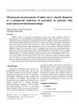
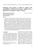
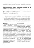
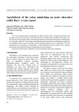
Light microscopy showed that, after a total incubation period of 14 days in the presence of 30 lm silicic acid and 10 lm Fe(III), many spicules were embedded in the thin rim region that surrounds the body of the primmorphs (Fig. 1C,D) and inside the 3D cell aggre- gates. In contrast, almost no spicules were found in primmorphs that had been cultured without additional silicic acid and 10 lm Fe(III) (Fig. 1A,B).
The spicules harbor in their center an organic axial fil- ament in an (cid:1) 1 lm wide canal. In this study we show that selenium ⁄ selenite induces genes which lead to the synthesis of proteins associated with silicatein fibers in the axial filament. These studies with selenium were trig- gered by observations that this element is required for the growth of sponge cells [9]. Serine undergoes chemical conversion into selenocysteine [10]. In eukaryotes as well as prokaryotes [11], selenium is biochemically incorpor- ated into proteins through selenocysteine, a process dur- ing which a tRNA–EF complex is delivered to a codon that would normally be read as a stop.
We used differential display to further
The formation of spicules (monactinal tylostyles) in primmorphs was also demonstrated by transmission electron microscopy. Cuts through primmorphs that had been incubated in the absence of silicic acid and Fe(III) did not show any spicules or cells forming spicules. This is in contrast with primmorphs that had been incubated for longer than 7 days in the presence of 30 lm silicic acid and 10 lm Fe(III), under conditions described in Experimental procedures.
identify genes ⁄ proteins in the siliceous demosponge S. domun- cula that may be involved in biosilicification in sponges.
A
B
C
D
Fig. 1. Light microscopic images of prim- morphs, grown for 14 days (7 days in RPMI medium ⁄ seawater and an additional 7 days in RPMI ⁄ DMEM ⁄ seawater) in the absence (A and B) or presence of 30 lM silicate and 10 lM ferric citrate (C and D). Spicules can be seen in the rim surrounding the body of the primmorphs (>).
FEBS Journal 272 (2005) 3838–3852 ª 2005 FEBS
3839
Effect of selenium on spicule formation
W. E. G. Mu¨ ller et al.
A
B
C
D
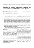
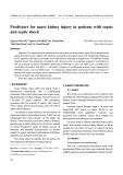
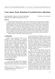
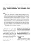
Fig. 2. Formation of spicules in primmorphs (transmission electron microscopic images). Primmorphs cultured for 14 days were ana- lyzed. (A, B) Section through a primmorph, showing the formation of an axial filament (af), a process that proceeds intracellularly. At higher magnification (B), the 15-nm round fibrils (fi) adjacent to the axial filament (af) become visible. (C) A small spicule that is still intracellularly located is shown in a sclerocyte. (D) A more mature spicule sp, now present extracellularly, is shown which surrounds the axial filament with its siliceous material.
in activity even when selenium was added to the primmorphs for 72 h at the indicated concentration ranges: 7.1 ± 1.1 UÆmg)1 at 0.1 lm selenium; 7.9 ± 1.0 UÆmg)1 at 1 lm selenium; 9.1 ± 1.8 UÆmg)1 at 10 lm selenium.
Effect of selenium on spicule formation in primmorphs
extracellular
into the the diameters of
Cross-sections showed either cells (sclerocytes) in the process of forming the axial filaments of the spicules or regions with extracellularly located spicules. In the primordial stage, the axial filaments are synthesized intracellularly in vesicles (Fig. 2A,B); the filaments vary in size between 0.3 and 1.5 lm in diameter. It should be highlighted that during formation of the axial filaments, characterized by rods filled with highly electron-dense material, these filaments are clo- sely associated with (cid:1) 15-nm round fibrils (Fig. 2B). These fibrils may be involved in the extrusion of space. During the spicules maturation, the axial filaments decrease to 0.4 lm (Fig. 2D); in parallel the siliceous spicules are formed around the filaments (Fig. 2D). Before the spicules are extruded they grow to lengths of 8 lm; Fig. 2C shows an intracellular spicule 1.5 lm in length.
In comparison,
the A semiquantitative determination revealed that amount of polymerized silica in primmorphs cultured in the absence of additional silicon was low, amount- ing to <0.4 mgÆ(g wet weight of primmorphs))1. When 30 lm silicic acid [and 10 lm Fe(III)] was added for 7 days to RPMI medium, the concentration increased to 13 ± 5 mgÆg)1. When it was added for 7 days in RPMI medium and then 7 days in RPMI ⁄ Dulbecco’s modified Eagle’s medium (DMEM), the concentration increased to 15 ± 7 mgÆg)1. the amount of silica in the tissue of adult sponge speci- mens is 74 ± 18 mgÆ(g wet tissue))1.
Effect of selenium on glutathione peroxidase activity
the silica concentration was
If 10 lm sodium selenite was added together with silicic acid and Fe(III) to the primmorphs, a doubling of seen after 7 days (32 ± 12 mgÆg)1), but no further increase was meas- ured after 14 days (34 ± 12 mgÆg)1). If sodium selenite was added alone, without additional silicic acid and Fe(III), no significant change in the amount of silica was found in the primmorphs.
First 14-day-old primmorphs were exposed to 0.1 lm, 1 lm and 10 lm sodium selenite for 0 h (control) or 72 h. Then extracts were prepared and glutathione per- oxidase activity was determined as described in Experi- mental procedures. In the absence of selenium (time zero), the enzyme activity was found to be 8.2 ± 1.2 UÆ(mg protein))1. There was no significant change
FEBS Journal 272 (2005) 3838–3852 ª 2005 FEBS
3840
Effect of selenium on spicule formation
W. E. G. Mu¨ ller et al.
Selenium in spicules
spicules were
may be suppressed and (very likely) used for the inser- tion of selenocysteine. The complete protein, with a calculated molecular mass of 13 918 Da (comprising the 123-amino-acid ORF) shares the highest sequence similarity with the 15-kDa selenoprotein M from humans (accession number NP_536355M) [15]. There- fore, the sponge molecule was termed selenoprotein M (SelM_SUBDO) and its cDNA SDSelM. A sequence comparison revealed that the sponge selenoprotein M has the highest similarity to human selenoprotein M (‘expect value’ E of e)33); comparatively low is the rela- tionship to the Drosophila melanogaster putative pro- tein CG7484-PB (E ¼ 2e)11). There are only very distant – if at all – relationships to the Caenorhabditis elegans protein with a coiled coil domain (E ¼ 0.13), cerevisiae deduced polypeptide the Saccharomyces thaliana Yjl049wp (E ¼ 0.56), and the Arabidopsis putative protein At1g08340 (E ¼ 0.23).
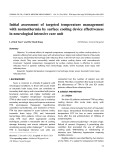
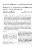
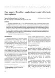
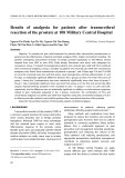
To clarify whether selenium is incorporated into spi- cules (axial filaments and the surrounding silica) of S. domuncula, primmorphs were incubated with 75Se, as described in Experimental procedures. After incu- bation for up to 3 days, isolated, mechanically disintegrated, and the radioactivity was assessed. At time zero, no activity was detected; after a 1-day incubation, 2500 c.p.m.Æ(5 mg solid mater- ial))1 was measured. The amount increased further to 5300 c.p.m.Æ5 mg)1 after the extended incubation period of 3 days. The extract from 75Se metabolically labeled spicules was subjected to SDS ⁄ PAGE. As shown in Fig. 3, the predominant band identified corresponds to a 14-kDa protein (lanes a and b). Weaker bands corresponding to sizes of 35 kDa and 23 kDa were also detected if larger amounts of pro- teins were analyzed (Fig. 3, lane a).
Cloning of the cDNA for the spicule-associated protein
Cloning of the cDNA encoding selenoprotein M from S. domuncula by differential display
Among the differentially expressed transcripts, one was found to encode selenoprotein M. The 652-nucleotide cDNA (accession number AJ875186) contained one ORF, spanning nucleotides 94–96 to 463–465(stop) (Fig. 4). One TGA stop codon exists at nucleotides 187–189, which can also function as a codon for seleno- cysteine [14]. As outlined below, the TGA stop codon
A further differentially expressed transcript was char- acterized, the cDNA encoding a sponge-specific pro- tein which was termed spicule-associated protein (SPIaP_SUBDO). The cDNA, SDSPIaP, is 860-bp long and comprises one ORF at nucleotides 4–6 to nucleo- tides 757–759 (stop) (accession number AJ872182). The 251-amino-acid polypeptide (Fig. 5A) has a calcu- lated molecular mass of 25 602 Da. This protein dis- played no striking homology to any protein reported in the database.
M
a
b
revealed a-helical
90 -
62 - 45 - 30 -
20 - 14 -
10 distinct
Fig. 3. SDS ⁄ PAGE analysis of 75Se-labeled protein(s) in the spi- cules. Labeled protein(s) were isolated from ground spicules as described in Experimental procedures. After electrophoresis, the dried gel was exposed to the film. Positions of the molecular mass markers are shown on the left; the arrowhead points to the 14-kDa protein. Extracts from 10 mg (lane a) and 1 mg (lane b) solid mater- ial were applied to the gel.
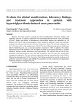
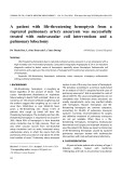
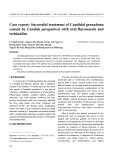
One selection criterion used to study this protein the spicule-associated was the existence of repeats; protein has high structural regularity (Fig. 5A). Sec- ondary-structure analysis regions at the C-terminus and N-terminus of the protein, and the central part of the molecule has extended stret- ches regularly interspersed with predicted turns and coil conformations. Very interesting with respect to the amino-acid sequence is the existence of six highly similar segments of 20 amino acids (60–79; 80–99; 100–119; 120–139; 140–159; 160–179; Fig. 5A). A closer analysis of the distribution of polar ⁄ nonpolar amino acids and calculation of the hydropathicity within the molecule revealed that the borders of the spicule-associated protein display highly hydrophobic (potential transmembrane) regions (Fig. 5B). Further- more, in the central part of the deduced protein, the hydropathicity plot regular indicates hydrophobic regions of approximately nine amino acids. The dominant domain consensus sequence (as in region number 4) reads (Q)TVNVTATPS, with
FEBS Journal 272 (2005) 3838–3852 ª 2005 FEBS
3841
Effect of selenium on spicule formation
W. E. G. Mu¨ ller et al.
Fig. 4. S. domuncula selenoprotein M. From the S. domuncula nucleotides sequence (SDSelM), selenoprotein M (SelM_SUBDO) is predic- ted and aligned with the human selenoprotein M precursor (SelM_HUMAN, NP_536355 [2]); the human sequence was shortened between amino acids 20–35 and 119–124, indicated by square brackets []). Residues conserved (similar or related with respect to their physicochemi- cal properties) in the two sequences are shown in white on black. The TGA triplet that encodes selenocysteine (U) is underlined.
A
B
50
40
30
20
10
0
-10
1
3
TM
TM
5
7
9
-20
-30
1
50
100
150
200
250
Fig. 5. Spicule-associated protein SPIaP_ SUBDO. (A) The deduced protein was ana- lyzed for the predicted secondary structure as described by Garnier et al. [50]; the helical conformation (X), the extended conformation (–), the turn (>) and the coil conformation (w) are indicated. The six highly similar segments of 20 amino acids are marked in white on black or are underlined. In addition, the 10 hydrophobic regions, present in the six 20-amino-acid blocks, are indicated and numbered (#). (B) Hydropathicity plot of the S. domuncula SPIaP_SUBDO; the calculation was per- formed by the method of Kyte and Doolittle [51]. The horizontal axes show the amino acid numbers along the protein vs. the cor- responding hydropathicity. The dotted lines at the )5 value divide hydrophobic regions (above) from hydrophilic regions (below). The 10 distinct hydrophobic regions are consecutively labeled. The two highly hydrophobic (potential transmembrane; TM) regions were identified [52]; they range from amino acids 32–55 and 204–237.
Recombinant proteins and antibodies
the amino acids glutamine, lysine and histidine at the N-terminus and the nonpolar amino acid proline and the hydroxy amino acids serine and threonine at the C-terminal border.
To identify selenoprotein M and the spicule-associated protein in sponge extract, the respective recombinant
FEBS Journal 272 (2005) 3838–3852 ª 2005 FEBS
3842
Effect of selenium on spicule formation
W. E. G. Mu¨ ller et al.
proteins were prepared. In parallel, the distribution of silicatein was analyzed with the tools available [16].
blotting and found to react with the 35-kDa protein (Fig. 6C, lane b). In control assays, it was established that the adsorbed polyclonal antibodies PoAb-SelM and PoAb-SPaP did not react with any protein on the filter (data not shown).
Protein expression of selenoprotein M after exposure to selenium
The cDNAs, SDSelM and SDSPIaP, were cloned into the expression vector pBAD ⁄ gIII as described in Experimental procedures. After induction with arabi- nose, the proteins could be identified in the bacterial lysate (Fig. 6A, lane a vs. b). The recombinant protein was purified (Fig. 6A, lane c) and used to raise anti- bodies in rabbits. The polyclonal antibody against SelM (PoAb-SelM) was found to react with the puri- fied recombinant fusion selenoprotein M (Fig. 6A, lane d). The size of this fusion protein was 17 kDa, which is in accordance with the size of the selenoprotein M fragment of 84 amino acids expressed (10 kDa) together with the protein stretch covering the myc epitope and polyhistidine.
the antibodies against
In a first series of experiments, 12-day-old primmorphs (7-day-old primmorphs were incubated for additional days in RPMI ⁄ DMEM ⁄ seawater) were exposed to 10 lm sodium selenite for 24 or 72 h (Fig. 7, lanes a and b). Primmorphs incubated in seawater ⁄ medium in the absence of selenium were used as a control. Extracts were prepared and subjected to western blot in the analysis. The blotting results revealed that, absence of selenium, the 14-kDa selenoprotein M is missing from the extract (not shown). After incubation for 24 h (lane a) and 72 h (lane b) with sodium selen- ite, the 14-kDa band, reflecting selenoprotein M, is clearly present on the immunoblot.
Similarly, the antibodies against the spicule-associ- ated protein (PoAb-SPaP) were tested. Antibodies were prepared against the 31-kDa recombinant fusion pro- tein (Fig. 6B, lane a); they reacted in the western blot assay with the recombinant protein (Fig. 6B, lane b). the recombinant In parallel, silicatein (Fig. 6C, lane a) were subjected to western
Gene expression studies
A
B
C
SelM c b
M a
d
SAP SILIC b a
b
a
90 - 62 -
45 -
35 31
30 -
The effect of selenium on the expression of the genes encoding selenoprotein M, spicule-associated protein, and silicatein was studied (Fig. 8). In the absence of sodium selenite, almost no transcripts were detected during the 72-h incubation period for selenoprotein M (the probe SDSelM was used) and spicule-associated protein (SDSPIaP), whereas a large number of silicatein (SDSILIC) transcripts could be detected by northern blotting. However, during the 72-h incubation with sodium selenite, within the concentration range 10–
20 -
17
M
a
b
14 -
-
+
+ arab
35
25
and SDSILIC (silicatein)
15 10
24
72 hrs
Fig. 6. Recombinant selenoprotein M, spicule-associated protein and silicatein. Sponge SDSelM (selenoprotein M), SDSPIaP (spi- cDNA was cule-associated protein) expressed in E. coli. (A) Expression of selenoprotein M (SelM): lane a, PAGE analysis (10% gels) of bacterial lysate obtained from E. coli grown in the absence of arabinose (– arab); lane b, lysate from bacteria that had been induced with arabinose (+ arab); lane c, affinity-purified fusion protein; molecular mass 17 kDa; lane d, western blot analysis of purified fusion protein using PoAb-SelM. (B) Spicule-associated protein (SAP). The purified fusion protein (lane a) was used to raise antibodies. The resulting PoAb-SPaP were found to react with the 31-kDa protein in the western blot assay (lane b). (C) The recombinant silicatein (SILIC) was used (lane a) to prepare antibodies (PoAb-SILIC); they recognized the 35-kDa recombinant protein (lane b). The size markers (M) are given.
Fig. 7. Formation of selenoprotein M in primmorphs after exposure to selenium. Twelve-day-old primmorphs, which had been incuba- ted for 7 days in RPMI ⁄ seawater and 5 days in RPMI ⁄ DMEM ⁄ sea- water were exposed to 10 lM sodium selenite for 24 h (lane a) or 72 h (lane b). Then the 3D cell aggregates were extracted and the cleared extract (30 lg per lane) was size separated by SDS ⁄ PAGE (15% gel). Blotting was performed; the blots were incubated with PoAb-SelM as described in Experimental procedures. In lane M, size markers were separated.
FEBS Journal 272 (2005) 3838–3852 ª 2005 FEBS
3843
Effect of selenium on spicule formation
W. E. G. Mu¨ ller et al.
SAP
SILIC
M Ext AF
SelM Ext AF
M Ext AF AF
0.7
SDSelM
75
150 100 75
45
0.9
SDSAP
37
50 37
26
30 20
24
14
1.4
SDSILIC
25 20
25 20
10
15
0
10 100 1000 µM Se
WB
WB
PAGE
WB
Fig. 8. Effect of selenium on the expression of selenoprotein M, spicule-associated protein and silicatein. Fourteen-day-old prim- morphs were exposed to 0–1000 lM sodium selenite for 72 h. Sub- sequently, RNA was isolated and equal amounts (5 lg) were size-separated, transferred, and probed with labeled selenopro- tein M (SDSelM), spicule-associated protein (SDSPIaP) or silicatein (SDSILIC) cDNA.
Fig. 9. Identification of selenoprotein M and spicule-associated pro- tein in the axial filaments by western blotting. Primmorphs (12 days old) were incubated with 10 lM sodium selenite for 72 h and then extracted. The cleared extract was analyzed by SDS ⁄ PAGE (PAGE) and then by western blotting (WB), using PoAb-SelM, PoAb-SPaP and PoAb-SILIC. In parallel, axial filaments were prepared and ana- lyzed in the same way. The size separated protein pattern of the extract (Ext) and the axial filament (AF) is shown for the analysis of silicatein (SILIC). Western blots ⁄ SDS ⁄ PAGE studies from left to right show the results for selenoprotein M (SelM), spicule-associ- ated protein (SAP) and silicatein (SILIC). Signals for selenopro- tein M (14-kDa band on the blot) and spicule-associated protein (26 kDa) are observed in the axial filament and to a large extent in the extracts, whereas silicatein (24 kDa) is predominantly found in the axial filament. Markers were separated in parallel (lane M).
1000 lm, considerable up-regulation of the expression of the genes for selenoprotein M and spicule-associated protein was detected (Fig. 8). On the basis of the data obtained, the steady-state expression was almost identi- cal within the selenium concentration range chosen. The expression level of silicatein remained almost unchanged during the exposure to selenium (Fig. 8).
Localization of selenoprotein M and spicule- associated protein by immunofluorescence analysis
From these data, we conclude that the steady con- centration of selenoprotein M and spicule-associated protein in primmorphs is controlled by selenium at the transcriptional level, whereas the expression of silica- tein is independent of the presence of sodium selenite.
The proteins were localized in tissue from S. domuncula using the antibodies PoAb-SILIC, PoAb-SelM and PoAb-SPaP. Sections were cut through tissue from which the spicules had not been removed, and incuba- ted with the antibodies.
Identification of selenoprotein M, spicule- associated protein and silicatein in total extract or in axial filaments by western blotting
The fluorescence images obtained with antibodies against silicatein show that primarily ⁄ exclusively the surfaces of the 5–7-lm thick and up to 450-lm long monactinal tylostyles and to a smaller extent the diac- tinal oxeas as well as the axial filaments are recognized by PoAb-SILIC (Fig. 10A); the corresponding Nomar- sky interference image is shown in parallel (Fig. 10B). This finding is interesting, as it indicates that silicatein is not restricted to the axial filament but also exists around the spicules.
To clarify whether selenoprotein M and spicule-associ- ated protein are associated with the axial filaments of the spicules from S. domuncula, western blot ⁄ antibody studies were performed. Extracts from sodium selenite- treated primmorphs and axial filaments from spicules were separated by size and subjected to western blot experiments (Fig. 9). The blots were incubated with antibodies against selenoprotein M (PoAb-SelM), spi- cule-associated protein (PoAb-SPaP), or silicatein (PoAb-SILIC).
It was shown that selenoprotein M (the 14-kDa band) exists in large amounts in the soluble extracts and to a small extent also in the axial filaments. Like- wise, the spicule-associated protein was identified in the total extracts and also in lower amounts in the axial filament (26-kDa band). Silicatein exists predom- inantly in the axial filaments (24-kDa protein) (Fig. 9).
The images obtained with the antibodies raised against selenoprotein M (Fig. 10C) and spicule-associ- ated protein (Fig. 10E) surprisingly revealed strong immunoreaction not only on the surfaces of the spicules but also in their canals which harbor the axial filaments. Parallel Nomarsky interference images (Fig. 10D,F) show that, in addition to these structures, areas in the mesohyl of the tissue are decorated by the antibodies.
FEBS Journal 272 (2005) 3838–3852 ª 2005 FEBS
3844
Effect of selenium on spicule formation
W. E. G. Mu¨ ller et al.
A
B
C
D
E
F
Fig. 10. Immunofluorescence analysis of sili- catein, selenoprotein M and spicule-associ- ated protein in tissue from S. domuncula. The slices were stained with PoAb-SILIC (A), PoAb-SelM (C) and PoAb-SPaP (E). In parallel, the corresponding Nomarsky interference images (B, D and F) are given; some spicules are marked in the paired images (>). One axial filament (af) in the center of a spicule is marked in (F).
Effect of selenium on silica formation in vitro
mixture, the sizes of the aggregates reached values of 50–100 lm (Fig. 11D).
Discussion
From earlier studies [3,4] it is known that silicatein, also in its recombinant form, catalyzes silica formation in vitro; in these studies the colorimetric molybdate assay was applied.
the
selenium concentration
Selenium is a trace element which is essential for meta- zoans from humans [11] to sponges [9]. In the marine environment, varies between 10 and 100 nm [17]. It is well established that selenium is found in naturally occurring proteins in two forms, either – rarely – it is inserted post-trans- lationally as a dissociable cofactor into molybdenum- containing enzymes [18], or cotranslationally into proteins as the amino acid selenocysteine [14].
The experiments described here show that exposure of primmorphs to selenium results in a significant increase in spicule formation. After a 7-day exposure to selenium, a substantial increase in biosilica content of the primmorphs was measured. The formation of new spicules in primmorphs can be monitored in
In the present study, the effect of selenium on silica formation was elucidated using the fluorescence dye Rhodamine 123 as an indicator. As outlined in Experi- mental procedures, the reaction was performed under controlled conditions, using recombinant silicatein. In the absence of the tetraethoxysilane substrate, only small aggregates, < 3 lm, were observed under fluor- escence light (Fig. 11A). If tetraethoxysilane was added to the assays with recombinant silicatein, the size of the aggregates increased to 10 lm after an incubation period of 15 min (Fig. 11B). Longer incubation for 3 h resulted in further growth of the aggregates to 30– 50 lm (Fig. 11C). If during this 3 h incubation period 1 lm sodium selenite was present in the reaction
FEBS Journal 272 (2005) 3838–3852 ª 2005 FEBS
3845
Effect of selenium on spicule formation
W. E. G. Mu¨ ller et al.
A
B
C
D
Fig. 11. Influence of selenium on the size of silica aggregates, formed in vitro. Recombin- ant silicatein was incubated in the standard reaction assay, in the absence of the tetra- ethoxysilane substrate for 3 h (A). If tetra- ethoxysilane was added to the reaction for 15 min (B) or 3 h (C), the size of the aggre- gates increased. (D) Larger aggregates of silica were formed if, during the 3 h incubation period, 1 lM sodium selenite was present in the reaction mixture. The silica formed was stained with Rhodamine 123 as described in Experimental procedures and inspected by fluorescence microscopy.
synthesis of small
tRNA population exists
in the
selenoproteins is regulated at the level of translation [20]. In S. domuncula, the increase in selenoprotein M expression after exposure to 10 lm selenium is large. The immunochemical analysis shows that a seleno- cysteine sponge, because translation to the full-size protein occurs.
parallel. It is shown that the axial filament, which con- sists of silicatein [2–4], is formed intracellularly in spe- cial cells termed sclerocytes [19]. After the initial intracellular spicules (cid:1) 8 lm in length and 1.5 lm in diameter, the spicules are prob- ably extruded into the bulky extracellular space. Then synthesis is completed extracellularly, again through the enzymic activity of silicatein, by appositional growth, as demonstrated here by immunohistochemical analysis. The antibodies against silicatein not only reacted with the silicatein of the axial filaments but also with proteins present on the surface of the spicules.
The biological role of selenoprotein M in higher metazoan phyla is not known in detail. In the zebrafish, selenoprotein M is expressed in the notochord, the somites, the spinal cord and the axial fin fold [21]. These data interestingly show that the expression pattern of the different selenoproteins in the fish is region-specific. To obtain an insight into the potential function of selenoprotein M in S. domuncula, antibodies were raised. Surprisingly the subsequent immunohistochemi- cal analysis revealed that selenoprotein M is localized in the axial filament and on the surface of the spicules. Metabolic labeling experiments with 75Se revealed that a 14-kDa protein also exists in the spicules.
It had previously been shown that selenocysteine in selenoproteins participates in redox reactions, especi- ally if selenocysteine has a close cysteine partner [11]. Exactly this constellation exists in selenoprotein M; one cysteine residue is present three amino acids along from selenocysteine towards the N-terminus. This intriguing finding may suggest that selenoprotein M functions as an enzyme.
Exposure of primmorphs to selenium results in the expression of two proteins which were shown to be selenoprotein M associated with spicule formation; and the spicule-associated protein. Selenoprotein M has, until now, only been described in metazoa and the sponge selenoprotein M is the phylogenetically oldest member. The size of selenoprotein M, deduced from the cDNA, is 13 918 Da. Western blotting studies per- formed here show that the native protein has a size of 14 kDa, suggesting that only a small signal peptide exists at the sponge protein, if at all. A phylogenetic analysis revealed the closest similarity of the sponge molecule to the human selenoprotein M, whereas the proteins of invertebrates (D. melanogaster and C. ele- gans) are only distantly related.
Electron microscopic images document that forma- tion of siliceous spicules starts with the synthesis of silica of 90–260-nm silica granules
[22]. Granular
The finding that the steady-state concentration of selenoprotein M is regulated at the level of transcrip- tion was surprising. In vertebrates the expression of all
FEBS Journal 272 (2005) 3838–3852 ª 2005 FEBS
3846
Effect of selenium on spicule formation
W. E. G. Mu¨ ller et al.
factors, e.g.
hydrophobic amino acids surrounded by the polar lysine, serine and threonine. amino acids glutamine, Taking into account this polar ⁄ hydrophobic ⁄ polar composition, this protein can be expected to form a tight association with membranes. This sponge-specific protein was termed spicule-associated protein because it exists in the axial filament and also on the surface of the spicules. The expression of this protein is not affec- ted by a change in the concentration of silicic acid in the surrounding milieu (not shown). In contrast, as demonstrated here, the expression of the silica-poly- merizing enzyme silicatein is not regulated by selenium but by silicic acid [4]. Hence, it becomes evident that morphogenesis of sponges is to a considerable extent dependent on outside inorganic factors. Both elements, silicon and selenium, can be considered morphogenetic factors which control spicule formation in sponges and in turn skeleton formation in these animals.
exactly this size, 70–300 nm, has recently been synthes- ized in vitro, using recombinant silicatein from S. do- muncula [23]. It is still unclear by which process(es) these silica granules are assembled into silica layers. Based on experimental data, it has been proposed that several units of silicatein a form a regular repeating structure of 5–8 nm [24] or 17 nm [2] periodicity. This useful information explains the 2D orientation of the growth of the silica granules ⁄ sheet. For the next phase in spicule production, forming and shaping has to occur. Additional low molecular mass organic molecules or larger size polypeptides, must be postulated that guide the packaging of the silica gran- ules into concentric layers. In elegant studies in diatoms it was shown that the sophisticated construction of the diatom shell is the result of high molecular mass pro- teins, frustulins [25], forming a coat around the dia- toms, and lower molecular mass proteins present in the silica shell, the silaffins [26]. The latter 4 to 17-kDa pro- teins are post-translationally modified at their lysines and serines. The serine units carry the phosphate groups, and the lysines are modified at their a-amino groups by methylpropylamine units [27].
The final interesting finding for the biotechnological application of silicatein, especially with respect to the understanding of biosilica formation in sponges, is that, in the presence of selenium, the size of the poly- merized silica particles formed from recombinant sili- catein is substantially larger.
Taken together, the data reported show that selen- ium has a stimulatory effect on formation of siliceous spicules in sponges. They may also shed new light on the factors involved in biosilica formation in metazoa. Experiments to elucidate potential catalytic effects of both free and protein-bound selenium in the polymer- ization process of silica are in progress. Our hypothesis is that selenium is not only involved in protein ⁄ silica- tein-controlled silica formation in sponges but func- tions also as a novel morphogenetic factor during body plan formation in this oldest metazoan phylum.
Experimental procedures
Chemicals and enzymes
inorganic
It must be stressed that the silica formation in diatoms proceeds nonenzymatically, in contrast with sponges which form the spicules enzymatically using silicatein. However, up until now, nobody has been able to iden- tify the proteins within the silica sheets in the spicules. In fact, published data strongly suggest that the thicken- ing of the spicules is performed enzymatically by apposi- tion [16]. The immunohistological data presented here show that silicatein is also present at the surface of the spicules, supporting this assumption. As the axial fila- ments of the spicules are not homogeneous and, especi- ally intracellularly, are associated with membranes and fibrils, a high-resolution protein analysis of the axial fila- ments is the only way to further identify (non)enzymatic proteins involved in spicule synthesis. Here we used differential display of transcripts to identify the axial filament-associated protein. The element selenium was chosen as inducer, because of its essential role in the growth of animals ⁄ sponges, its quasi-enzy- matic function, and its chemical property of existing in different valences (II, IV and VI). The transition of the different co-ordination states allows incorporation into organic molecules [28].
Sponges
The sources of chemicals and enzymes used were given pre- viously [4,29]. Natural sterile filtered seawater and sodium metasilicate were obtained from Sigma-Aldrich (Taufkir- chen, Germany). Sodium selenite (Na2SeO3) came from Sigma (Taufkirchen, Germany). Na2[75Se]O3 was from Amersham Corp. [Little Chalford, Buckinghamshire, UK; 370 MBqÆ(mg Se))1] or Polatom (Otwock Swierk, Poland; 1500 MBqÆmg)1).
Using differential display of transcripts, we identified a second protein that is up-regulated in primmorphs after incubation with selenium. The deduced protein shares no significant sequence similarity to any protein in the database. The characteristic feature of this poly- peptide is the presence of 10 highly similar repeats, composed of nine amino acids. In the center are
FEBS Journal 272 (2005) 3838–3852 ª 2005 FEBS
3847
Live specimens of S. domuncula (Porifera, Demospongiae, Hadromerida) were collected near Rovinj (Croatia) and
Effect of selenium on spicule formation
W. E. G. Mu¨ ller et al.
Preparation of spicules and axial filaments
Silica content of primmorphs
grids and analyzed with a Tecnai 12 microscope (FEI Elec- tron Optics, Eindhoven, the Netherlands). then kept in aquaria in Mainz (Germany) for more than 2 years before their use.
Differential display technique
Formation of primmorphs, incubation conditions and extracts
To determine semiquantitatively the silica content in prim- morphs, these 3D cell aggregates were disintegrated with sodium hypochlorite (10% for 24 h at room temperature). The particles, mainly spicules and their precursors, were obtained by centrifugation, treated with NaOH, and the amount of released silicic acid determined colorimetrically by the molybdate assay [3], using the ‘Silicon Test’ as des- cribed by the manufacturer (Merck, Darmstadt, Germany). Spicules and their axial filaments were prepared as des- cribed [30]. Briefly, tissue samples were treated first with H2SO4 ⁄ HNO3 and then with butan-1-ol ⁄ water ⁄ SDS. The spicules were collected by sedimentation. The axial fila- ments were isolated from the spicules by dissolving the sil- ica with hydrofluoric acid (2 m HF ⁄ 8 m NH4F; pH 5) at room temperature overnight. The axial filaments were collected, dialyzed against distilled water, and collected by centrifugation [2].
These studies were performed with either primmorphs kept for 7 days in the absence of additional selenium in the RPMI 1640 ⁄ seawater medium or primmorphs that had been cultivated for 72 h in RPMI 1640 ⁄ seawater, supple- mented with 10–1000 lm sodium selenite.
The procedure for the formation of primmorphs (3D cell cultures) from single cells was as described previously [12,13]. Starting from single cells, primmorphs of 3–7 mm are formed after 5 days; they were cultivated in natural sea- water, supplemented with 1% RPMI 1640 medium and in 30 lm silicate (as sodium metasilicate) and 10 lm ferric cit- rate [31]. Seven-day-old, nonattached, 3D cell cultures were transferred into natural seawater, supplemented with 0.25% RPMI 1640 ⁄ 0.75% DMEM and 30 lm silicate and 10 lm Fe(III); incubation proceeded for the indicated periods of time. In this medium the primmorphs attached to the glass slide after 1 day. Sodium selenite was added as indicated; in one series of experiments, silicate and iron were omitted from the medium. An inverted-stage Olympus IX70 micro- scope was used for images.
inhibitor
Transmission electron microscopy
Extracts from primmorphs were prepared by homogeni- zation in lysis buffer [1· Tris-buffered saline, pH 7.5, 1 mm EDTA, 1% Nonidet P40, protease cocktail (Roche, Nutley, NJ, USA; one tablet per 10 mL)], centri- fuged, and the supernatants subjected to western blot ana- lysis.
FEBS Journal 272 (2005) 3838–3852 ª 2005 FEBS
3848
The technique of differential display was applied to iden- tify the changes in the expression levels of transcripts as described [32,33]. Total RNA (1 lg) from controls and selenium-treated samples was reverse-transcribed using an equimolar oligonucleotide mixture of dT23N (Sigma; N rep- resents G, C or A) as 3¢ primer in a reaction with the Mol- oney murine leukemia virus reverse transcriptase (Promega, Madison, WI, USA) at a reaction temperature of 37 (cid:1)C. The resulting cDNA was used as template for the subse- quent PCR with the 5¢-IRD 800 infrared labeled arbitrary primer 5¢-AGTGAATGCG-3¢ and one of the dT23N prim- ers in the assay. To avoid DNA-based contamination from the reagents, the assay was incubated (before the addition of the template) with the restriction enzyme Sau3AI for 20 min at 37 (cid:1)C followed by a final inactivation step at 72 (cid:1)C for 10 min. The PCR parameters used were: 94 (cid:1)C for 3 min, 45 cycles of 94 (cid:1)C for 30 s, 40 (cid:1)C for 2 min, and 72 (cid:1)C for 30 s, with an additional extension step at 72 (cid:1)C for 10 min. After amplification, the labeled fragments were separated on a 6% polyacrylamide sequencing gel using a DNA sequenator (Li-Cor 4000S). The sequencing run was stopped after 3 h, and the gel was transferred to millimeter paper and vacuum dried. The differences in the banding pattern on the gel were detected by an infrared scanning device (Odyssey; LiCor, Lincoln, NE, USA). The major DNA bands differentially seen in the assays with selenite were excised and diluted in distilled water. The resulting fragments were subsequently re-amplified, subcloned in pCRII-Topo vector (Invitrogen, Carsbad, CA, USA), and sequenced using the ‘Thermo-Sequenase Fluorescent Labe- led Primer Cycle Sequencing Kit’ (Amersham ⁄ Pharmacia, Little Chalfont, Buckinghamshire, UK) and the sequencing device (LongReadIR 4200; Li-Cor). For transmission electron microscopic analyses, the sponge samples were cut into pieces (2 mm3), incubated in 0.1 m phosphate buffer (supplemented with 2.5% glutaraldehyde, 0.82% NaCl, pH 7.4) and washed in 0.1 m phosphate buf- fer (1.75% NaCl) at room temperature. After the samples had been treated with 1.25% NaHCO3, 2% OsO4 and 1% NaCl, they were dehydrated with ethanol. The dried sam- ples were incubated with propylene oxide, fixed in propyl- ene oxide ⁄ araldite (2 : 1, w ⁄ w), covered with pure araldite, and hardened at 60 (cid:1)C for 2 days before being cut into 60-nm ultrathin slices (Ultracut S; Leica, Wetzlar, Germany). The samples were transferred to coated copper
Effect of selenium on spicule formation
W. E. G. Mu¨ ller et al.
Raising of antibodies
complete ORF of the SDSPIaP cDNA was used; again the cDNA was inserted into the vector at the same restriction sites, NcoI and HindIII. Expression and purification proce- dures were the same as for selenoprotein M.
Sequence analysis
More than 30 sequences were obtained; among them three partial cDNAs were isolated, the deduced polypep- tides of which showed sequence similarity to selenopro- tein M and two partial cDNAs that showed very low homology to human mucin. The complete cDNA was obtained by primer walking [34,35]. The selenoprotein M cDNA was 652 nucleotides long and was termed SDSelM. The second cDNA, the deduced polypeptide of which dis- played no striking homology to proteins reported in the databases, was termed spicule-associated protein, SDSPIaP, and had a size of 860 bp.
Western blotting
the long form of Polyclonal antibodies (PoAbs) were raised against recom- binant r-SelM and r-SPaP in female rabbits (New Zealand White) as described [41]; the PoAbs were termed PoAb- SelM and PoAb-SPaP, respectively. In control experiments, 100 lL PoAb-SelM or PoAb-SPaP was adsorbed to 20 lg r-SelM or r-SPaP (30 min; 4 (cid:1)C) before use. The prepar- ation of antibodies against recombinant silicatein was as described previously [16]; they were termed PoAb-SILIC and raised against the r-silicatein (35 kDa).
Preparation of recombinant proteins selenoprotein M and spicule-associated protein
Expression of selenoprotein M
The sequences were analyzed using computer programs blast and fasta. Multiple alignments were performed with clustal w version 1.6 [36]. Phylogenetic trees were construc- ted on the basis of amino-acid sequence alignments by neigh- bor-joining, as implemented in the ‘Neighbor’ program from the phylip package [37]. The distance matrices were calcula- ted using the Dayhoff PAM matrix model as described [38]. The degree of support for internal branches was further assessed by bootstrapping [37]. The graphic presentations of the alignments were prepared with genedoc [39].
Selenium in spicules
followed by one isolated Protein samples, extracts from primmorphs, and axial fila- ments were analyzed. Samples of 5 lg protein (axial fil- ament) or 30 lg extract were dissolved in loading buffer (Roti-Load; Roth, Karlsruhe, Germany), boiled for 5 min and subjected to SDS ⁄ PAGE (10% polyacrylamide and 0.1% SDS) [40]. The gels were stained with Coomassie Bril- liant Blue. In parallel, the proteins were transferred and the poly(vinylidene difluoride) membranes (Millipore-Roth) were incubated with PoAb-SILIC, PoAb-SelM or PoAb- SPaP (1 : 500 dilution each); the immune complexes were visualized by incubation with anti-rabbit IgG (alkaline staining with phosphatase conjugated), 4-chloro-1-naphthol. Multi-Tag-Markers (Roche, Mann- heim, Germany) were used as size markers.
sodium selenite incorporation of
Spicule-associated protein
75Se, as The [370 MBqÆmg)1 (10 mCiÆmg)1)], into axial filaments of spicules was determined in primmorphs. Samples of 2 g each were cultured for 0, 1–3 days in seawater [42] supplemented with 200 nCiÆmL)1 75Se. Then spicules were isolated with H2SO4 ⁄ HNO3, as described above. They were washed thor- oughly with distilled water by centrifugation and mechanic- ally pulverized to separate the silica ‘shell’ and the axial filaments. The amount of 75Se-labeled material was deter- mined, and the c.p.m. obtained was correlated with 5 mg spicules, before the disintegration. In a second series of experiments, (2 g) were incubated for 3 days with 200 nCiÆmL)1 75Se. Then spicules were isolated, pulverized and extracted with lysis buffer. After centrifuga- tion, the 2000 g supernatant was collected, concentrated with the Micron-Amicon Centrifugal Filter Device System and subjected to (Millipore, Bedford, MA, USA),
the primmorphs A fragment of the sponge SDSelM sequence was expressed in Escherichia coli (TOP10). The ORF (nucleotides 210–462; corresponding to amino acids 40–123 of the mature protein) primer was forward by PCR using (5¢-CCATGGCAACCAAACCCTCTGGTCCTA-3¢ (the NcoI restriction site is underlined) and one reverse primer (Hin- (5¢-AAGCTTAGATACTTCTTGACTTTCACC-3¢) dIII). Hence, the TGA stop codon which is located at nucleo- tides 187–189 (amino acid 32 in the deduced protein) was not included. The 252-bp part was cloned into the expression vec- tor pBAD ⁄ gIIIA (Invitrogen), which contained at the 3¢-ter- minus the myc epitope and the polyhistidine region. The insert was expressed overnight at 30 (cid:1)C in the presence of 0.0002% l-arabinose. The fusion protein was extracted and purified with the Histag purification kit (Novagen, Madison, WI, USA). The purity of the material was checked by electro- phoresis on 10% polyacrylamide gels containing 0.1% SDS as described by Laemmli [40]. The recombinant selenopro- tein M, r-SelM, was dialyzed against 25 mm Tris ⁄ HCl buffer (pH 7.2), supplemented with 10 mm dl-dithiothreitol.
FEBS Journal 272 (2005) 3838–3852 ª 2005 FEBS
3849
For the preparation of the recombinant protein of spicule- the same vector with the associated protein, r-SPaP,
Effect of selenium on spicule formation
W. E. G. Mu¨ ller et al.
SDS ⁄ PAGE (10% gel ⁄ 0.1% SDS). The dried gel was exposed to Kodak X-Omat XAR X-ray film for 2 days at )80 (cid:1)C.
RNA preparation and northern blot analysis
Analytical ⁄ biochemical techniques
reaction. Rhodamine 123 (Sigma) was used to stain silica aggregates formed, as described [47]. Finally, the specimens were analyzed with an Olympus AHBT3 microscope ⁄ AH3- RFC light fluorescence using the excitation light wavelength 490 nm.
Immunohistochemistry
RNA was extracted from liquid-nitrogen pulverized tissue with Trizol reagent (Gibco-BRL, Grand Island, NY, USA) as described [43]. Then 5 lg total RNA was electrophoresed and blotted on to Hybond-N+ nylon membrane (Amer- sham). Hybridization was performed with a 250-nucleotide segment of the SDSelM cDNA, a 300-nucleotide segment of SDSPIaP and a 400-nucleotide fragment from the silica- tein-A gene SDSILIC (accession number AJ272013 [4]). The probes were labeled with the PCR-DIG probe synthesis kit (Roche). After being washed DIG-labeled nucleic acid was detected with anti-DIG FAB fragments and visualized by chemiluminescence technique using CDP (Roche). Protein concentrations were determined as described by Lowry et al. [48] using BSA as standard. Glutathione per- oxidase activity was determined as described [49]. In brief, primmorphs were extracted with 10 mm Tris ⁄ HCl buffer (containing 2 mm EDTA, 100 mm NaCl, pH 7.2). After centrifugation (10 000 g; 10 min; 4 (cid:1)C), the supernatant was collected and subjected to the coupled NADPH oxida- tion enzyme reaction. Hydrogen peroxide was used as a substrate. The enzyme unit was defined as 1 nmol GSH oxidized per min. Five parallel experiments were performed, and mean ± SD values are given.
Acknowledgements
We thank Ms. E. Sehn (Zoological Institute, Univer- sity of Mainz) for valuable technical assistance. This work was supported by grants from the Deutsche Fors- chungsgemeinschaft, the Bundesministerium fu¨r Bildung und Forschung Germany (project: Center of Excellence BIOTECmarin) and the International Human Frontier Science Program.
References
1 Lowenstam HA (1981) Minerals formed by organisms. Science 211, 1126–1131.
2 Shimizu K, Cha J, Stucky GD & Morse DE (1998) Sili- catein alpha: cathepsin 1-like protein in sponge biosilica. Proc Natl Acad Sci USA 95, 6234–6238. 3 Cha JN, Shimizu K, Zhou Y, Christianssen SC, Chm-
Staining of polymerized silica with Rhodamine 123
Fresh sponge tissue from the Adriatic Sea was fixed in 2% paraformaldehyde [44]; after dehydration, the samples were embedded in Technovit 8100 [45], according to the instruc- tions of the manufacturer as outlined in Pancer et al. [46]. Sections of 8 lm thickness were prepared. The sponge tis- sue was deliberately not treated with HF ⁄ NaHF solution so as not to remove the siliceous spicules. The sections were blocked with 1% BSA in NaCl ⁄ Pi and incubated with PoAb-SelM, PoAb-SPaP or PoAb-SILIC overnight at 4 (cid:1)C. The antibodies were used at a 1 : 200 dilution in 0.5% BSA in NaCl ⁄ Pi buffer. Rhodamine-conjugated goat anti-rabbit immunoglobulin (Dako, Carpinteria, CA, USA) was used as a secondary antibody. The preimmune rabbit serum was used as a control; it did not react with any structures in the sections. For light microscopic studies (immunofluorescence analysis), an Olympus AHBT3 microscope was used. The fluorescence studies were performed with an AH3-RFC reflected light fluorescence attachment using the excitation light wave-length 334 ⁄ 365 nm. In parallel, the nonstained sections were inspected directly using Nomarsky interfer- ence contrast optics. elka BF, Stucky GD & Morse DE (1999) Silicatein fila- ments and subunits from a marine sponge direct the polymerization of silica and silicones in vitro. Proc Natl Acad Sci USA 96, 361–365.
4 Krasko A, Batel R, Schro¨ der HC, Mu¨ ller IM & Mu¨ ller WEG (2000) Expression of silicatein and collagen genes in the marine sponge Suberites domuncula is controlled by silicate and myotrophin. Eur J Biochem 267, 4878–4887. 5 Mu¨ ller WEG (2005) Spatial and temporal expression
patterns in animals. In Encyclopedia of Molecular Cell Biology and Molecular Medicine (Meyers, RA, ed.). Wiley-VCH GmbH, Weinheim. (in press)
FEBS Journal 272 (2005) 3838–3852 ª 2005 FEBS
3850
6 Mu¨ ller WEG, Wiens M, Adell T, Gamulin V, Schro¨ der HC & Mu¨ ller IM (2004) The bauplan of the Urmeta- zoa: the basis of the genetic complexity of Metazoa Enzymatic silica formation was studied using r-silicatein [31]. The reactions were performed on a glass slide in a vol- ume of 100 lL. The reaction mixture contained 10 lg r-sili- catein in NaCl ⁄ Pi (pH 7.2) containing 5 mm Fe(III) and 1 lm ZnCl2. As substrate, 4.5 mm tetraethoxysilane (Sigma) was used. The reaction was performed at 22 (cid:1)C for up to 3 h; the mixture was then washed three times with NaCl ⁄ Pi. Where mentioned, 1 lm sodium selenite was added to the
Effect of selenium on spicule formation
W. E. G. Mu¨ ller et al.
using the siliceous sponges [Porifera] as living fossils. Int Rev Cytol 235, 53–92.
20 Sunde RA (2001) Regulation of selenoprotein expres- sion. In Selenium: its Molecular Biology and Role in Human Health (Hatfield, DL, ed.), pp. 81–96. Kluwer Academic Publishers, Boston. 7 Weissenfels N (1989) Biologie und Mikroskopische Anat- omie der Su¨ßwasserschwa¨mme (Spongillidae). Gustav Fischer Verlag, Stuttgart. 8 Simpson TL, Gil M, Connes R, Diaz JP & Paris J
21 Thisse C, Degrave A, Kryukov GV, Gladyshev VN, Obrecht-Pfumio S, Krol A, Thisse B & Lescure A (2003) Spatial and temporal expression patterns of sele- noprotein genes during embryogenesis in zebrafish. Gene Expr Patterns 3, 525–532. (1985) Effects of germanium (Ge) on the silica spicules of the marine sponge Suberites domuncula: transforma- tion of the spicule type. J Morphol 183, 117–128. 9 Willoughby R & Pomponi SA (2000) Quantitative
22 Pisera A (2003) Some aspects of silica deposition in Lithistid demosponge desmas. Microsc Res Tech 62, 312–326. assessment of marine sponge cells in vitro: development of improved growth medium. In Vitro Cell Dev Biol 36, 194–200. 23 Tahir MN, The´ ato P, Mu¨ ller WEG, Schro¨ der HC, 10 Wu ZP & Hilvert D (1989) Selenosubtilisin as a glu-
tathione peroxidase mimic. J Am Chem Soc 112, 5647– 5648. Janshoff A, Zhang J, Huth J & Tremel W (2004) For- mation of biosilica by immobilization of silicatein on novel functionalized surfaces. Chem Commun (Camb) 24, 2848–2849.
11 Hatfield DL & Gladyshev VN (2002) How selenium has altered our understanding of the genetic code. Mol Cell Biol 22, 3565–3576.
24 Croce G, Frache A, Milanesio M, Marchese L, Causa` M, Viterbo D, Barbaglia A, Bolis V, Bavestrello G, Cerrano C, et al. (2004) Structural characterization of siliceous spicules from marine sponges. Biophys J 86, 526–534. 25 Kro¨ ger N & Sumper M (2000) The biochemistry of
12 Custodio MR, Prokic I, Steffen R, Koziol C, Borojevic R, Bru¨ mmer F, Nickel M & Mu¨ ller WEG (1998) Prim- morphs generated from dissociated cells of the sponge Suberites domuncula: a model system for studies of cell proliferation and cell death. Mech Ageing Dev 105, 45–59. silica formation. In Biomineralization from Biology to Biotechnology and Medical Application (Ba¨ uerlein E, ed.), pp. 151–170. Wiley-VCH, Weinheim.
13 Mu¨ ller WEG, Wiens M, Batel R, Steffen R, Borojevic R & Custodio MR (1999) Establishment of a primary cell culture from a sponge: primmorphs from Suberites domuncula. Mar Ecol Progr Series 178, 205–219. 26 Kro¨ ger N, Lorenz S, Brunner E & Sumper M (2002) Self-assembly of highly phosphorylated silaffins and their function in biosilica morphogenesis. Science 298, 584–586. 27 Kro¨ ger N, Deutzmann R & Sumper M (2001) Silica- 14 Low SC & Berry MJ (1996) Knowing when not to stop: selenocysteine incorporation in eukaryotes. Trends Bio- chem Sci 21, 203–208. 15 Korotkov KV, Novoselov SV, Hatfield DL & Glady- precipitating peptides from diatoms: the chemical struc- ture of silaffin-1a from Cylindotheca fusiformis. J Biol Chem 276, 26066–26070.
shev VN (2002) Mammalian selenoprotein in which sele- nocysteine (Sec) incorporation is supported by a new form of Sec insertion sequence element. Mol Cell Biol 22, 1402–1411. 28 Bo¨ ck A (2001) Selenium metabolism in bacteria. In Selenium: its Molecular Biology and Role in Human Health (Hatfield, DL, ed.), pp. 8–32 Kluwer Academic Publishers, Boston. 16 Mu¨ ller WEG, Krasko A, Le Pennec G, Steffen R,
29 Kruse M, Mu¨ ller IM & Mu¨ ller WEG (1997) Early evo- lution of metazoan serine ⁄ threonine- and tyrosine kinases: identification of selected kinases in marine sponges. Mol Biol Evol 14, 1326–1334.
Ammar MSA, Wiens M, Mu¨ ller IM & Schro¨ der HC (2003) Molecular mechanism of spicule formation in the demosponge Suberites domuncula: silicatein – collagen – myotrophin. Prog Mol Subcell Biol 33, 195–122. 17 Schroeder RA, Orem WH & Kharaka YK (2002) Chemical evolution of the Salton Sea, California: nutrient and selenium dynamics. Hydrobiologia 473, 23–45.
30 Kaluzhnaya OV, Belikov SI, Schro¨ der HC, Rothenber- ger M, Zapf S, Kaandorp JA, Borejko A, Mu¨ ller IM & Mu¨ ller WEG (2005) Dynamics of skeletal formation in the Lake Baikal sponge Lubomirskia baicalensis. Part I. Biological and biochemical studies. Naturwissenschaften 92, 128–133.
18 Gladyshev VN, Khangulov SV & Stadtman TC (1994) Nicotinic acid hydroxylase from Clostridium barkeri: electron paramagnetic resonance studies show that sele- nium is coordinated with molybdenum in the catalyti- cally active selenium-dependent enzyme. Proc Natl Acad Sci USA 91, 232–236. 19 Uriz MJ, Turon X & Becerro MA (2000) Silica deposi- 31 Krasko A, Schro¨ der HC, Batel R, Grebenjuk VA, Stef- fen R, Mu¨ ller IM & Mu¨ ller WEG (2002) Iron induces proliferation and morphogenesis in primmorphs from the marine sponge Suberites domuncula. DNA Cell Biol 21, 67–80.
FEBS Journal 272 (2005) 3838–3852 ª 2005 FEBS
3851
tion in Demospongiae: spiculogenesis in Crambe crambe. Cell Tissue Res 301, 299–309. 32 Mu¨ ller WEG, Krasko A, Skorokhod A, Bu¨ nz C, Grebe- njuk VA, Steffen R, Batel R, Mu¨ ller IM & Schro¨ der
Effect of selenium on spicule formation
W. E. G. Mu¨ ller et al.
HC (2002) Histocompatibility reaction in the sponge Suberites domuncula on tissue and cellular level: central role of the allograft inflammatory factor 1. Immuno- genetics 54, 48–58.
42 Bo¨ hm M, Schro¨ der HC, Mu¨ ller IM, Mu¨ ller WEG & Gamulin V (2000) The mitogen-activated protein kinase p38 pathway is conserved in metazoans: cloning and activation of p38 of the SAPK2 subfamily from the sponge Suberites domuncula. Biol Cell 92, 95–104.
33 Mu¨ ller WEG, Klemt M, Thakur NL, Schro¨ der HC, Aiello A, D’Esposito M, Menna M & Fattorusso E (2003) Molecular ⁄ chemical ecology in sponges: evidence for an adaptive antibacterial response in Suberites domuncula. Mar Biol 144, 19–29.
43 Grebenjuk VA, Kuusksalu A, Kelve M, Schu¨ tze J, Schro¨ - der HC & Mu¨ ller WEG (2002) Induction of (2¢-5¢) oligo- adenylate synthetase in the marine sponges Suberites domuncula and Geodia cydonium by the bacterial endo- toxin lipopolysaccharide. Eur J Biochem 269, 1382–1392. 44 Romeis B (1989) Mikroskopische Technik. Urban ⁄ Schwarzenberg, Mu¨ nchen. 45 Beckstead JH (1985) Optimal antigen localization in
human tissues using aldehydefixed plastic-embedded sec- tions. J Histochem Cytochem 9, 954–958.
34 Ausubel FM, Brent R, Kingston RE, Moore DD, Smith JA, Seidmann JG & Struhl K (1995) Current Protocols in Molecular Biology. John Wiley and Sons, New York. 35 Wiens M, Koziol C, Hassanein HMA, Batel R, Schro¨ - der HC & Mu¨ ller WEG (1998) Induction of gene expression of the chaperones 14–3)3 and HSP70 induced by PCB 118 (2,3¢,4,4¢,5-pentachlorobiphenyl) in the marine sponge Geodia cydonium: novel biomarkers for polychlorinated biphenyls. Mar Ecol Prog Series 165, 247–257.
46 Pancer Z, Kruse M, Scha¨ cke H, Scheffer U, Steffen R, Kova´ cs P & Mu¨ ller WEG (1996) Polymorphism in the immunoglobulin-like domains of the receptor tyrosine kinase from the sponge Geodia cydonium. Cell Adhes Commun 4, 327–339.
47 Li CW, Chu S & Lee M (1989) Characterizing the sili- con deposition vesicle of diatoms. Protoplasma 151, 158–163. 36 Thompson JD, Higgins DG & Gibson TJ (1994) CLUS- TAL W: improving the sensitivity of progressive multi- ple sequence alignment through sequence weighting, positions-specific gap penalties and weight matrix choice. Nucleic Acids Res 22, 4673–4680. 37 Felsenstein J (1993) Phylip, version 3.5. University of Washington, Seattle. 48 Lowry OH, Rosebrough NJ, Farr AL & Randall RJ (1951) Protein measurement with the folin phenol reagent. J Biol Chem 193, 265–275.
38 Dayhoff MO, Schwartz RM & Orcutt BC (1978) A model of evolutionary change in protein. In Atlas of Protein Sequence and Structure (Dayhoff, MO, ed.), pp. 345–352. National Biomedical Research Foundation, Washington, DC.
49 Cheng WH, Ho YS, Ross DA, Han Y, Combs GF & Lei XG (1997) Overexpression of cellular glutathione peroxide does not affect expression of plasma glu- tathione peroxide or phospholipids peroxide glutathione peroxide in mice offered diets adequate or deficient in selenium. J Nutr 127, 405–417.
39 Nicholas KB & Nicholas HB Jr (1997) Genedoc: a Tool for Editing and Annotating Multiple Sequence Align- ments, Version 1.1.004. Distributed by the author; http://www.psc.edu/biomed/genedoc. 40 Laemmli UK (1970) Cleavage of structural proteins 50 Garnier J, Osguthorpe DJ & Robson B (1978) Predict- ing the secondary structure of globular proteins. J Mol Biol 120, 97–120.
during the assembly of the head of bacteriophage T4. Nature 227, 680–685. 51 Kyte J & Doolittle RF (1982) A simple method for dis- playing the hydrophobic character of a protein. J Mol Biol 157, 105–132. 41 Schu¨ tze J, Krasko A, Diehl-Seifert B & Mu¨ ller WEG
FEBS Journal 272 (2005) 3838–3852 ª 2005 FEBS
3852
(2001) Cloning and expression of the putative aggrega- tion factor from the marine sponge Geodia cydonium. J Cell Sci 114, 3189–3198. 52 Rao J & Argos P (1986) A conformational preference parameter to predict helices in integral membrane pro- teins. Biochim Biophys Acta 869, 197–214.