44
Journal of Medicine and Pharmacy, Volume 9, No.3/2019
ANTEROLATERAL THIGH FLAP IN LOWER LIMB
RECONSTRUCTION
Le Hong Phuc1, Tran Thiet Son2, Le Nghi Thanh Nhan1
(1) Hue University of Medicine and Pharmacy, Hue University; (2) Hanoi Medical University
Abstract
Introduction: Anterolateral thigh flap is one of the most researched and widely used perforator flaps in
the recent decades in plastic surgery as a whole and in limb reconstruction, especially in cases with complex
deflects, in particular. This report aimed to evaluate anterolateral thigh flap in reconstruction of complex
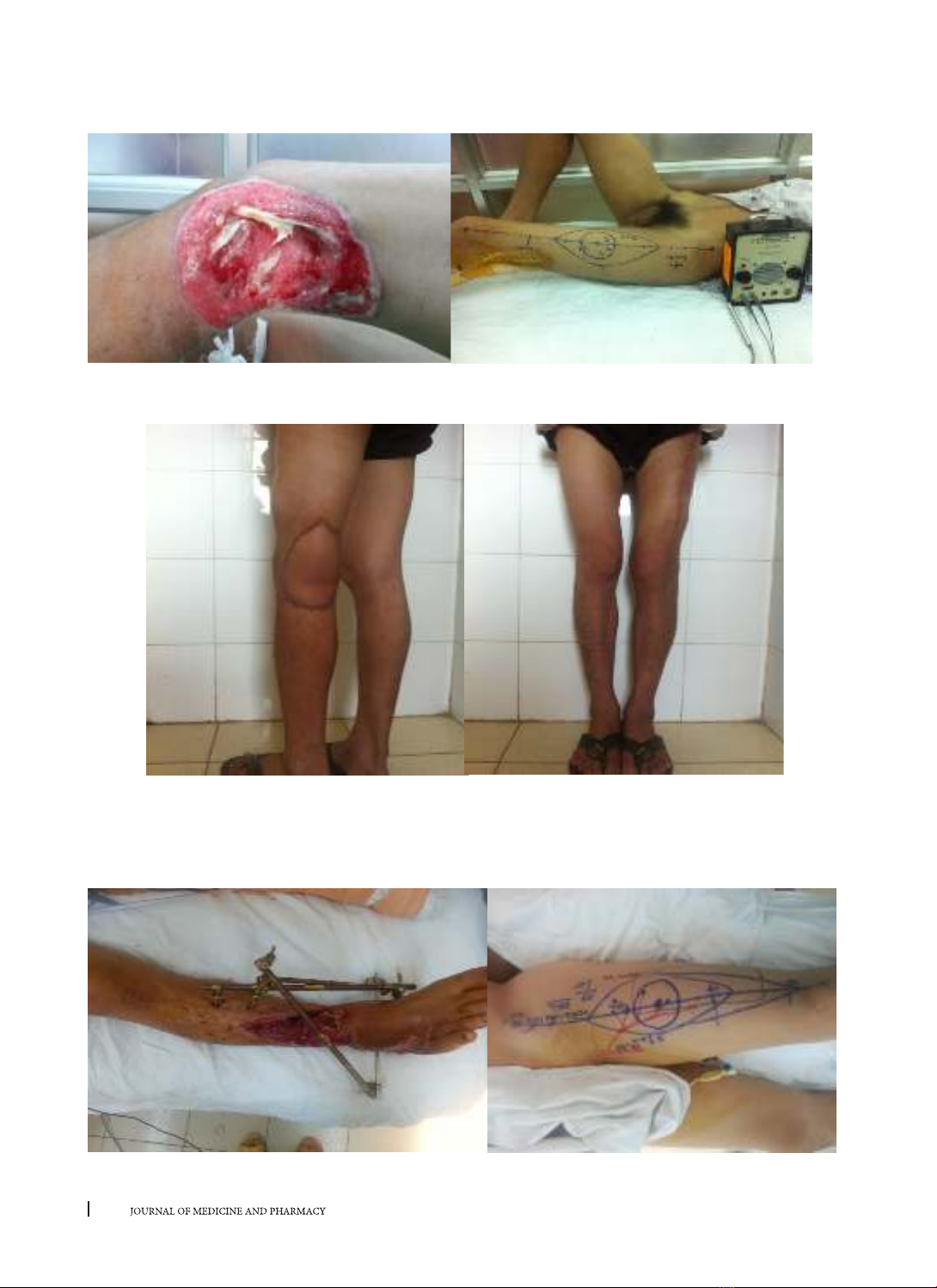
lower limb soft tissue defects. Subjects and methods: From August 2014 to August 2015, at Hue University of
Medicine and Pharmacy Hospital, 12 cases with complex soft tissue defects in lower limb were reconstructed
and covered with ALT flaps: two distal based pedicle ALT flaps for popliteal and around knee joint defects and
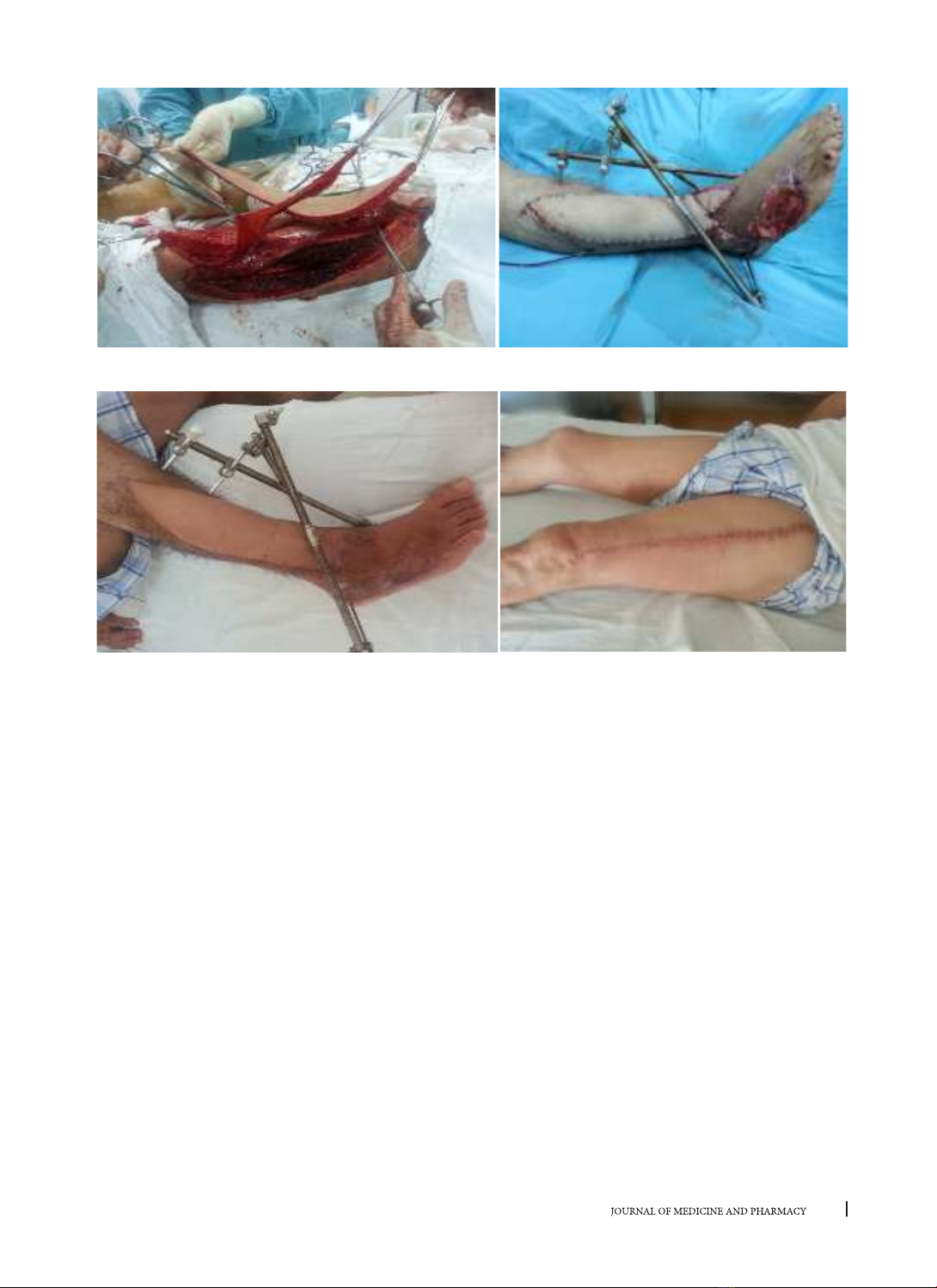
10 composite ALT free flaps for lower leg reconstruction. Results: Twelve flaps used included: two peripheral
pedicled fasciocutaneous flaps, ten complex free flaps (01 complex myo-fasciocutaneous flap providing
muscle for deep space filled, fascial for tendon reconstruction and surface covering of the defect; 05 vastus
lareralis myocutaneous flaps providing muscle for dead space filling and covering; 04 fasciocutaneous flaps
involving the fascia lata for fascial reconstruction and covering). The size of flaps ranged from 8 to 27cm
in length and from 6 to 13cm in width. The largest flap was 240cm2,the smallest was 50cm2. All 12 flaps
survived. Short-term results at one month after surgery were consideredas good in eleven patients and fair
in one patient. There were no special complications at donor sites. Paresthesia at the donor site was noted
in two cases. Conclusion: ALT flap with its versatility as peripheral pedicled flap or free flap can be used in
lower limb reconstruction with high success rate of 100% (12/12). Preoperative skin perforator mapping by
Doppler was highly accurate (12/12) compared with intraoperative findings. Complex free ALT flap is suitable
for reconstruction of major defects involving different type of tissue in lower limb with satisfied results.
Key words: Anterolateral thigh flap, limb, ower limb
Corresponding author: Le Hong Phuc, email: phucbstmhue@gmail.com DOI: 10.34071/jmp.2019.3.6
Received: 2/7/2018, Resived: 11/1/2019; Accepted: 4/6/2019
1. BACKGROUND
Anterolateral thigh flap (ALT) is one of the
perforator flaps regularly studied and widely used
today. Song et al. published a report on the first flap
in 1984 using a flap based on perforator from the
descending branch of the circumflex femoral pedicle
to treat burn scars in head and neck region. Since
then, it has been increasingly used in plastic Surgery.
In particulary, the flap has many advantages such
as long and relatively constant pedicle with large
diameter, large volume of tissue which can be
harvested for bulking and covering and associated
low rate of complications of donor site [4]. Due to
the flexible use of the flap, its use is increasingly
expanded to provide tissue for deep or dead space
filler, covering or reconstruction of defects in
different organs. Flap can be used indifferent forms:
peripheral pedicled flap (based on the collateral
circulation from genicular artery) or central pedicled
flap (descending branch of the circumflex femoral
artery). In addition, other commonly used forms
nowadays are classic free flap or thinned, chimeric
or composite flaps. The flap has many advantages
in reconstructive surgery for complex defectsand
helps reduce the number of surgery and allows early
functional and anatomical recovery The purpose of
this study was to assess the initial results of ALT flap
in the treatment of the lower limb defects.
2. SUBJECTS AND METHODS
2.1. Subjects: Twelve patients (12 soft tissue
defects), aged 21-62, male/female 4/7, were
operated using different types ALT flaps from 8/2014
to 8/2015.
2.2. Methods
Study design: uncontrolled descriptive
prospective clinical study
Research protocol: thorough clinical exam,
clinical and radiographic evaluation of the lesions,
reconstruction planning, flap selection, surgery,
follow-up and evaluation.
Results were evaluated based on the following
criteria: flap survival, wound healing, functional and
esthetic results at donor and recipient sites. We
classified short- and long-term results using a 4-level
scale.
45
Journal of Medicine and Pharmacy, Volume 9, No.3/2019
We assessed post-operative flap survival which
was also short-term results of surgery using the
following criteria:
+ Flap color
+ Flap cappilary refill
+ Doppler ultrasound signal
- With the forementioned criteria, we classified
the results as:
+ Good: Flap is evenly pink with good capillary
refill and good doppler signal of vascular pulsation.
+ Fair: Flap is mostly pink with a only a small blue
part at the periphery of the flap (< 20% flap surface),
good capillary refill, good doppler signal of vascular
pulsation.
+ Average:
Flap is slightly blue or part of the flap is blue (<
30% surface), bad capillary refill, audible but unclear
and irregular doppler signal.
+ Bad: > 50% surface of the flap is blue, bad
capillary refill, difficultly audible or non-audible
doppler signal.
- Evaluation time:
+ Postoperative day 1, 2 and 3
+ 1 week post-op
- Long-term results were assessed by: flap color,
flap quality, scar around the flap, scar formation,
functional and esthetic results of donor and
recipient sites. It was further classified using also a
4-level scale:
Good: soft but pressure-resistant flap,
goodesthetic results (flap shape fits recipient site),
no ulcer, no inflammation or infection; soft scar at
donor site, normal contraction-relaxation activity of
quadriceps, normal joint mobility.
Fair: Good scar formation at recipient site, bulky
flap, excessive size of flap is of minor degree and
acceptable by patients, hypertrophic but soft scar at
donner site, normal contraction-relaxation activity
of quadriceps, normal joint mobility.
Bad: Infection, inflammation, retracting scar
which limit mobility and necessitate an additional
procedure; hypertrophic scar, reduced contraction-
relaxation activity of quadriceps, slightly reduced
joint mobility.
Failure: Flap is necrotic or sclerosed,
hypertrophic, ulcerated, or suffered from persistant
inflammation which require re-operation;
hypertrophic or incorrect scar at donor site, reduced
contraction-relaxation activity of quadriceps,
abnomal joint mobility.
Evaluation time: 1 month, 3 months, 6 months
after the operation.
3. RESULTS
3.1. General characteristics
- Causes: The majority of the lesions was the
result of trauma (7/12). Other causes included burn
(1 case), osteomyelitis (1 case), firearm injury (1
case), vegatato-ulcerative lesion of upper third leg
stump (1 case) and fasciocutaneous and tendons
necrosis following snake bite (1 case).
- Location: around the knee joint (2 cases), upper
third leg (2 cases), lower third leg (4 cases), heel (2
cases) and dorsal foot (2 cases).
- Characteristics of the defect: simple soft tissue
defect (2 cases), associated with osteo-articulation
infections (4 cases), associated with fascial lesions
(3 cases), associated with bony lesions (2 cases) and
leg stump ulcer (1 case).
3.2. Characteristics of flaps: 2 cases with simple
cutaneous distal based pedicled flap for simple
defects; 1 case with myo-fascio-cutaneous pedicled
flap providing muscle for deep space filling, fascia for
flexor tendon reconstruction and cutaneous paddle
for surface coverage after grade IIIB the lower third
leg opened fracture, 6 cases with vastus lareralis
myocutaneous flaps for deep space and surface
combined defect coverage following osteomyelitis
treatment, 4 cases with fasciocutaneous flap for
surface defect coverage.
- Pedicle composition: 1 artery and 2 venea
commitantes (11/12 cases), 1 artery and 1 vena
commitante (1/12 case)
- Type of perforator: Septocutaneous perforator
of descending branch of lateral circumflex femoral
artery (2/12 cases), musculocutaneous perforator
(10/12 cases). In all cases, preoperative mapping
of perforator was compatible with intraoperative
findings.
3.3. Treatment results: Flap survival (12/12).
Short-term results after one month: classified
as good (11/12) and average (1/12). No special
complications was noticed at donor site. 2/12 cases
showed slight paresthesia at donor site. Primary
closure at donor site was possible in all 12 cases.
Some illustrating images:
46
Journal of Medicine and Pharmacy, Volume 9, No.3/2019
Case 1. Nguyen Xuan Ch, YOB 1994.
Figure 1.1. Fasciocutaneous defect around the knee joint, perforator mapping with Doppler and
peripheral pedicled flap planning
Figure 1.2. Follow-up after 3 months of a patient with distal based pedicled ALT flap showed good
results at donor and recipient sites
Case 2. Nguyen Van T, 49T, soft tissue defect following grade IIIB open fracture
Figure 2.1. Defect associated with bone loss, lower third leg open fracture and ALT designed
47
Journal of Medicine and Pharmacy, Volume 9, No.3/2019
Figure 2.2. Composite ALT flap being harvested and post-operative results of reconstruction
Figure 2.3. Follow-up after 3 months with good results at donor and recipient sites
4. DISCUSSION
Considering peripheral pedicled flap, ALT flap
does not only receive blood supply from perforator
of descending branch of lateral circumflex femoral
artery but also from laterosuperior genicular artery
distally in case of lesions from lower third thight to
upper third leg, especially defects around the knee
joint. The application of ALT flap in the treatment
of lesions from lower third thigh to upper third leg
has been reported in a number of studies of authors
from Taiwan, China, Japan and Korea. Chen CY (2007)
reported treatment of post-burn defects in the knee
region by retrograde flap. This type of flap was used
by Zhao Y and other Vietnamese authors to repair
scar in the knee joint and upper third leg region.
This distal based pedicled ALT flap was even used
to cover the defect at donor site after harvesting
of a large ALT flap for reconstruction of other body
parts. In our study, we permanently clamped the
central pedicle to test the blood supply capacity of
peripheral pedicle before harvesting. This clamping
trial showed satisfied blood supply, therefore, we
didnot perform microvalcular anastomosis with
proximal pedicle. Esthetic and functional results
were correct at both sides.
ALT flap is also used by plastic surgeon for soft
tissue defects in the leg and foot. The indication
for flap in this region is more limited compared to
head and neck region [5,6]. The defect is usually due
to burn, tumor and especially trauma. Moreover,
as the poor-blood-suply skin is the only barrier
to underlying bone and fascia, lesions in this area
expose the patient to a significant risk of hard-to-
heal, easy-to-be-infected defects.
Therefore, a large and well-vascularized flap
is necessary to cover the infectious lesion. In the
majority of cases, free flap is used. Its versality
is useful in one-stage reconstructive procedure.
Particularly with complex defects, complex flap is
required to cover and to provide muscle for deep/
dead space filler of various tissue losses (fascia,
bone). Min Jea Lee (2012) used ALT under the form
of myocutaneous flap with the success rate of more
than 90%. When compared with fasciocutaneous
48
Journal of Medicine and Pharmacy, Volume 9, No.3/2019
flap, there was a lower risk of perforator injury but
the difference was not statistically significant [9]. In
our study, we used 5 vastus lateralis myocutenous
flap to fill dead space and cover the missing surface
after debridement of infected bone with good results.
In 2000, Lee J. W et al reported 3 cases of
reconstruction of Achilles tendon and soft tissue
defect using ALT flap combined with fascia lata
showing good outcomes. Jeng Seng-Feng et al also
published 2 similar cases with a follow-up time
9 months-2 years with satisfied results, reduced
hospitalization time and good recovery of limb
function [6, 10, 11]. In 2012, Wong Chin-Ho et al
published a series of 7 cases with reconstruction
of large soft tissue defects associated with bone
lesion using vastus lateralis myocutenous flap with
good long-term results [12]. We applied the same
techniques used by Lee J.W. (2000) in our 4 cases
[9]. Furthermore, Houtmeyer Ph et al reported the
results of reconstruction of Achilles tendon, tendons
of extensor digitorium and soft tissue defect using
pedicled ALT flap combined with fascia lata in 6
cases. The successful application of complex flap
in the repair of complicated soft tissue defects can
help reduce the number of surgery, hospitalization
length, facilitate quickly recovery of the anatomy
and function of the limb. It is also the current trend
in plastic surgery for complex defects.
Considering flap complications and treatment
results, a meta-analysis on 42 studies from different
centers and nations by Jessica Collins MD in 2012
showed the most frequent complication being
paresthesia at the donor site [7]; we had 2 cases with
this type of complication. However, more patients
with longer follow-up are needed to draw exact
conclusion. Besides, the conservation of femoral
nerve and muscles during dissection and not-too-
tight skin closure will help reduce complications
at donor site. In these 2 cases, we harvested large
flapsmeseared >20% of thigh circumference. The
extensive dissection can damage lateral femoral
cutaneous nerve. Excessive pressure during skin
closure also limited wound healing.
5. CONCLUSION
Free or pedicled ALT flap can be used very flexibly
with high success rate. Preoperative perforator
mapping can be performed with high accuracy
(12/12). Free complex ALT flap is a good solution for
major soft tissue defects in the lower limb.
REFERENCES
1. Phạm Thị Việt Dung, Trần Thiết Sơn (2011), “Tính linh
hoạt của vạt đùi trước ngoài trong phẫu thuật tạo hình”, Y
học thực hành, Số 8 (777), tr 8-11.
2. Trần Đăng Khoa, Trần Thiết Sơn, Phạm Đăng Diệu,
Phạm Thị Việt Dung (2010), “Nghiên cứu đặc điểm giải phẫu
ứng dụng động mạch đùi ngoài trên người Việt nam”,
Tạp chí Y học TP Hồ Chí Minh, Số 14, tr 163-173.
3. Nguyễn Đình Minh, Trần Thiết Sơn (2010), Tạo hình
các khuyết phần mềm chi dưới bằng vạt đùi trước ngoài”,
Tạp chí Y học Việt Nam, Số 2(274), tr. 432-435.
4. Chrisovalantis Lakhiani, Michael R. Lee, Michel
Saint-Cyr (2012), Vascular Anatomy of the Anterolateral
Thigh Flap: A Systematic Review”, Plastic and Reconstruc-
tion Surgery, 130(6), pp. 1254-1268.
5. Fu-chan Wei, Vivek Jain, Naci Celik, Hung-chi Chen,
David Chwei-Chin Chuang and Chih-hung Lin (2003), “Have
We Found an Ideal Soft-Tissue Flap? An Experience with
672 Anterolateral Thigh Flaps”, Plastic and Reconstructive
Surgery, 109 (7), pp. 2219-2226.
6. Houtmeyers Philippe et al (2012), “Reconstruction of
the Achille Tendon and overlying Soft Tissue by Free Com-
posite Anterolateral Thigh Flap with Vascularized Fascia
Lata”, Journal of Reconstr Microsurgery, 28, pp. 205-210.
7. J Collins, O Ayeni, A Thoma (2012), A systematic
review of anterolateral thigh flap donor site morbidity,
Can J Plast Surg, 20(1), pp. 17-23.
8. Wolff K.D, Hölzle. F. (2005), Anterolateral Thigh/Vas-
tus Lateralis Flap”, Raising of Microvascular Flaps, pp. 41-65.
9. Lee Min Jae, Yun In Sik, Rah Dong Kyun, Lee Won
Jai (2012), “Lower Extremity Reconstruction Using Vastus
Lateralis Myocutaneous Flap versus Anterolateral Thigh
Fasciocutaneous Flap”, Arch Plast Surg, 39, pp. 367-375.
10. Yur-Ren Kuo, Seng-Feng Jeng, Mei-Hui Kuo, Yi-
Tien, Liu, Ping-Wen Lai (2002), “Versatility of the Free
Anterolateral Thigh Flap for Reconstruction of Soft-Tissue
Defects: Review of 140 Cases”, Annals of Plastic Surgery,
48 (2), pp. 161-166.
11. Yur-Ren Kuo, Mei-Hui Kuo, Wen-Chieh Chou, Yi-
Tien, Liu, Barbara S. Lutz, Seng-Feng Jeng (2003), “One-
Stage Reconstruction of Soft Tissue and Achilles Tendon
Defects Using a Composite Free Anterolateral Thigh Flap
With Vascularized Fascia Lata: ClinicalExperience and
Functional Assessment”, Annals of Plastic Surgery, 50 (2),
pp. 149-155.
12. Wong Chin-Ho, Ong Siang Yee, Wei F.C (2012),
The Anterolatero thigh - Vastus lateralis conjoint flap for
complex defects of the lower limb”, Journal of Plastic, Re-
constructive and Aesthetic surgery, 65, pp. 235-239.