HUE JOURNAL OF MEDICINE AND PHARMACY ISSN 3030-4318; eISSN: 3030-4326HUE JOURNAL OF MEDICINE AND PHARMACY ISSN 3030-4318; eISSN: 3030-4326
72 73
Hue Journal of Medicine and Pharmacy, Volume 15, No.2/2025 Hue Journal of Medicine and Pharmacy, Volume 15, No.2/2025
The presence of bacteria on periodontal ligament cells in different
storage environments for teeth after displacement from the alveolar
bone: preliminary in vitro research results
Phan Anh Chi1*, Le Thi Thu Nga1, Tran Thi Thanh Ngan1, Le Thi Bao Chi2
(1) Faculty of Odonto-Stomatology, University of Medecine and Pharmacy, Hue University
(2) Department of Microbiology, University of Medicine and Pharmacy, Hue University
Abstract
Introduction: Teeth displaced from their alveolar bone can be replanted if properly handled and stored in
suitable environments. To help minimize the failure of reimplantation, teeth should be preserved in a storage
environment that is initially free of bacteria. Research objective: Evaluating the morphology of bacteria
on the periodontal ligament cells in different storage environments at various time points after the teeth
have been removed from the alveolar bone using Gram staining. Materials and Methods: An in vitro study
without a control group was conducted using 48 premolars and third molars after extraction, which were
stored in four environments: Dulbecco’s modified Eagle’s medium (DMEM), fresh whole milk (unsweetened),
saline solution, and electrolyte-replenishing drink. The presence of bacteria was assessed at four time
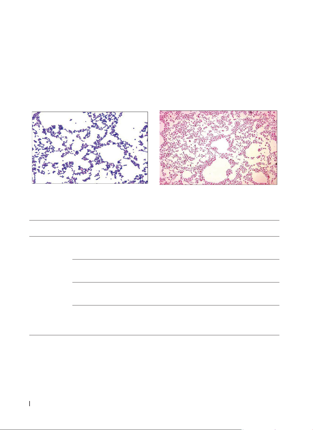
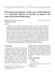
points: 30 minutes, 1 hour, 2 hours, and 24 hours. Results: In DMEM and fresh whole milk, Gram-positive
cocci, Gram-positive bacilli, and Gram-negative bacilli were presented at 30 minutes, 1 hour, and 2 hours;
by 24 hours, Gram-negative cocci were also presented. In the saline solution, Gram-positive cocci and bacilli
were also presented at all time points; Gram-negative bacilli appeared at 2 and 24 hours. In the electrolyte-
replenishing drink, Gram-positive cocci were the only bacteria found at all time points. Conclusion: Bacteria
were presented on the periodontal ligament cells of the root surface in all four environments and at all four-
time points of storage. However, at different time points, the number of bacterial groups found in DMEM and
fresh whole milk was the highest and nearly equivalent, followed by the saline solution and the lowest count
in the electrolyte-replenishing drink.
Keywords: bacteria, periodontal ligament cells, alveolar bone.
*Corresponding Author: Phan Anh Chi. Email: pachi@huemed-univ.edu.vn
Received: 15/10/2024; Accepted: 20/3/2025; Published: 28/4/2025
DOI: 10.34071/jmp.2025.2.11
1. INTRODUCTION
Teeth that are displaced from the alveolar bone
account for approximately 0.5 - 16% of all dental
trauma cases [1]. This severe dental injury causes
damage to the periodontal ligament, severs the
neurovascular bundle at the root apex, and may lead
to pulp necrosis. Immediate reimplantation of the
tooth into the alveolar bone is considered ideal and
is recommended only for permanent teeth; however,
it may not always be feasible [2]. Teeth can be
replanted into the alveolar bone if properly handled
and stored in a suitable environment. This helps
prevent drying, reducing surface root resorption and
increasing the chances of survival for periodontal
ligament cells [3]. Two of the most critical factors
affecting the prognosis of a tooth displaced from the
alveolar bone after reimplantation are the duration
of external drying and the storage environment
of the tooth [4]. Preserving the tooth in a suitable
storage environment that can maintain the viability
of the remaining periodontal ligament cells on the
root surface for as long as possible is key to the
successful reimplantation of the tooth into the
alveolar bone [5].
Recent studies have diversified the options
for the best storage environment; however, no
single storage environment has all the necessary
characteristics for tooth preservation. The ideal
medium must be capable of maintaining the
viability of periodontal ligament and pulp cells, have
physiological osmolarity and pH, possess antioxidant
properties, contain minimal or no bacterial
contamination, and be readily available in locations
where accidents occur, such as playgrounds, sports
fields, homes, schools, and hospitals, all while being
cost-effective [6].
The storage environments mentioned include
Hank’s Balanced Salt Solution (HBSS), Minimum
Essential Medium (MEM), Dulbecco’s Modified
Eagle’s Medium (DMEM), milk, coconut water, saline
solution, Save-A-Tooth, propolis, egg white, green
tea, aloe vera, saliva, soy milk, probiotics, royal jelly,
rice water, electrolyte-replenishing drinks, oresol
solution, and Emdogain [7-11]. Several national
HUE JOURNAL OF MEDICINE AND PHARMACY ISSN 3030-4318; eISSN: 3030-4326HUE JOURNAL OF MEDICINE AND PHARMACY ISSN 3030-4318; eISSN: 3030-4326
72 73
Hue Journal of Medicine and Pharmacy, Volume 15, No.2/2025 Hue Journal of Medicine and Pharmacy, Volume 15, No.2/2025
and international studies have been conducted
on various tooth preservation media; however,
there are currently limited studies evaluating the
presence of bacteria on periodontal ligament cells
of teeth displaced from the alveolar bone in these
storage environments. Therefore, we conducted
this preliminary study to evaluate the morphology
of present bacteria on the periodontal ligament cells
in different storage environments at various time
points after the teeth have been removed from the
alveolar bone using Gram staining.
2. MATERIALS AND METHODS
2.1. Research subjects: The study was conducted
on 48 premolars and third molars scheduled for
extraction due to orthodontic treatment at different
dental clinics in Hue City.
2.1.1. Selection criteria
- Permanent teeth with fully formed apices and
intact roots.
2.1.2. Exclusion criteria
- Teeth with a history of trauma or fractures.
- Teeth with severe caries, periodontal disease,
pulp pathology, or periapical pathology.
- Patients with a history of systemic diseases or
those currently on medication.
- Patients who are uncooperative or do not
consent to participate in the study.
2.1.3. Duration and location of the research
- Duration: From 01/2023 to 12/2023
- Location: Skill lab of the Faculty of Odonto-
Stomatology and the Department of Microbiology,
Hue University of Medicine and Pharmacy.
2.2. Research methods
2.2.1. Study design: In vitro experimental study
without a control group.
2.2.2. Sample selection method:
48 premolars and third molars that met the study
criteria were selected and randomly assigned to four
storage environment groups:
- Group 1 (n = 12): DMEM
- Group 2 (n = 12): Fresh whole milk (unsweetened)
- Group 3 (n = 12): Saline solution
- Group 4 (n = 12): Electrolyte-replenishing drink
Within each group, the 12 teeth were further
divided into four subgroups, each containing three
teeth, to evaluate the presence of bacteria at
different storage time points (30 minutes, 1 hour, 2
hours, and 24 hours).
2.2.3. Research methods
2.2.3.1. Preparation of teeth and placement in
storage environment
- The tooth, freshly extracted from the patient’s
mouth, was grasped using a forceps at the crown
area. A #12 scalpel blade was used to carefully
remove 3 mm of the periodontal ligament from the
root’s surface at the cervical region to eliminate
damaged cells caused by the extraction process.
Subsequently, the tooth was placed immediately
into a Falcon tube (15 ml capacity) containing 10 ml
of storage environment. The time from when the
tooth was extracted from the alveolar bone through
the scraping process and placement in the storage
environment must not exceed 45 seconds to ensure
consistency among samples and minimize errors.
- The storage environment had to submerge
the tooth throughout the study entirely. All Falcon
tubes containing the tooth samples in the storage
environment were kept at room temperature
(approximately 25°C) for the entire testing period.
After the designated time had elapsed, the tooth
was carefully retrieved using forceps at the crown
area. The root and crown’s surface were cleaned
using a plastic pipette to irrigate the tooth with saline
solution twice, ensuring the storage environment
was thoroughly removed from the tooth’s surface.
2.2.3.2. Assessment of bacterial presence on
periodontal ligament cells of teeth after displacement
from the alveolar bone
After cleaning, the tooth was grasped with
forceps at the crown area. A #12 scalpel blade was
used to scrape the apical two-thirds of the root
surface to collect periodontal ligament cells. This
process ensured that a sufficient quantity of cells
was obtained for subsequent analysis. Then, the cell
debris collected solution was added to a centrifuge
tube (1 ml capacity) with 500 µl of PBS solution and
mixed thoroughly. After that, this mixture was used
for Gram staining.
The Gram staining procedure followed the
standard protocol of the Department of Microbiology
at Hue University of Medicine and Pharmacy. Firstly,
a slide was made from the debris solution collected
from the Eppendorf tube. Then, it was fixed by
passing over the flame 2 or 3 times. The slide needed
to be cooled down completely before processing the
staining steps. A few drops of gentian violet solution
were added to the surface of the slide and allowed to
sit for 01 minute. After removing the gentian violet,
the slide was fixed with Lugol’s solution (iodine) for
1 minute. The slide was continued gently, rinsing
under tap water to wash out the previous reagents
thoroughly. The slide was decolonized using 96%
ethanol within 30 seconds before being cleaned
with tap water. The final step was to apply safranin
counterstain for 01 minutes, washing and drying the
HUE JOURNAL OF MEDICINE AND PHARMACY ISSN 3030-4318; eISSN: 3030-4326HUE JOURNAL OF MEDICINE AND PHARMACY ISSN 3030-4318; eISSN: 3030-4326
74 75
Hue Journal of Medicine and Pharmacy, Volume 15, No.2/2025 Hue Journal of Medicine and Pharmacy, Volume 15, No.2/2025
slide, which could be ready for observation under
the microscope. Different Gram-staining bacteria
were reported at 1000X magnification under a light
microscope. The characteristic properties of those
bacteria were also examined and recorded.
2.3. Research variables and evaluation methods
The results from the debris solution collected
from storage media at different time points were
checked and reported for:
- Determine the presence of different Gram
bacteria throughout the entire field of view (Gram-
negative bacteria appeared in pink color, Gram-
positive bacteria in purple color) [12].
- Describe the bacterial morphology, which is
cocci or bacilli bacteria.
statistical analysis
2.4. Statistical analysis
Use Excel 2021 software to enter collected data.
Use SPSS 20 software to manage and process data
3. RESULTS
A B
Figure 1. The image of Gram staining from the Gram-Stained Bacterial Images [13]
A. Image of Gram-positive cocci. B. Image of Gram-negative bacilli.
Table 1. The presence and morphology of bacteria on periodontal ligament cells
on the root surface stored in DMEM at different time points.
Storage media Time points Presence
of bacteria Bacterial morphology
DMEM
30 minutes Yes
(1) Gram-positive cocci
(2) Gram-positive bacilli
(3) Gram-negative bacilli
1 hour Yes
(1) Gram-positive cocci
(2) Gram-positive bacilli
(3) Gram-negative bacilli
2 hours Yes
(1) Gram-positive cocci
(2) Gram-positive bacilli
(3) Gram-negative bacilli
24 hours Yes
(1) Gram-positive cocci
(2) Gram- negative cocci
(3) Gram-positive bacilli
(4) Gram-negative bacilli
When stored in DMEM, bacteria were presented on the periodontal ligament cells on the root surface at
all 4 time points.
- At 30 minutes, 1 hour, and 2 hours, there was the presence of Gram-positive cocci, Gram-positive bacilli,
and Gram-negative bacilli.
- At 24 hours, there was an additional presence of Gram-negative cocci.
HUE JOURNAL OF MEDICINE AND PHARMACY ISSN 3030-4318; eISSN: 3030-4326HUE JOURNAL OF MEDICINE AND PHARMACY ISSN 3030-4318; eISSN: 3030-4326
74 75
Hue Journal of Medicine and Pharmacy, Volume 15, No.2/2025 Hue Journal of Medicine and Pharmacy, Volume 15, No.2/2025
Table 2. The presence and morphology of bacteria on periodontal ligament cells on the root surface
stored in fresh whole milk (unsweetened) at different time points.
Storage media Time points Presence of bacteria Bacterial morphology
Fresh whole milk
(unsweetened)
30 minutes Yes (1) Gram-positive cocci
(2) Gram- negative cocci
1 hour Yes
(1) Gram-positive cocci
(2) Gram-positive bacilli
(3) Gram-negative bacilli
2 hours Yes
(1) Gram-positive cocci
(2) Gram-positive bacilli
(3) Gram-negative bacilli
24 hours Yes
(1) Gram-positive cocci
(2) Gram- negative cocci
(3) Gram-positive bacilli
(4) Gram-negative bacilli
When stored in fresh whole milk (unsweetened), bacteria were also present on the periodontal ligament
cells on the root surface at all 4 time points.
- At 30 minutes, Gram-positive cocci and Gram-negative bacilli were present.
- At 1 hour and 2 hours, both Gram-positive cocci and Gram-positive bacilli were observed, along with
Gram-negative bacilli.
- At 24 hours, there was an additional presence of Gram-negative cocci.
Table 3. The presence and morphology of bacteria on periodontal ligament cells on the root surface are
stored in a Saline solution at different time points
Storage media Time points Presence of bacteria Bacterial morphology
Saline solution
30 minutes Yes (1) Gram-positive cocci
(2) Gram-positive bacilli
1 hour Yes (1) Gram-positive cocci
(2) Gram-positive bacilli
2 hours Yes
(1) Gram-positive cocci
(2) Gram-positive bacilli
(3) Gram-negative bacilli
24 hours Yes
(1) Gram-positive cocci
(2) Gram-positive bacilli
(3) Gram-negative bacilli
When stored in a saline solution, bacteria were present on the periodontal ligament cells on the root
surface at all four time points.
- Gram-positive cocci and Gram-positive bacilli were presented at all time points in saline solution storage.
In addition, at 2 hours and 24 hours, Gram-negative bacilli bacteria were present.
Table 4. The presence and morphology of bacteria on periodontal ligament cells
on the root surface stored in an electrolyte-replenishing drink at different time points.
Storage media Time points Presence of bacteria Bacterial morphology
Electrolyte-
replenishing
drink
30 minutes Yes (1) Gram-positive cocci
1 hour Yes (1) Gram-positive cocci
2 hours Yes (1) Gram-positive cocci
24 hours Yes (1) Gram-positive cocci
When stored in an electrolyte-replenishing drink, bacteria were present on the periodontal ligament cells
on the root surface at all 4-time points. However, only Gram-positive cocci were observed.
HUE JOURNAL OF MEDICINE AND PHARMACY ISSN 3030-4318; eISSN: 3030-4326HUE JOURNAL OF MEDICINE AND PHARMACY ISSN 3030-4318; eISSN: 3030-4326
76 77
Hue Journal of Medicine and Pharmacy, Volume 15, No.2/2025 Hue Journal of Medicine and Pharmacy, Volume 15, No.2/2025
4. DISCUSSION
DMEM is an improved version of Eagle’s
Minimum Essential Medium (EMEM), supplemented
with additional nutrients. It is used as a cell culture
medium, helping to maintain cell survival and
promote superior cell proliferation.
Sterilized milk: Milk is significantly better
than other solutions in terms of its physiological
properties, including pH (6.5 - 7.2) and osmolarity
(270 mOsm/kg), which are compatible with cells. It
also contains essential amino acids, carbohydrates,
and vitamins. Milk is readily available, bacteria-
free, and contains epidermal growth factors that
stimulate cell proliferation and regeneration.
Saline solution: This solution contains 0.9%
NaCl with an osmolarity of 280 mOsm/kg. Although
it is compatible with cells (since cell survival and
growth occur at an osmolarity range of 230 - 400
mOsm/kg and a pH of 6.6 - 7.8), it lacks essential
nutrients necessary for cellular metabolism, such as
magnesium, calcium, and glucose. The saline solution
appears suitable for the short-term preservation of
avulsed teeth for under 2 hours.
Electrolyte-replenishing drinks are a potential
storage medium commonly found at sporting events.
They should only be used as a storage medium if no
better option is available.
From the results of Tables 1 to 4, it is evident
that all four storage environments exhibited the
presence of bacteria on the periodontal ligament
cells on the root surface at four different time
points. This can be explained by the growth and
dispersion of bacteria on the tooth’s surface into the
storage medium, which subsequently adheres to the
periodontal ligament cells on the root surface. Our
study collected periodontal cell samples from only
the apical two-thirds of the root surface; thus, the
bacteria found in our research are directly related to
those suspended in the storage medium.
Tables 1 and 2 indicate that both DMEM and
fresh whole milk (unsweetened) exhibited the
presence of multiple bacterial groups on the
periodontal ligament cells on the root surface. Even
at 30 minutes, 1 hour, and 2 hours, three bacterial
groups were presented, and later on, by 24 hours,
four groups of bacteria were identified. This can be
explained by the fact that DMEM and fresh whole
milk are nutrient-rich environments, providing ideal
conditions that support the survival of human cells
as well as promoting bacterial growth, particularly
oral bacteria. These bacteria proliferate rapidly
in these media, leading to a correspondingly high
number of bacteria adhering to the root surface.
Table 3 shows that the saline solution maintained
a consistent presence of two bacterial groups at all
four storage time points. Gram-negative bacilli could
also be present at 2 hours and 24 hours. While there
is currently a lack of research on the virulence of
these Gram-negative bacilli and other bacterial
groups contributing to increased complications such
as root resorption following tooth re-implantation,
some studies indicate a correlation between
certain bacterial groups and chronic periodontal
inflammation, similar to those found in periodontal
diseases. This highlights the potential risks associated
with bacterial presence in the context of dental re-
implantation [14].
Table 4 indicates that only one bacterial
group - Gram-positive cocci - was detected in the
electrolyte solution at all four storage time points.
The pH plays a crucial role in the homeostasis of
microorganisms, as many bacteria require a specific
pH for growth. Additionally, the redox potential is a
key physicochemical parameter characterizing the
growth state of microorganisms, and continuous
fluctuations can favor the development of various
bacterial groups. Changes in pH and concentrations
of reactive gases (O2, H2, and H2S) are considered
primary factors influencing microbial development
[15]. Thus, aside from the nutrient richness of DMEM
and milk that allows for rapid bacterial proliferation
and the more limited growth observed in saline, the
low pH, redox potential, and CO2 concentration in
the electrolyte solution effectively inhibit bacterial
growth compared to the other three environments.
Different environmental factors, such as
temperature, pH, redox potential, ion strength, and
osmotic pressure, significantly influence the growth
and metabolism of microorganisms [15]. Thus, when
the storage environment has optimal factors for
maintaining the viability of periodontal ligament
cells, it also creates opportunities for the growth of
various bacterial species and the rapid multiplication
of their numbers. It is important to note that the
primary purpose of the tooth storage medium is to
preserve the viability of periodontal ligament cells,
and bacterial contamination is a secondary concern
that needs to be considered [16]. However, when
re-implanting a tooth into the socket, if it is not
adequately cleaned and disinfected, it may carry
many pathogens due to various microorganisms
adhering to it, leading to root resorption and other
infectious diseases, particularly tetanus. During the
re-implantation process, systemic or local antibiotics
can effectively prevent and eliminate bacteria.
According to the International Association of Dental