
JOURNAL OF 108 - CLINICAL MEDICINE AND PHARMACY Vol. 19 - Dec./2024 DOI: https://doi.org/10.52389/ydls.v19ita.2525
117
Malignant otitis externa - a case report
Nguyen Thi Phuong Thao
1
*, Dao Trung Dung
2
,
Ho Chi Thanh1 and Nguyen Van Huu1
1108 Military Central Hospital,
2
Bach Mai Hospital
Summary
Malignant or necrotizing otitis externa (MOE) is a severe infectious condition of the external ear
canal. It is rare but has a high mortality rate due to complications such as osteomyelitis of the skull base
and intracranial involvement. The reported clinical case involves a 72-year-old male patient with a long
history of type 2 diabetes, who presented with severe ear pain, stenosis of the external ear canal, and no
response to medical treatment one month prior to hospitalization. The CT scan showed bone
destruction of the skull base and part of the external ear canal. Despite being treated aggressively with
radical mastoid surgery and broad-spectrum antibiotics, the patient's condition worsened, leading to
complications including cranial nerve paralysis, meningitis, and ultimately death. This report aims to
analyze the clinical and paraclinical characteristics and progression of the disease to draw lessons for the
diagnosis and treatment of this dangerous condition.
Keywords: Malignant otitis externa, the external auditory canal.
I. BACKGROUND
Malignant otitis externa (MOE) is a severe
infection that originates in the external auditory
canal and subsequently spreads to nearby
structures, particularly leading to skull base
osteomyelitis, and causes dangerous complications
such as meningitis, brain abscess, and septic
thrombophlebitis of the venous sinuses. Its
prevalence ranges from 0.221 to 1.19 cases per
100.000. The disease is primarily bacterial, especially
involving Pseudomonas aeruginosa, though in rare
cases it may be caused by fungi1.
MOE mainly affects elderly individuals with
compromised immune systems, such as
uncontrolled diabetes, HIV/AIDS, or those on
immunosuppressive medications (post-organ
transplant, etc)1. The condition was first recognized
in 1838 when Toulmouche reported a case of
temporal bone osteomyelitis2. In 1968, Chandler JR
introduced the term "malignant otitis externa" to
Received: 15 November 2024, Accepted: 28 December 2024
*Corresponding author: drthao108@gmail.com -
108 Military Central Hospital
highlight the disease’s dangerous nature, with a
mortality rate reaching approximately 50%3. Later,
the term "necrotizing otitis externa" was suggested
to avoid confusion with true malignant diseases.
Both terms are still used interchangeably today.
Common symptoms of MOE include ear pain,
otorrhea, edema, granulation tissue observed in
the exteranal auditory canal and a sensation of
fullness in the ear, often leading to misdiagnosis as
acute otitis externa, especially in the early stages.
Despite advances in treatment, MOE is still a
potential devastating condition that continues to
pose a diagnostic and therapeutic challenge. This
report describes a case of MOE, analyzing the
clinical, paraclinical features, and disease course to
draw lessons for diagnosis and treatment.
II. CASE PRESENTATION
T.D.T - A 72-year-old male presented to the
hospital with a 20-year history of diabetes treated
with insulin injections. He had suffered a stroke one
year prior, resulting in residual right hemiparesis and
left peripheral facial paralysis. The patient also had a
history of endoscopic surgery for fungal maxillary
sinusitis one month before admission, which had
JOURNAL OF 108 - CLINICAL MEDICINE AND PHARMACY Vol. 19 - Dec./2024 DOI: https://doi.org/10.52389/ydls.v19ita.2525
118
resolved (No nasal discharge and a clean middle
nasal meatus on endoscopy). The main reasons for
presentation was his left-sided severe headache,
accompanied by ear pain and foul-smelling ear
discharge on the same side for one month. He had
been prescribed with oral and topical antibiotics at a
local clinic, but the symptoms persisted.
At the time of admission, the patient was alert,
afebrile, without signs of meningitis, and had left
peripheral facial paralysis grade IV due to previous
history of stroke (according to House-Brackman
Classification), with no clear signs of propression as
reported by the patient's family. He experienced
severe pain on the right side of his head, right side
of his face, and the entire right ear, with a VAS score
of 7-8. On examination, the right auricle showed
edema and moist inflammation, the left external ear
canal was narrowed, preventing visualization of the
tympanic membrane while the skin over the
mastoid was normal, mild trismus and dysphagia.
The tympanic membrane on the contralateral ear
appeared to be intact and thickened. Blood tests
revealed an increased white blood cell count of
14.7G/L, neutrophils at 75%, CRP at 114mg/L,
glucose level at 24.2mmol/L, and HbA1c at 13.5%.
Figure 1. Imflammation of the left auricular
Initially diagnosed was inflammation and
stenosis canal of the external auditory canal in a
patient with a history of stroke. The patient was
treated aggressively with intravenous levofloxacin
750mg per day and daily ear toilet. Intravenous and
oral paracetamol- tramadol and subcutaneous 20UI
insulin 3 times/day were prescribed additionally.
After treatment with insulin, glucose in blood in
within the normal limits. However, after two weeks,
the patient continued to experience severe pain,
and the canal stenosis remained unchanged and the
tympanic membrane could not been observed. A
temporal bone CT scan showed a fully stenose ear
canal, fluid and inflamed tissue in the mastoid and
middle ear, a thickended left side nasopharynx
mucosa, extensive bone erosion at the tegmen and
the temporomandibular joint. MRI showed irregular
thickening of the nasopharyngeal on the left side.
The patient underwent radical mastoidectomy
in the third week of admission, revealing ulcerative
inflammatory lesions and fungal involvement
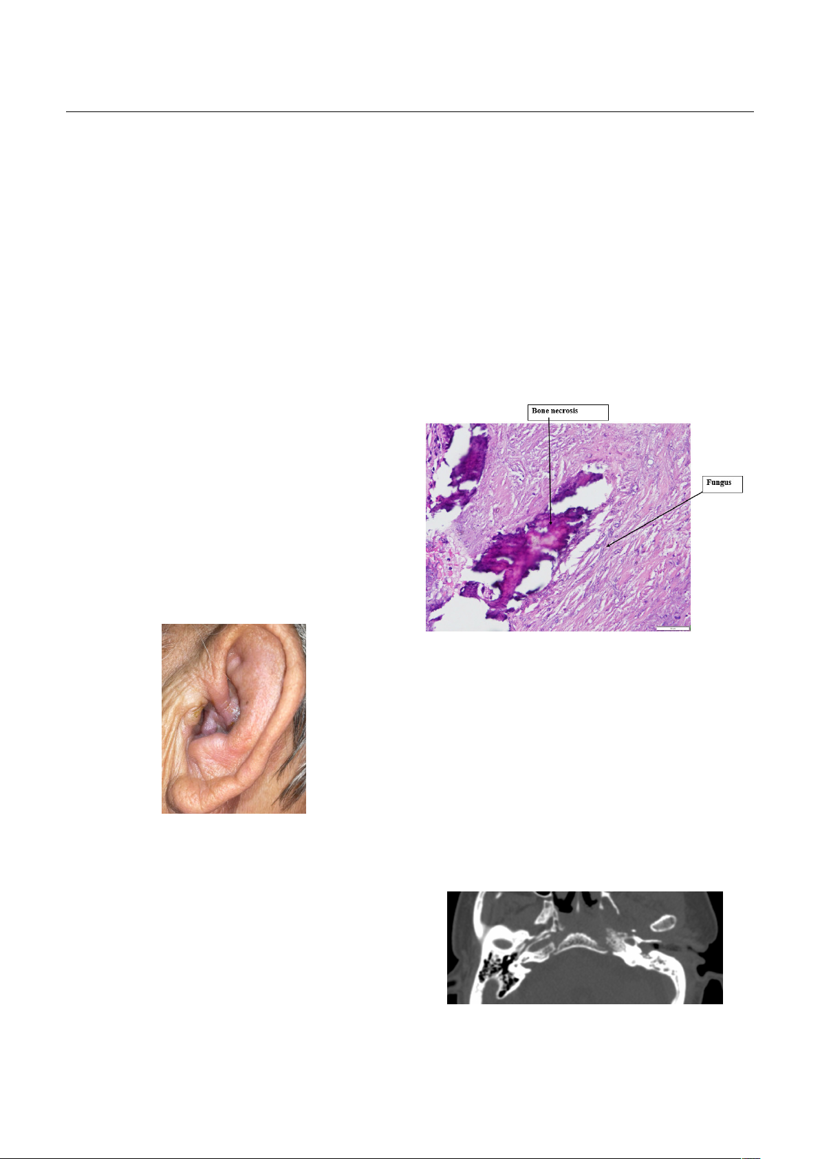
confirmed by Grocott staining of middle ear tissues.
Figure 2. Histopathology of middle ear tissue
Despite treatment with intravenous ceftazidime
6g/day, pain management, and continued blood
glucose control, the patient’s condition deteriorated
after 10 days with increasing dysphagia and
multiple cranial nerve palsies. CRP increased at the
level of 290mg/L. The CT scan showed difuse brain
edema corresponding to the blood supply regions
of the anterior and middle cerebral artery on both
sides. The patient then developed pneumonia,
bacterial meningoencephalitis, recurrent stroke, and
ultimately died at the 30th day of hospitalization.
Figure 3. Preoperative temporal bone CT scan

JOURNAL OF 108 - CLINICAL MEDICINE AND PHARMACY Vol. 19 - Dec./2024 DOI: https://doi.org/10.52389/ydls.v19ita.2525
119
III. DISCUSSION
Malignant otitis externa is a rare disease with a
high mortality rate that requires timely diagnosis
and treatment. A systematic review by Gonzalez and
colleagues indicates that common symptoms
include severe ear pain unresponsive to standard
pain relief, ear discharge, hearing loss,
temporomandibular joint pain, and headaches.
However, due to the symptoms resembling those of
other common external ear infections, the diagnose
is ofter delayed, especially when neurological
symptoms become pronounced. Consequently,
Cohen and Friedman proposed definitive diagnostic
criteria, emphasizing key indicators such as
disproportionate pain relative to the clinical
findings, edematous external ear canal, discharge,
granulation tissue, numerous micro-abscesses
observed during surgery, and bone scans with Tc-99
MDP showing strong uptake in lesions, which do not
respond to local treatment lasting over a week.
Additional criteria may include diabetes, cranial
nerve palsies, bone erosion on imaging results,
cachexia, and old age4. Symptoms of cranial nerve
palsies, particularly facial nerve involvement, are
complications of the disease and are considered
poor prognostic factors5.
Our case aligned with the epidemiological
characteristics described in the literature,
demonstrating that in elderly patients with
uncontrolled diabetes who present with acute
external otitis symptoms such as ear pain and
discharge, malignant otitis externa should be
considered.
In this patient, due to presenting at a late stage,
the characteristic sign granulation tissue found at
the junction of the cartilaginous and bony ear
canal—was replaced by a stenosed ear canal. In
addition, the main symptoms such as severe
headache, ear discharge, and swelling of the
external ear canal had not improved with local
treatment and had been present for about a month
before hospitalization. Because of the diffuse nature
of the pain along with the coexisting sinus
pathology, the patient was initially diagnosed with
headache due to sinusitis and was referred for
endoscopic sinus surgery. However, the symptoms
did not improve afterward.
Among the other symptoms of the disease,
notable is the condition of cranial nerve paralysis. In
this patient, due to the sequelae of a previous
stroke, there were signs of peripheral paralysis of the
VII cranial nerve on the same side as the lesion, as
well as pre-existing cranial nerve damage. As a
result, this group of neurological symptoms was not
grouped with the newly appearing ear symptoms.
This comorbid factor made the diagnosis more
difficult and delayed. Signs of small abscesses were
found during the radical mastoidectomy. Here, due
to the severity and fairly typical nature of the
disease, the patient had not undergone a bone scan
using Tc-99m MDP.
According to the consensus of the British ENT
Association in 2020, a patient is diagnosed with
malignant otitis externa (MOE) if all of the following
criteria are met: Ear pain with ear discharge,
granulation tissue or external ear canal
inflammation, histopathological results excluding
malignant pathology, CT scan showing bone
destruction of the external auditory canal, soft tissue
involvement of the external ear canal, or MRI
showing edema of the temporal bone marrow.
Clinically, this patient met all the diagnostic criteria
and diagnose wass confirm6.
In MOE, imaging diagnosis is a useful tool for
both diagnosis and prognosis. Peleg and colleagues
classified MOE into two forms: “severe” and “non-
severe” by scoring the imaging signs on CT scans7.
However, the drawback of this scale is that it does
not evaluate the progression of the disease. Later,
Tengku, based on the results of temporal CT scans
and bone scans, divided MOE into five stages8. Stage
I involves localized inflammation in the soft tissue of
the external ear canal, stage II includes inflammatory
bone lesions confined to the mastoid bone, stage III
shows invasive inflammatory lesions penetrating
inward, infiltrating the petrous bone and the
temporomandibular joint, with or without
accompanying tissue in the oropharyngeal region.

JOURNAL OF 108 - CLINICAL MEDICINE AND PHARMACY Vol. 19 - Dec./2024 DOI: https://doi.org/10.52389/ydls.v19ita.2525
120
In stage IV, lesions extends to the nasopharyngeal
area, with or without abscesses. Finally, in stage V,
the inflammatory process spreads to the
contralateral skull base. According to Tengku’s
grading scale, this patient presented to us in stage
IV, where the CT scan showed destruction of the
petrous bone, the body of the sphenoid bone, the
left lateral skull base, and infiltrative inflammation in
the nasopharyngeal area on the same side. This is a
severe stage with a high mortality rate.
The causative agents of MOE can be bacteria or
fungi. Among them, Pseudomonas aeruginosa is the
primary pathogen, accounting for over 95% of cases.
Identifying the causative microorganisms plays an
important role in conducting antibiotic
susceptibility tests, which helps to adjust the
appropriate antibiotics. However, in practice,
approximately 13% of bacterial cultures may be
negative9. In this patient, the negative result could
be due to the prolonged use of antibiotics and local
ear drops prior to the culture, which may have
prevented the identification of the causative
bacteria. The patient was also completely ruled out
for suspected cancerous lesions when the
postoperative pathology results indicated acute
inflammatory ulcers and the biopsy of the ipsilateral
mucosa showed no malignant cells. A biopsy sample
from the middle ear was then stained with Grocott,
resulting in a positive finding for Aspergillus.
Treating MOE is always a challenge for
clinicians, even in developed countries. In principle,
the treatment regimen needs to be comprehensive,
involving systemic and local interventions, blood
glucose strictly controlled, supportive care, and
functional rehabilitation10. Surgery is also indicated
in cases where medical treatment is ineffective,
primarily to remove necrotic tissue, drain abscesses,
and collect infected bone for pathology test. In
patient T.D.T, we recommended surgery due to the
worsening local ear inflammation despite intensive
medical treatment. Additionally, surgery would
allow for the removal of the pathological tissue and
help exclude malignant conditions. After
undergoing a radical surgery, there were significant
improvements in clinical symptoms, such as
reduced headache and less ear discharge. The
antibiotic regimen used for the patient in the early
stages, given the negative microbial culture results,
was based on general recommendations:
Ceftazidime 6g/day in combination with
intravenous levofloxacin. Due to the patient's severe
condition, exhaustion, and limited experience in
treating fungal diseases, we did not administer
antifungal medications. Additionally, the patient's
blood glucose levels were closely monitored and
adjusted in collaboration with the endocrinology
department, along with continuous supportive care
using nutritional solutions. After an initial period of
relative improvement, the patient quickly
deteriorated, especially when experiencing
gastroesophageal reflux episodes leading to
aspiration pneumonia, and showing signs of
meningitis such as fever, neck stiffness, and a
positive Rivalta reaction on cerebrospinal fluid
analysis. The patient's condition then worsened
rapidly, resulting in death when lung function was
lost, along with diffuse brain edema due to
extensive bilateral cerebral infarction and occlusion
of the feeding vessels of the anterior circulation and
both middle cerebral arteries.
It can be said that this patient belongs to the
severe disease group with a poor prognosis right
from the first day of hospitalization, based on the
criteria in the literature. Specifically, according to
Tengku's imaging diagnostic criteria, at the time of
diagnosis, the patient was in stage IV-V with
multiple lesions at the ipsilateral skull base and
beginning to affect the contralateral side. At the
time of admission, signs of VII cranial nerve paralysis
and other cranial nerves such as IX and X causing
aspiration and difficulty swallowing were also
indicators of a severe prognosis8. Additionally,
according to Stevens, the presence of fungi in the
affected tissue tends to lead to rapid deterioration
of the disease, increasing the mortality rate from 0%
in the group without fungi to 46% in the group
where fungi were found. In this patient, the fungus
identified in the tissue was Aspergillus11. The fact

JOURNAL OF 108 - CLINICAL MEDICINE AND PHARMACY Vol. 19 - Dec./2024 DOI: https://doi.org/10.52389/ydls.v19ita.2525
121
that antifungal antibiotics had not yet been used
could also be a factor contributing to the rapid
progression of the disease.
IV. CONCLUSION
Malignant otitis externa is a severe and
dangerous condition of the external auditory canal.
The diagnosis should be considered when severe
ear pain and headaches are disproportionate to
clinical findings and unresponsive to treatment,
particularly in high-risk patients (elderly,
immunocompromised). Imaging in cluding CT-scan,
MRI and Tc-99m are crucial for diagnosis and staging
as. Prognostic factors include comorbidities such as
diabetes and liver/kidney dysfunction, and
neurological or endocrine complications should be
carefully monitored. Comprehensive,
multidisciplinary care is essential for favorable
outcomes in this challenging disease.
REFERENCES
1. González JLT, Suárez LLR, de León JEH (2021)
Malignant otitis externa: An updated review.
American Journal of Otolaryngology 42(2):
102894.
2. Kumar S, Singh U (2015) Malignant otitis externa: A
review. J Infect Dis Ther 3(204): 2332-
0877.1000204.
3. Chandler JR (1968) Malignant external otitis. The
Laryngoscope 78(8): 1257-1294.
4. Cohen D, Friedman P (1987) The diagnostic criteria
of malignant external otitis. The Journal of
Laryngology 101(3): 216-221.
5. Marina S, Goutham M, Rajeshwary A, Vadisha B,
Devika T (2019) A retrospective review of 14 cases of
malignant otitis externa. Journal of otology 14(2):
63-66.
6. Hodgson SH, Khan MM, Patrick-Smith M et al
(2023) UK consensus definitions for necrotising otitis
externa: A Delphi study. BMJ open 13(2): 061349.
7. Peleg U, Perez R, Raveh D, Berelowitz D, Cohen D
(2007) Stratification for malignant external otitis.
Otolaryngology Head Neck Surgery 137(2): 301-305.
8. Kamalden TMIT, Misron K (2021) A 10-year review
of malignant otitis externa: Anew insight. European
Archives of Oto-Rhino-Laryngology: 1-8.
9. Takata J, Hopkins M, Alexander V et al (2023)
Systematic review of the diagnosis and management
of necrotising otitis externa: Highlighting the need
for high‐quality research. Clinical Otolaryngology
48(3): 381-394.
10. Al Aaraj MS, Kelley C (2023) Necrotizing (Malignant)
Otitis Externa. StatPearls [Internet]. StatPearls
Publishing.
11. Stevens SM, Lambert PR, Baker AB, Meyer TA
(2015) Malignant otitis externa: A novel stratification
protocol for predicting treatment outcomes.
Otology & Neurotology 36(9): 1492-1498.


![Bài giảng Cập nhật vấn đề hồi sức bệnh tay chân miệng nặng [mới nhất]](https://cdn.tailieu.vn/images/document/thumbnail/2025/20250920/hmn03091998@gmail.com/135x160/23301758514697.jpg)