TẠP CHÍ Y häc viÖt nam tẬP 545 - th¸ng 12 - sè 1 - 2024
149
risk factors and 18F-fluorodeoxyglucose positron
emission tomography/computed tomography
parameters". 36 (5), pp. 425-432.
3. Ciappuccini R. et al. (2020), "Tumor burden of
persistent disease in patients with differentiated
thyroid cancer: correlation with postoperative risk-
stratification and impact on outcome". 20, pp. 1-12.
4. Gay S. et al. (2022), "2-[18F] FDG PET in the
Management of Radioiodine Refractory
Differentiated Thyroid Cancer in the Era of
Thyrosin-Kinases Inhibitors: A Real-Life
Retrospective Study". 12 (2), pp. 506.
5. Luo Y. et al. (2020), "Clinical, pathological, and
molecular characteristics correlating to the
occurrence of radioiodine refractory differentiated
thyroid carcinoma: a systematic review and meta-
analysis". 10, pp. 549882.
6. Manohar P. M. et al. (2018), "Prognostic value
of FDG-PET/CT metabolic parameters in
metastatic radioiodine-refractory differentiated
thyroid cancer". 43 (9), pp. 641-647.
7. Robbins R. J. et al. (2006), "Real-Time Prognosis
for Metastatic Thyroid Carcinoma Based on 2-
[18F]Fluoro-2-Deoxy-d-Glucose-Positron Emission
Tomography Scanning", The Journal of Clinical
Endocrinology & Metabolism. 91 (2), pp. 498-505.
8. Roy M. et al. (2022), "Using 18F-FDG-PET/CT
Metrics to Predict Survival in Ra-Dio-Iodine
Refractory Thyroid Cancers". 12 (10), pp. 2381.
9. Santhanam P. et al. (2018), "The relationship
of BRAFV600E mutation status to FDG PET/CT
avidity in thyroid cancer: a review and meta-
analysis". 24 (1), pp. 21-26.
10. Wang H. et al. (2021), "Investigating 18F-FDG
PET/CT parameters as prognostic markers for
differentiated thyroid cancer: A systematic
review". 11, pp. 648658.
BÁO CÁO CA LÂM SÀNG DỊ VẬT HỐC MẮT
TRÊN BỆNH NHÂN CHẤN THƯƠNG XUYÊN THỦNG NHÃN CẦU
Nguyễn Thanh Nam1, Biện Thị Cẩm Vân1, Tôn Tường Trí Hải2
TÓM TẮT36
Chẩn đoán và xử trí dị vật hốc mắt phức tạp trên
bệnh nhân chấn thương xuyên thủng nhãn cầu. Chúng
tôi báo cáo ca lâm sàng khó về dị vật kim loại lớn,
nằm trong xoang sàng sau, cạnh ống thị giác trong
hốc mắt bên trái trên một bệnh nhân bị chấn thương
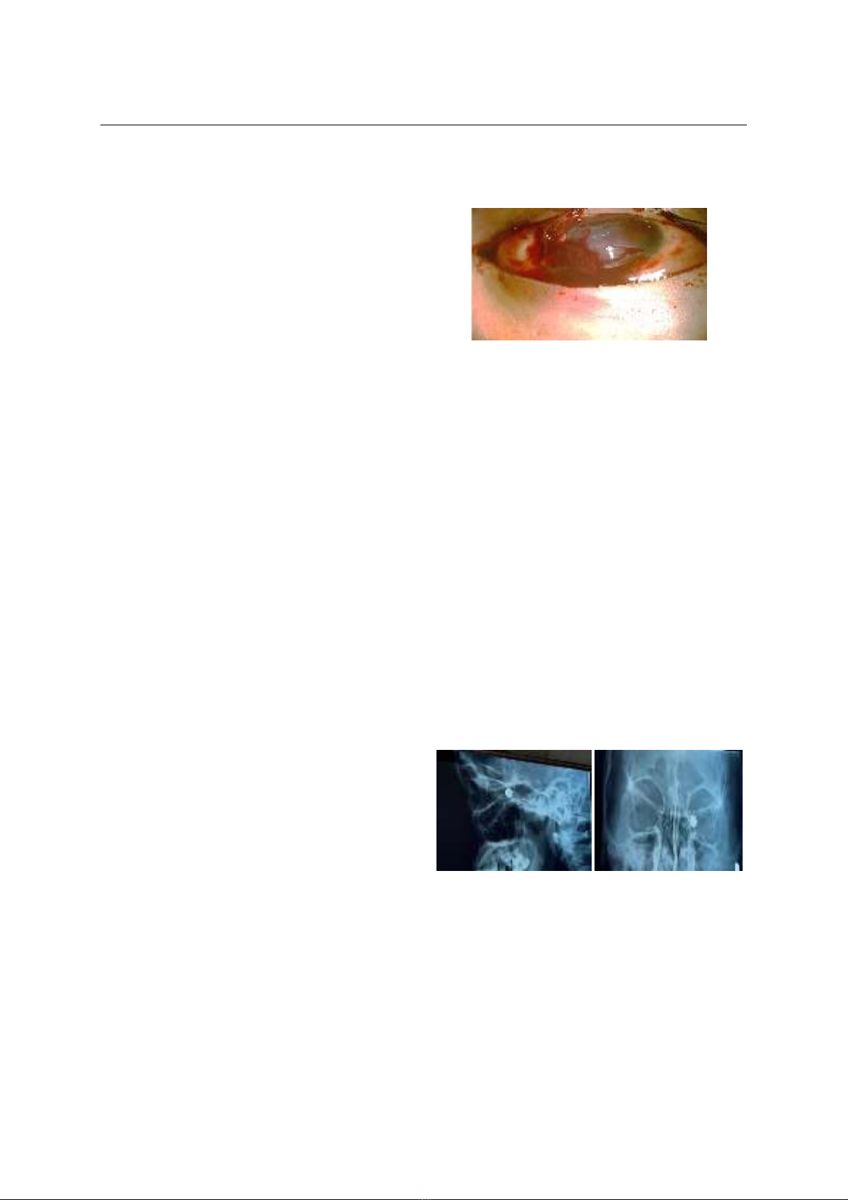
nhãn cầu do ná bắn. Bệnh nhân nữ 22 tuổi, thị lực
mắt trái sáng tối âm tính, được chẩn đoán vỡ nhãn
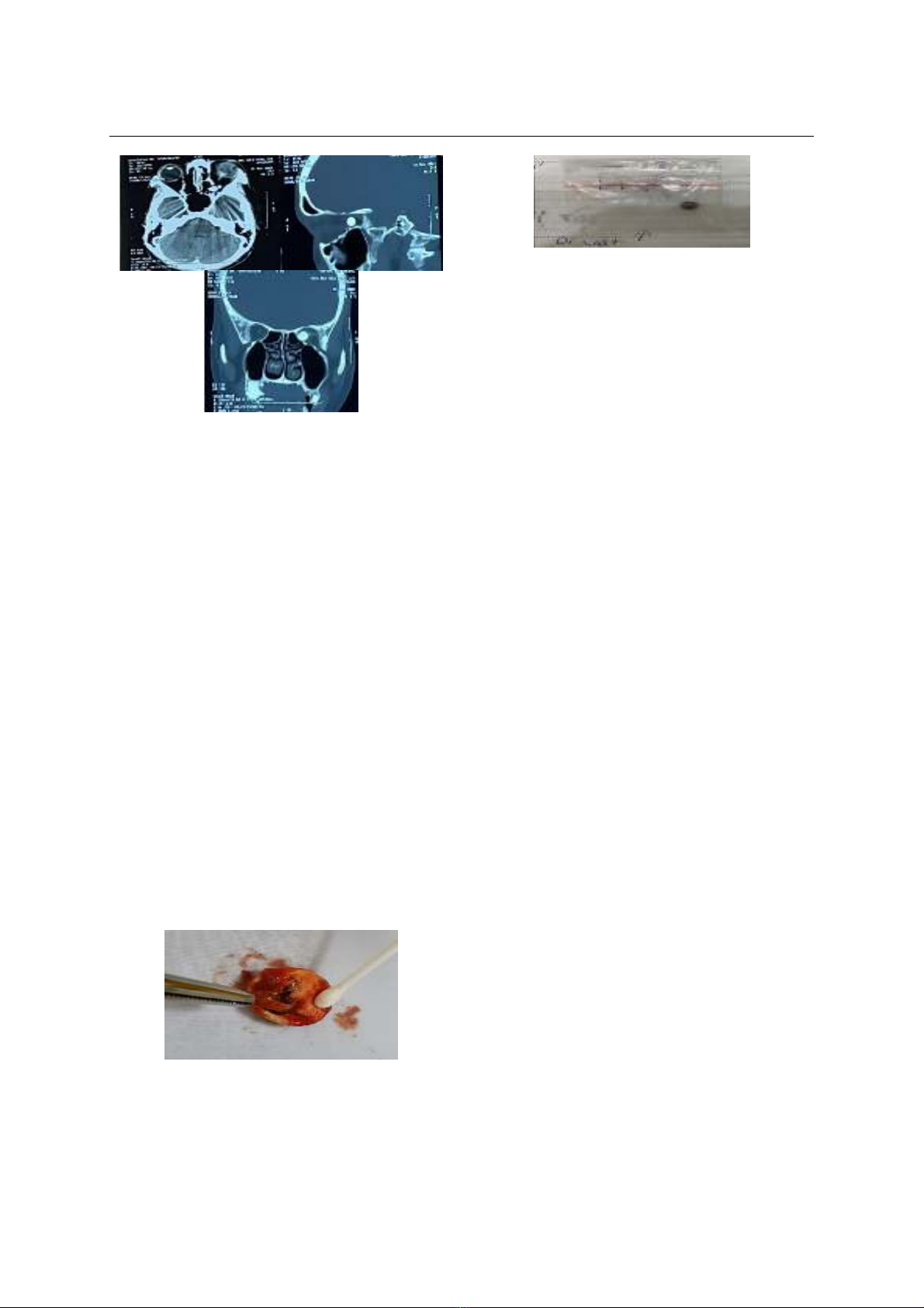
cầu bên trái do ná bắn. CT Scan phát hiện dị vật kích
thước lớn, nằm trong xoang sàng sau, cạnh ống thị
giác. Bệnh nhân được điều trị với kháng sinh phổ rộng
và corticosteroid đường tĩnh mạch. Vì mắt trái có lỗ
rách lớn ở cực sau không thể bảo tồn, bệnh nhân
được cắt bỏ nhãn cầu kèm lấy dị vật hốc mắt. Sau 15
ngày điều trị, hậu phẫu không nhiễm trùng, không
phát hiện dị vật còn sót.Hầu hết các chấn thương
xuyên nhãn cầu đều để lại dị vật, do đó thái độ nghi
ngờ là điều cần thiết trong chẩn đoán phát hiện. Việc
khai thác bệnh sử, cơ chế chấn thương và kết hợp CT
Scan hốc mắt là lựa chọn đầu tay.
Từ khóa:
Dị vật
hốc mắt, chấn thương xuyên nhãn cầu
SUMMARY
CASE REPORT ORBITAL FOREIGN BODY IN
ASE OF OCULAR PERFORATING TRAUMA
Diagnosis and management of complex
intraorbital foreign body in a patient with perforating
ocular injury. We report the challenging case of a
1Bệnh viện Mắt TP. Hồ Chí Minh
2Trường Đại học Y khoa Phạm Ngọc Thạch
Chịu trách nhiệm chính: Nguyễn Thanh Nam
Email: drnam49@yahoo.com
Ngày nhận bài: 12.9.2024
Ngày phản biện khoa học: 22.10.2024
Ngày duyệt bài: 22.11.2024
large metallic foreign body located in the posterior
ethmoid sinuses, near the optic canal of left orbital in
a patient following perforating ocular injury caused by
slingshot. A 22-year-old female patient, presented to
us with no light perception in the left eye caused by
slingshot. Orbital CT Scan showed a large intraorbital
foreign body located in the posterior ethmoid sinuses,
near the optic canal of left orbital. She was treated by
intravenous broad spectrum antibiotics and
corticosteroids. The large laceration at the posterior
pole could not be closed, the patient underwent eye
enucleation and foreign body removal. She was
discharged from hospital after 15 days of treatment
with no signs of postoperative infection. Most
perforating ocular injuries involving with forgein
bodies. Thus, suspicions are crucial to defining the
diagnosis. An accurate and detailed history, trauma
mechanism as well as CT Scan of the orbit, which are
the first – choices.
Keywords:
Intraorbital foreign
body, perforating ocular injury
I. ĐẶT VẤN ĐỀ
Chấn thương nhãn cầu kèm với dị vật hốc
mắt có thể đưa đến những tổn thương nghiêm
trọng về mặt cấu trúc và chức năng của nhãn
cầu cũng như tổ chức hốc mắt1. Một nghiên cứu
hồi cứu 713 bệnh nhân chấn thương nhãn cầu có
dị vật được thực hiện tại bệnh viện Getúlio
Vargas ở Brazil cho thấy nam giới chiếm 96,21%,
phần lớn các trường hợp là do tai nạn lao động
(61,29%) và dị vật bằng kim loại chiếm nhiều
nhất (75,17%)2. Dị vật hốc mắt có thể được
phân loại dựa vào cầu tạo: a) Dị vật kim loại
chẳng hạn như sắt; b) Dị vật không phải kim loại
bao gồm dị vật vô cơ như thủy tinh; c) dị vật