Can Tho Journal of Medicine and Pharmacy 10(7) (2024)
170
IMMUNOGLOBULIN E IN PEDIATRIC ASTHMA:
ADVANCES IN UNDERSTANDING AND MANAGEMENT
Huynh Hoang Khang, Nguyen Dinh Nguyen Chuong, Tran Cong Ly*
Can Tho University of Medicine and Pharmacy
*Corresponding author: tcly@ctump.edu.vn
Received: 24/03/2024
Reviewed: 16/05/2024
Accepted: 22/05/2024
ABSTRACT
Asthma, a pervasive chronic inflammatory ailment of the respiratory system, remains a
global health conundrum. The Global Burden of Disease Study (GBD) of 2019 underscores its
widespread impact, revealing that asthma afflicts 262 million individuals worldwide, translating
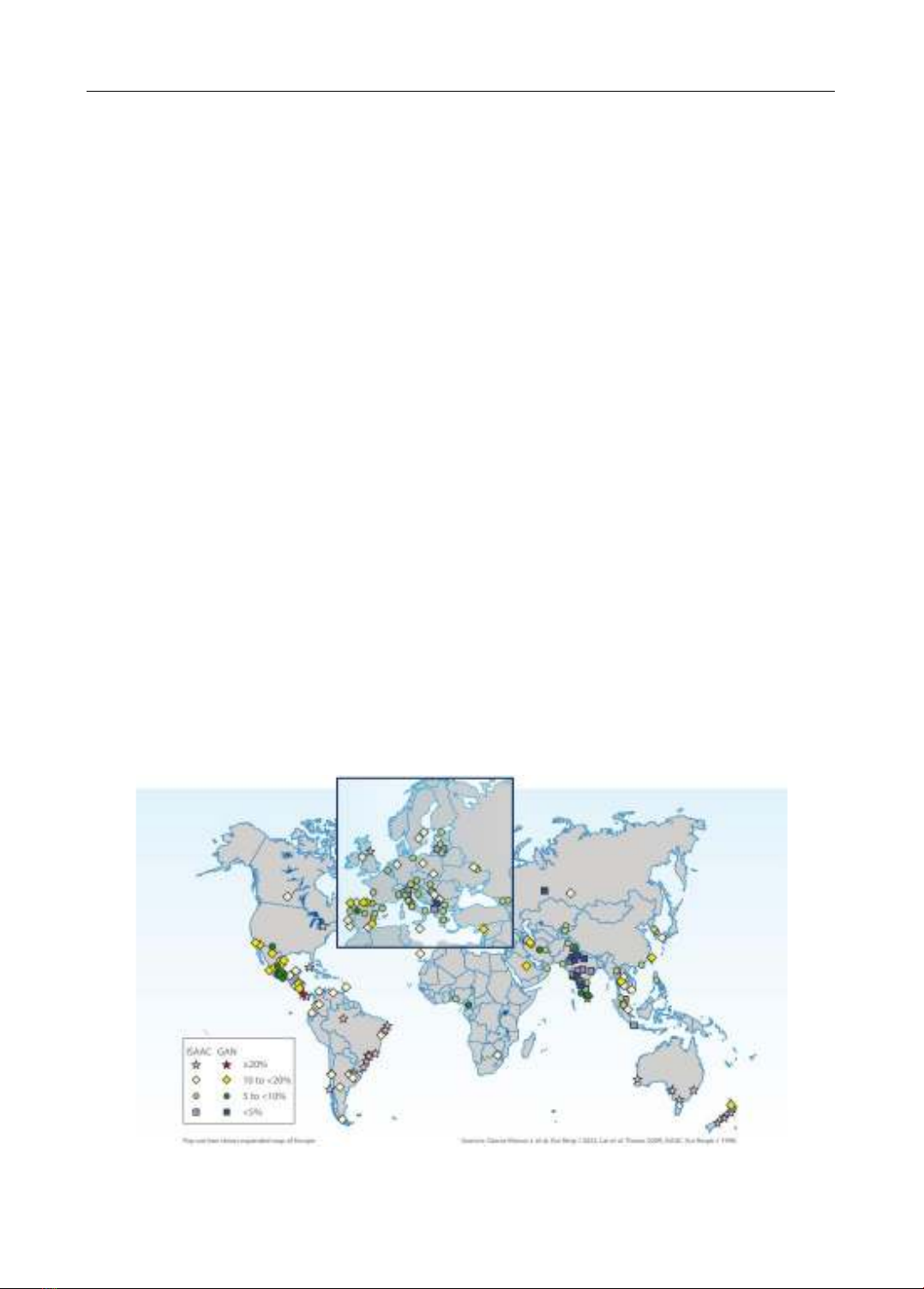
into an age-standardized prevalence of 3,416 per 100,000 population. The incidence among
children is particularly alarming, with nearly 14% of the global pediatric population diagnosed
with the condition. This statistic positions asthma as the foremost chronic respiratory disease among
children, a trend that is on the rise, especially across Asia and Europe, as evidenced by the
International Study of Asthma and Allergies in Childhood (ISAAC). Characterized by variable
airflow limitation, bronchial hyperresponsiveness, excessive mucus production, and airway
inflammation leading to airway constriction, asthma’s multifaceted nature complicates its
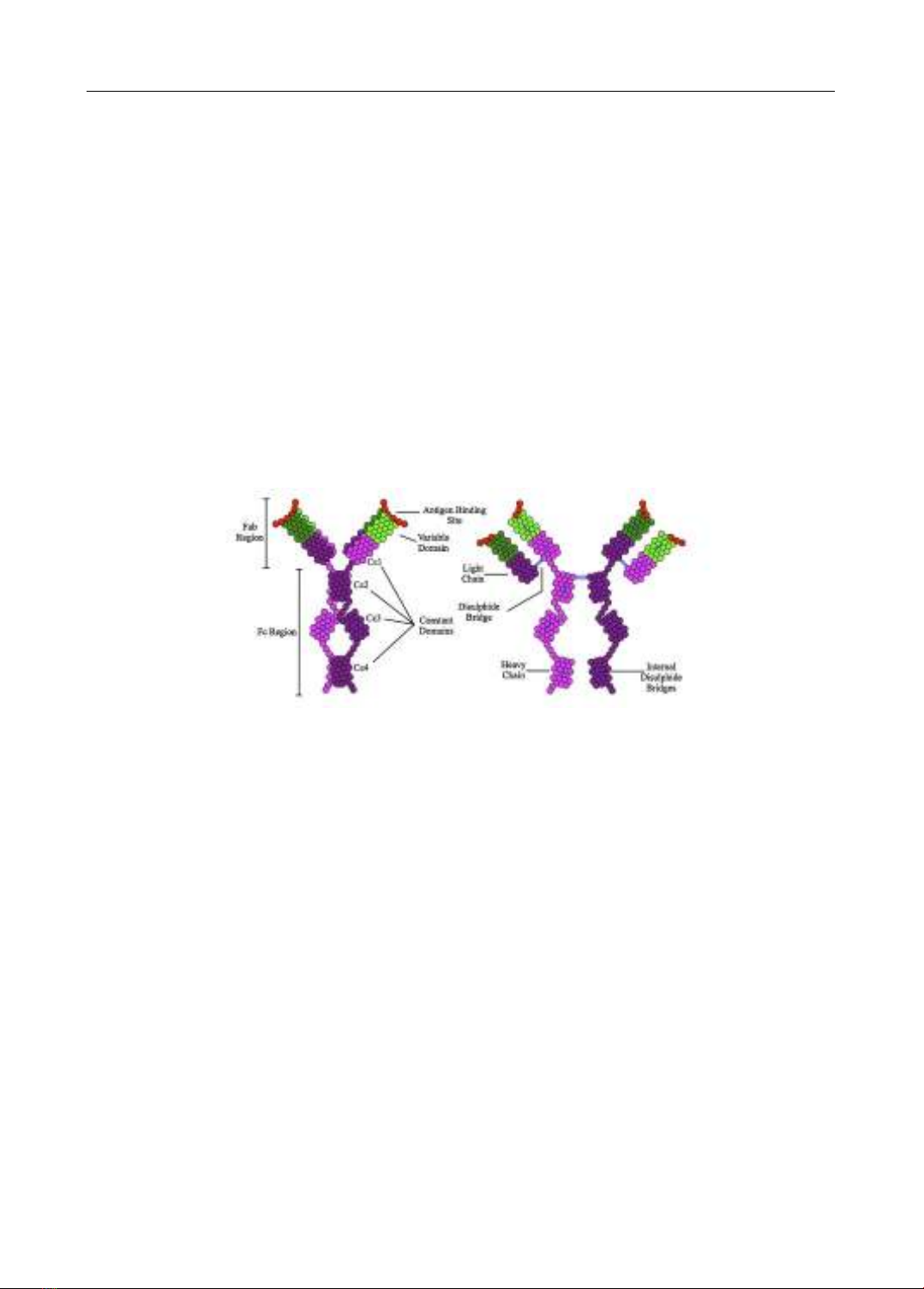
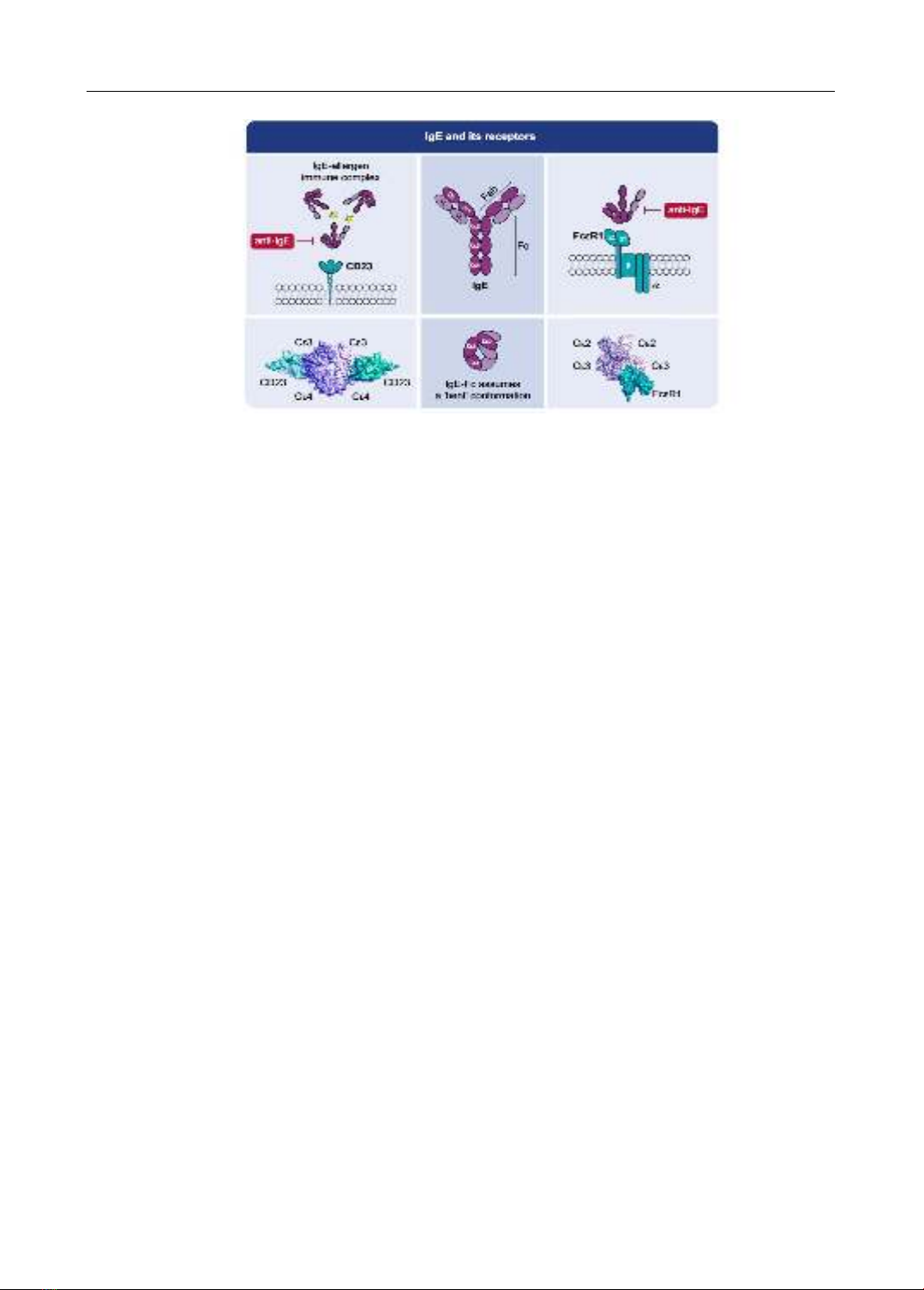
management. In the realm of immunology, Immunoglobulin E (IgE) has been identified as a pivotal
player. Recognized officially as the fifth class of serum immunoglobulins during the 1968 WHO
International Reference Center for Immunoglobulins conference in Lausanne, IgE’s crucial role in
the pathophysiology of asthma has been rigorously studied. Serum IgE levels, both total and specific,
have been proven instrumental in the diagnosis, treatment, and prevention of pediatric asthma. The
landmark approval of Omalizumab by the US Food and Drug Administration in 2003 heralded a
Can Tho Journal of Medicine and Pharmacy 10(7) (2024)
171
new era in the biologic management of asthma, targeting children aged six and above. This was
followed by the development of Ligelizumab and Quilizumab, innovative anti-IgE medications
currently under investigation for their potential to alleviate symptoms and decelerate the disease’s
progression. The integration of Allergy Immunotherapy (AIT) alongside monoclonal antibody
therapies like Omalizumab, Ligelizumab, and Quilizumab signifies a monumental shift toward
personalized medicine in asthma care. These advances promise not only to ameliorate the quality
of life for pediatric asthma patients but also to redefine the landscape of asthma management.
Nonetheless, the quest for enhanced treatment modalities for young asthmatics necessitates further
in-depth research. The burgeoning field of anti-IgE therapy, in concert with AIT, is poised to set
new benchmarks in pediatric asthma management, steering us towards a future where asthma’s grip
on children’s health is significantly loosened.
Keywords: IgE, allergy, pediatric asthma, Omalizumab, Allergen Immunotherapy (AIT).
I. INTRODUCTION
Asthma stands as a formidable adversary among non-communicable diseases,
significantly diminishing the quality of life for countless individuals worldwide. It ranks as
the sixteenth leading cause of years lived with disability globally. With over 300 million
people currently grappling with asthma, projections indicate that by 2025, an additional 100
million may be afflicted [1]. The Global Burden of Disease Study (GBD) in 2019
highlighted that asthma affected 262 million individuals globally, resulting in an age-
standardized prevalence of 3416 per 100,000 people. Notably, while children experience
higher rates of asthma incidence and prevalence, adults suffer from increased morbidity and
mortality associated with the condition [2].
A staggering 14% of children worldwide have been diagnosed with asthma,
positioning it as the most common chronic respiratory disease among this demographic [3].
An upward trend in the incidence of asthma among children has been observed across Asia
and Europe, as documented by the International Study of Asthma and Allergies in
Childhood. In a study encompassing 11 countries, the incidence rates in Asia were found to
range from 10 to 30 percent [4].
Figure 1. Current asthma symptom prevalence in 6-7 year old children [2]
Can Tho Journal of Medicine and Pharmacy 10(7) (2024)
172
Allergies emerge from an intricate immune response involving TH2 cells, mast cells,
eosinophils, and Immunoglobulin E (IgE) against non-microbial environmental antigens or
allergens. There exists a discernible genetic predisposition to atopic diseases, including
asthma, urticaria, hay fever, and eczema, along with an identifiable IgE-mediated response
post-allergen exposure. Key allergens include house dust mites, fungal spores, pollens, and
animal dander [5]. Although total IgE serves as a broad measure for allergic conditions, it
lacks specificity. Conversely, specific IgE presents a highly specific marker for diagnosing
allergic conditions [4]. However, elevated total IgE levels may also be indicative of other
conditions, including parasitic infestations, and hence are not solely attributable to allergic
disorders. The limitations of testing methods and allergen selection mean that a negative
specific IgE result cannot definitively exclude allergen sensitivity [5].
Asthma is a complex condition, varying widely in its etiology, triggers, clinical
presentation, and crucial treatment responsiveness [6]. Epidemiological evidence strongly
supports the link between IgE antibodies (both specific and total) and asthma, underscoring the
allergic underpinnings of the disease. As Sporik et al. articulated, asthma is predominantly
allergic and invariably associated with IgE-mediated reactions. Numerous studies corroborate
the significant association between allergen sensitivity and asthma, as evidenced through skin
tests or the detection of specific IgE in the blood [7]. The presence of allergen-specific IgE
antibodies is a common trait among children with chronic and persistent asthma, establishing it
as a critical factor for the onset of lifelong asthma [8]. The NIH Asthma Outcomes Task Force
advises that aeroallergen sensitivity be evaluated as a vital biomarker for asthma classification,
in addition to eosinophilia and antigen-specific IgE [9],[10].
This review aims to elucidate the intricate role of IgE in pediatric asthma, thereby
contributing to a deeper understanding of its pathophysiology and aiding in the development
of more effective diagnostic and therapeutic strategies.
II. CONTENT
2.1. Structure and function of IgE
The history of the discovery of IgE
Immunoglobulin E (IgE) stands as the most recently identified member of the
immunoglobulin family, historically associated with the expulsion of worms and the onset
of allergies [11]. The phenomenon of anaphylaxis, deriving from the Greek words “ana”
(against) and “phylaxis” (protection), was first observed by Portier and Richet in 1902 in
dogs following exposure to sea anemone toxin. This type of hypersensitive reaction was
later observed in humans after injections of horse serum for passive immunity against
diseases like tetanus and diphtheria [12]. Von Pirquet in 1906 succinctly described this
phenomenon as “supersensitivity without immunity” [13]. The landmark Prausnitz-Küstner
test, developed in 1921 by Prausnitz and Küstner, facilitated the passive sensitization of
healthy individuals’ skin, transmitting positive skin test results [14].
Coca and Growe, in 1925, delved into the skin-sensitizing factor present in the sera
of individuals suffering from ragweed hay fever, coining the term “atopic regains” for these
skin-sensitizing antibodies. The breakthrough came in 1967 when Teruko Ishizaka,
alongside her husband Kimishige Ishizaka, identified the reagin antibody IgE. Concurrently,
Swedish scientists Hans Bennich and S.G.O. Johansson at Uppsala University isolated IgE
from a multiple myeloma patient, labeling the discovery “IgND” after the patient’s initials.
This discovery linked IgND to inhibition of the Prausnitz-Küstner (P-K) test, a connection
Can Tho Journal of Medicine and Pharmacy 10(7) (2024)
173
further strengthened by their findings of elevated serum IgND levels in individuals with
asthma, differentiating between those with and without allergies. The 1968 WHO
International Reference Center for Immunoglobulins conference in Lausanne officially
recognized IgE as the fifth class of serum immunoglobulins [12].
IgE: A key biomarker in allergic asthma
Extensive research has consistently shown that individuals with asthma tend to have
elevated levels of total serum IgE [15]. While the EGEA study did not find a direct
correlation between total IgE levels and clinical severity scores, it did observe a positive
relationship with a history of hospitalizations for asthma and the use of inhaled
corticosteroids (ICS) in the preceding year. Another study pointed out that children with
severe asthma exhibited significantly higher blood IgE levels compared to those with mild
to moderate asthma. Given that asthma attacks are often triggered by environmental
allergens, specific IgE antibodies against certain allergens signify sensitization and are
associated with allergic asthma [16]. Thus, IgE levels are indicative of allergy presence in
asthma cases [17].
Structure of immunoglobulin E
Figure 2. The structure of immunoglobulin E [12]
Similar to other immunoglobulins, IgE consists of two heavy chains and two light
chains, with the heavy chain’s constant (epsilon) region conferring isotype specificity [12].
Each chain is structured into immunoglobulin domains, approximately 110 amino acids in
length. These chains are bonded covalently through disulfide links, with the variable regions
pairing to form two antigen-binding sites.
The high-affinity IgE receptor (FcεRI), part of the immunoglobulin (Ig) superfamily,
is prominently expressed on mast cells and basophils as an αβγ2 tetramer, hosting around
200,000 molecules per cell. It enhances allergen presentation to CD4+ T cells when
expressed trimerically on monocytes, dendritic cells (DCs), and Langerhans cells. FcεRI’s
efficiency in allergen uptake is vastly superior to endocytosis or pinocytosis. Cross-linking
of tetrameric FcεRI on basophils and mast cells triggers cell activation, leading to the release
of mediators that amplify the allergic response [18].
CD23, the low-affinity IgE receptor or FcεRII, is crucial for IgE-mediated immune
complex presentation and antigen transport, in addition to regulating IgE synthesis by B
cells. This 45 kDa type II integral membrane protein belongs to the C-type lectin
superfamily and comprises a lectin "head" domain connected to the membrane by an N-
terminal stalk, which likely forms leucine-zipper type oligomers, generally as trimers [18].
Can Tho Journal of Medicine and Pharmacy 10(7) (2024)
174
Figure 3. IgE and its receptor [19]
2.2. The impact of total and specific IgE in pediatric asthma
Asthma, a leading non-communicable disease, profoundly affects the quality of life
across the globe [1]. This chronic respiratory condition exhibits a spectrum of symptoms
due to varying degrees of airflow limitation, bronchial hyperresponsiveness, mucus
hypersecretion, and airway inflammation. Such factors lead to airway constriction,
manifesting as wheezing, dyspnea, and chest tightness in patients [20]. Asthma’s
complexity stems from an interplay of host factors, environmental influences, and genetic
predispositions, encompassing obesity, nutritional deficiencies, infections, allergic
sensitization, air pollution, pollens, molds, other aeroallergens, weather conditions, and
asthma-predisposing genes [9].
Pathophysiology of asthma and the role of IgE
The hallmark of asthma is airway obstruction, primarily due to a reduction in airway
diameter [21]. This condition is further characterized by the thickening of the airway walls,
which is due to airway smooth muscle hyperplasia, increased extracellular matrix
deposition, thickening of the basement membrane lamina reticularis, and enhanced
submucosal gland production [10]. Chronic inflammation leads to the narrowing of the
airways, fueled by the infiltration and activation of immune cells, including eosinophils,
lymphocytes, neutrophils, mast cells, and innate lymphoid cells (ILCs) [21].
IgE antibodies play a pivotal role in the biological cascade following allergen
exposure, promoting the development of additional IgE antibodies and compromising innate
antiviral immune responses [8]. Allergic (or atopic) asthma triggers a chronic Th2-type
inflammatory response upon exposure to specific inhaled allergens, activating dendritic
cells (DCs) and the airway epithelium to produce targeted IgE antibodies [22]. Th2 cells
facilitate IgE synthesis through IL-4 and IL-13 cytokines. Immunologically, IgE contributes
to asthma by binding to cells displaying the high-affinity IgE receptor (FcεRI), leading to
the release of pro-inflammatory mediators and the onset of asthma symptoms [23]. The
discovery of FcεRI receptors on airway smooth muscle cells indicates a role for the IgE
pathway in airway remodeling, suggesting that targeting IgE could be an effective strategy
to prevent or mitigate allergic responses [7], [24].