188
JOURNAL OF MEDICAL RESEARCH
JMR 184 E15 (11) - 2024
Corresponding author: Nguyen Van Hung
Hanoi Medical University Hospital
Email: dr.hungnguyen.hmu@gmail.com
Received: 20/09/2024
Accepted: 25/10/2024
I. INTRODUCTION
MANAGEMENT OF PNEUMONITIS POST-CONCURRENT
CHEMORADIATION AND IMMUNOTHERAPY IN PATIENTS
WITH ADVANCED LUNG CANCER: CASE REPORT
AND LITTERATURE REVIEW
Nguyen Van Hung1,2,, Nguyen Nhat Tan2
Pham Duy Manh1,2, Tran Trung Bach1,2,3, Trinh Le Huy1,2
1Hanoi Medical University Hospital
21Hanoi Medical University
3K Hospial
4Military Hospital 103
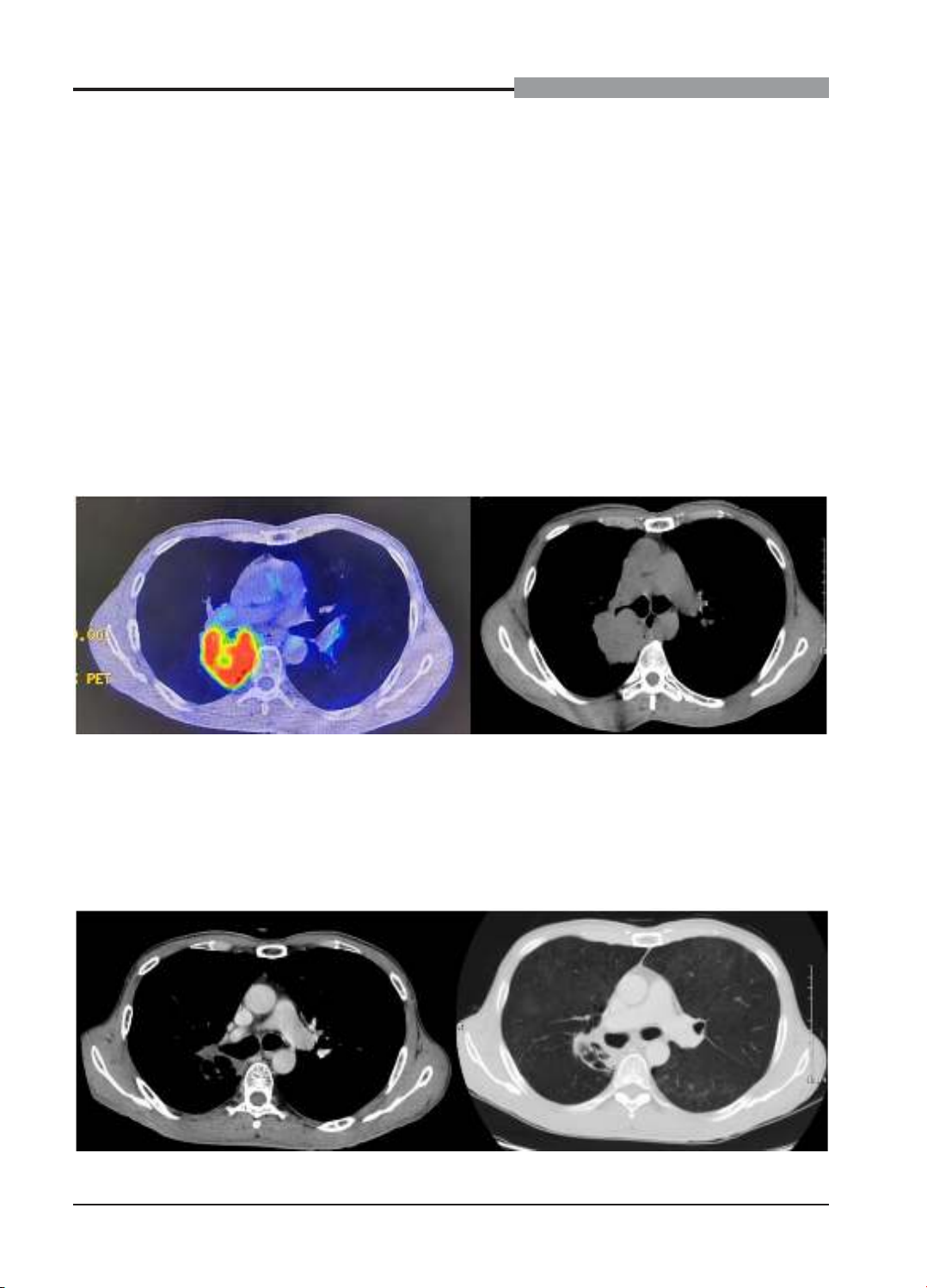
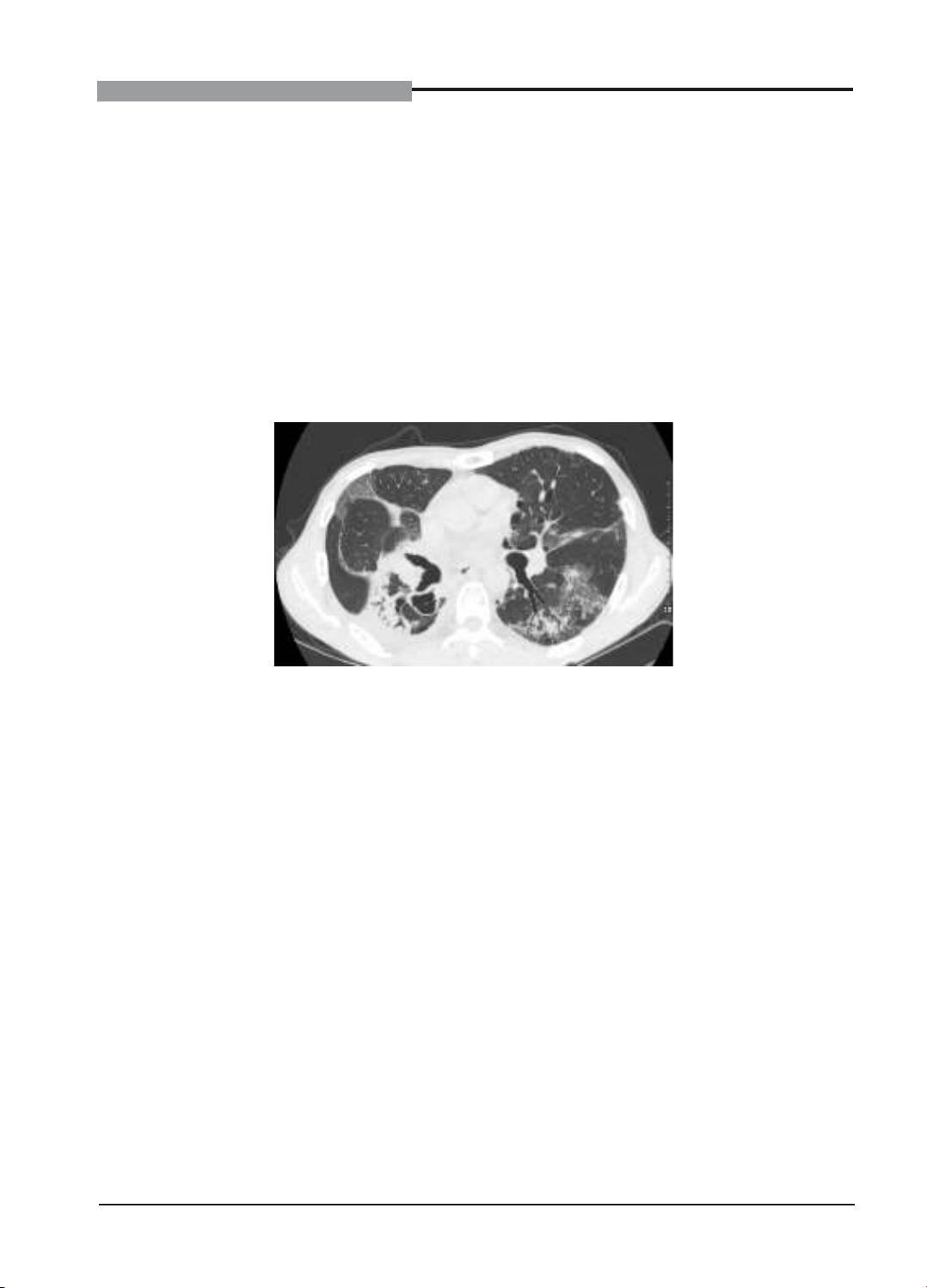
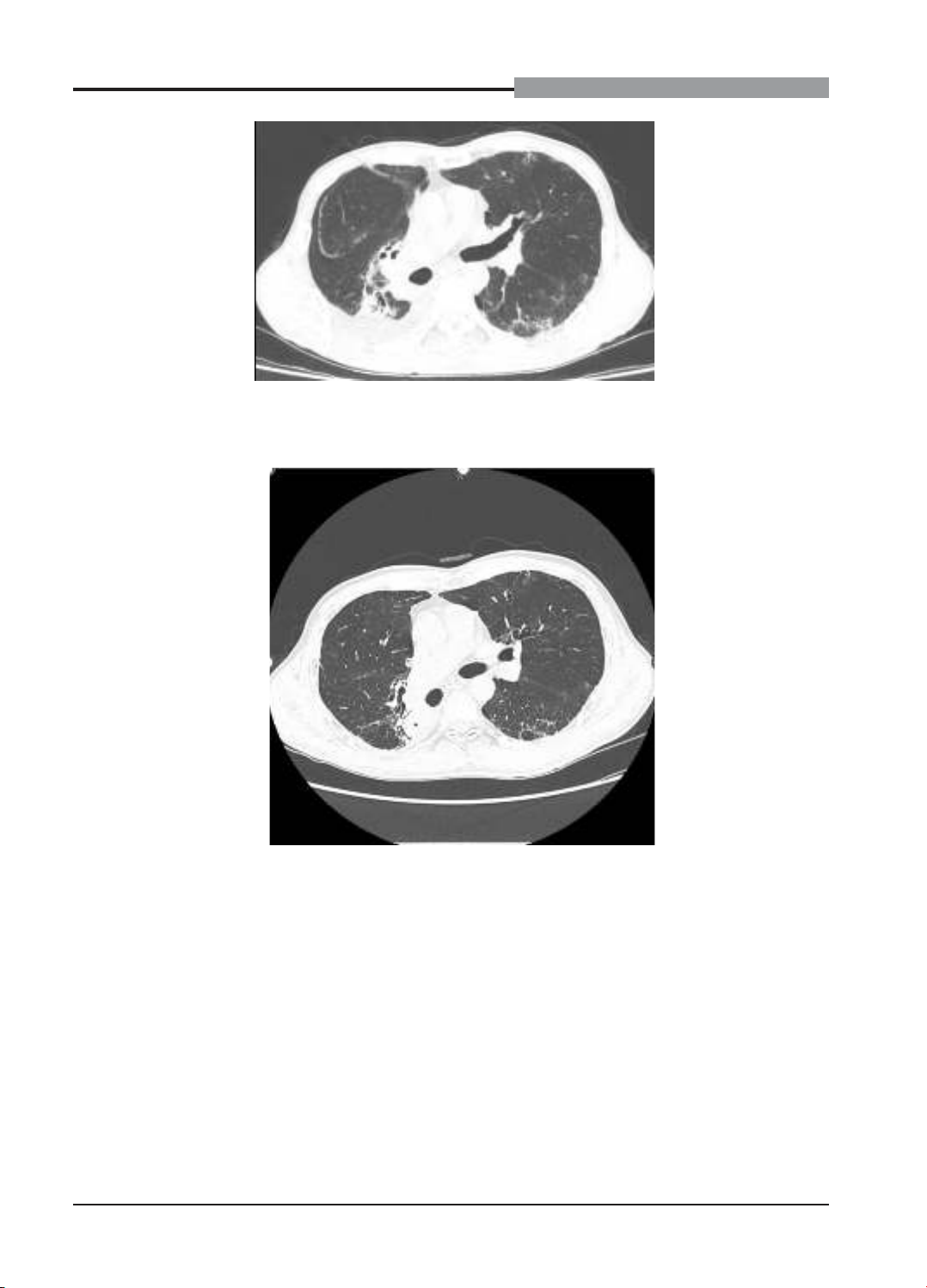
Immune-related pneumonitis is a rare but serious complication in patients with non-small cell lung
cancer (NSCLC) treated with durvalumab after concurrent chemoradiotherapy (CRT). We encountered a
61-year-old male with stage III non-small cell lung cancer (NSCLC) receiving concurrent chemoradiotherapy
followed by durvalumab maintenance therapy. After 1 cycle of durvalumab treatment, he developed
progressive dyspnea, dry cough, and low-grade fever. Chest CT scans revealed diffuse ground-glass
opacities in both lungs, indicative of immune-related pneumonitis. Durvalumab was discontinued, and
the patient was treated with high-dose corticosteroids and antibiotics. After one month, he fully recovered.
Durvalumab was cautiously resumed under close monitoring without recurrence of pneumonitis. Conclusion:
Early identification and management of immune-related pneumonitis are essential in NSCLC patients
receiving durvalumab. Reintroducing durvalumab after recovery may be safe with vigilant monitoring.
Keywords: Non-small cell lung cancer, durvalumab, immune-related pneumonitis, chemoradiotherapy,
immunotherapy rechallenge.
Non-small cell lung cancer (NSCLC) in the
locally advanced stage (stage III) accounts for
approximately 30% of newly diagnosed lung
cancer cases.1 Concurrent chemoradiation
(CRT) is the standard treatment for
unresectable stage III NSCLC patients.2 The
addition of durvalumab, a monoclonal antibody
targeting PD-L1, after definitive concurrent
chemoradiation (dCRT) has significantly
improved progression-free survival (PFS)
and overall survival (OS) in stage III NSCLC
patients.3,4
However, the use of durvalumab post-dCRT
is associated with risks of immune-related
pneumonitis (IRP) and radiation pneumonitis
(RP).5 Differentiating and managing these
two complications present significant clinical
challenges. We report a case of a stage III NSCLC
patient who developed IRP after durvalumab
treatment and subsequently resumed
durvalumab therapy after recovery, aiming to
discuss the diagnosis, treatment, and feasibility
of immunotherapy rechallenge. This study aims
to present a case of pneumonitis in a patient
189
JOURNAL OF MEDICAL RESEARCH
JMR 184 E15 (11) - 2024
treated with concurrent chemoradiotherapy
(cCRT) followed by durvalumab, focusing on the
clinical manifestations, management strategies,
and the implications for medical practice.
II. CASE REPORTS
A 61-year-old male with a 20 pack-year
smoking history presented with persistent
dry cough, chest pain, and dyspnea. Chest
computed tomography (CT) revealed a large
tumor in the right upper lung lobe invading the
pleura and mediastinal lymph nodes (Figure 1).
Transbronchial biopsy and histopathological
results confirmed pulmonary adenocarcinoma.
The patient underwent a PET-CT scan, which
revealed a mass with increased FDG uptake in
the right pulmonary hilum region involving all
three lobes consistent with a primary malignant
lesion, accompanied by several satellite
nodules in the right lower lobe. Imaging also
showed several lymph nodes with increased
FDG uptake in the mediastinum and right
pulmonary hilum, consistent with secondary
regional lymphadenopathy. The patient’s brain
MRI results did not detect any secondary
lesions. The patient was diagnosed with stage
cT4N2M0 NSCLC according to the 8th edition
of the TNM classification.6
Figure 1. PET -CT and CT chest images before treatment
The patient underwent concurrent
chemoradiotherapy (CRT) with weekly
Paclitaxel – Carboplatin combined with
radiotherapy at 60Gy in 30 fractions. After
completing CRT, a re-evaluation showed a
partial response (Figure 2), and he continued
maintenance therapy with Durvalumab at
1500mg every 4 weeks.
Figure 2. CT chest image after CRT
190
JOURNAL OF MEDICAL RESEARCH
JMR 184 E15 (11) - 2024
Three weeks after the first cycle of treatment
with Durvalumab, the patient developed
progressively worsening dyspnea, dry cough,
and low-grade fever (37.8°C). Physical
examination revealed bilateral crackles upon
auscultation. Chest CT showed diffuse ground-
glass opacities in both lungs, especially
in the peripheral regions, not confined to
the irradiated areas (Figure 3). Blood tests
indicated normal white blood cell counts and
mildly elevated C-reactive protein (CRP).
Pulmonary function tests showed decreased
lung function compared to pre-durvalumab
treatment (FEV1 decreased from 80% to 65%).
Blood tests indicated normal white blood cell
counts and mildly elevated high-sensitivity
C-reactive protein (hs-CRP) at 5.8 ng/mL,
while Procalcitonin was low at 0.05 ng/mL. The
patient underwent blood cultures from both
arms to identify bacteria and rapid tests to rule
out influenza and COVID-19, which were all
negative.
Figure 3. CT chest images after concurrent chemoradiotherapy and 1 cycle of durvalumab
Based on the patient’s clinical condition,
which did not exhibit clear signs of infection
despite declining respiratory function, and after
thorough consultations with radiologists and
respiratory and infectious disease specialists,
we excluded other causes of pneumonia and
diagnosed as immune-related pneumonitis.
Durvalumab was discontinued, and the patient
was hospitalized with a suspected diagnosis of
grade 2 immune-related pneumonitis.7 He was
treated with intravenous methylprednisolone at
1 mg/kg/day and antibiotics with levofloxacin
500 mg/day, twice a day to infection
prophylactic. Supplemental oxygen via nasal
cannula was provided, and vital signs were
closely monitored.
After 48 hours of treatment, the patient’s
clinical symptoms improved significantly, with
reduced dyspnea and cough. After one month
of treatment, a follow-up chest CT after 30 days
showed a marked reduction of inflammatory
lesions (Figure 4), respiratory symptoms
did not recur, and pulmonary function tests
returned close to baseline levels. We decided
to discontinue prophylactic antibiotics and
continue tapering the corticosteroid therapy to
complete a total of eight weeks.
Following careful assessment and weighing
of risks and benefits, durvalumab was reinitiated
with close monitoring. During subsequent
treatment, the patient did not experience
recurrence of pneumonitis and continued
durvalumab according to the treatment
schedule.
191
JOURNAL OF MEDICAL RESEARCH
JMR 184 E15 (11) - 2024
Figure 4. CT chest images after 1 month of treatment with corticosteroids and prophylactic
antibiotics
Figure 5. CT chest image after continuing with 8 cycles of Durvalumab
III. DISCUSSION
Immune-related pneumonitis (IRP) is a rare
but serious complication in patients with non-
small cell lung cancer (NSCLC) treated with
PD-1/PD-L1 inhibitors.8 In the PACIFIC trial, the
incidence of IRP was 4.4% in the durvalumab
group compared to 3.8% in the placebo group.4
Clinical reports indicate that IRP can be fatal if
not promptly diagnosed and treated.5 IRP often
presents with nonspecific symptoms, making it
easily confused with radiation pneumonitis (RP)
or pulmonary infections. It can occur at any time
during immunotherapy and typically manifests
with symptoms such as cough, dyspnea, and
low-grade fever. Radiation pneumonitis (RP)
is a more common complication, occurring in
approximately 15 - 20% of patients following
thoracic radiotherapy.9,10 RP usually arises
between 4 and 12 weeks after completing
192
JOURNAL OF MEDICAL RESEARCH
JMR 184 E15 (11) - 2024
radiotherapy, with pulmonary lesions typically
confined to the irradiated fields. This contrasts
with IRP, where lesions are often more
widespread and not limited to the radiation
fields. Imaging of RP may show ground-glass
opacities or fibrosis confined to the irradiated
lung, whereas IRP often presents with interstitial
patterns diffusely affecting both lungs.11,12
In this case, the patient’s respiratory
symptoms emerged 3 weeks after the first cycle
durvalumab initiation, with chest CT revealing
diffuse bilateral pulmonary inflammation. This
presentation aligns with IRP, whereas RP is
usually confined to the irradiated area. Excluding
infections and other causes is crucial. The
diagnosis of IRP was made based on criteria
from the American Society of Clinical Oncology
(ASCO) and the Society for Immunotherapy of
Cancer (SITC).13
Treatment of IRP involves discontinuation of
immunotherapy and administration of high-dose
corticosteroids. Corticosteroids are considered
the standard therapy, helping to reduce
inflammation and alleviate symptoms.13,14 In this
case, the patient was also treated with empirical
prophylactic antibiotics to prevent and manage
potential infections.7 He responded well to
treatment, with symptom improvement after 48
hours and significant reduction of pulmonary
lesions after one week.
Rechallenging with durvalumab after
IRP is a complex decision, requiring careful
consideration of the risk of recurrence versus
therapeutic benefits. Current guidelines
suggest that resuming immunotherapy may be
considered in patients with mild to moderate IRP
who have fully recovered and are under close
monitoring.13 In this case, after the patient fully
recovered, we decided to resume durvalumab
at a reduced dose with vigilant monitoring. The
patient did not experience recurrence of IRP
during subsequent treatment. This result is
consistent with some studies that have reported
that rechallenging with immunotherapy after
IRP can be safe in certain patients, although the
risk of recurrent complications remains. Close
monitoring and educating the patient about
warning signs are essential.15,16
IV. CONCLUSION
This clinical case presents an instance
of pneumonitis following concurrent
chemoradiotherapy and immunosuppressive
treatment, highlighting the challenges in
diagnosing and managing complications,
particularly post-treatment pneumonitis. It
highlights that with appropriate treatment and
monitoring, patients may successfully resume
immunotherapy, potentially improving their
overall outcomes and quality of life.
REFFERENCES
1. Bray F, Laversanne M, Sung H, et al.
Global cancer statistics 2022: GLOBOCAN
estimates of incidence and mortality worldwide
for 36 cancers in 185 countries. CA: A Cancer
Journal for Clinicians. 2024;74(3):229-263.
doi:10.3322/caac.21834
2. Postmus PE, Kerr KM, Oudkerk M, et
al. Early and locally advanced non-small-cell
lung cancer (NSCLC): ESMO Clinical Practice
Guidelines for diagnosis, treatment and follow-
up†. Annals of Oncology. 2017;28:iv1-iv21.
doi:10.1093/annonc/mdx222
3. Sj A, A V, D D, et al. Durvalumab after
Chemoradiotherapy in Stage III Non-Small-
Cell Lung Cancer. The New England journal
of medicine. 2017;377(20). doi:10.1056/
NEJMoa1709937
4. Antonia SJ, Villegas A, Daniel D, et
al. Overall Survival with Durvalumab after
Chemoradiotherapy in Stage III NSCLC. N Engl
J Med. 2018;379(24):2342-2350. doi:10.1056/