RESEARC H Open Access
Detection of EGFR mutations with mutation-specific
antibodies in stage IV non-small-cell lung cancer
Sara Simonetti
1
, Miguel Angel Molina
1
, Cristina Queralt
2
, Itziar de Aguirre
2
, Clara Mayo
1
, Jordi Bertran-Alamillo
1
,
José Javier Sanchez
3
, Jose Luis Gonzalez-Larriba
4
, Ulpiano Jimenez
5
, Dolores Isla
6
, Teresa Moran
2
, Santiago Viteri
1
,
Carlos Camps
7
, Rosario Garcia-Campelo
8
, Bartomeu Massuti
9
, Susana Benlloch
1
, Santiago Ramon y Cajal
1,10
,
Miquel Taron
1,2*
, Rafael Rosell
1,2
Abstract
Background: Immunohistochemistry (IHC) with mutation-specific antibodies may be an ancillary method of
detecting EGFR mutations in lung cancer patients.
Methods: EGFR mutation status was analyzed by DNA assays, and compared with IHC results in five non-small-cell
lung cancer (NSCLC) cell lines and tumor samples from 78 stage IV NSCLC patients.
Results: IHC correctly identified del 19 in the H1650 and PC9 cell lines, L858R in H1975, and wild-type EGFR in
H460 and A549, as well as wild-type EGFR in tumor samples from 22 patients. IHC with the mAb against EGFR with
del 19 was highly positive for the protein in all 17 patients with a 15-bp (ELREA) deletion in exon 19, whereas in
patients with other deletions, IHC was weakly positive in 3 cases and negative in 9 cases. IHC with the mAb
against the L858R mutation showed high positivity for the protein in 25/27 (93%) patients with exon 21 EGFR
mutations (all with L858R) but did not identify the L861Q mutation in the remaining two patients.
Conclusions: IHC with mutation-specific mAbs against EGFR is a promising method for detecting EGFR mutations
in NSCLC patients. However these mAbs should be validated with additional studies to clarify their possible role in
routine clinical practice for screening EGFR mutations in NSCLC patients.
Background
Non-small-cell lung cancer (NSCLC) is one of the most
frequent human malignancies, constituting about 80% of
all lung tumors. NSCLC can be divided into genetic
subsets on the basis of the activating mutations that
they harbor; each of these subsets may correspond to
patient cohorts that are likely to benefit from treatment
with specific inhibitors [1].
Activating mutations in the epidermal growth factor
receptor (EGFR), affecting hotspots within exons that
code for the tyrosine kinase domain, can be found in
10-40% of NSCLC patients, mostly in adenocarcinomas,
with the higher frequency observed in Asian patients
[1,2]. About 50% of mutated patients harbor in-frame
deletions in exon 19, (around codons 746 to 750) and
35-45% show the substitution of leucine 858 by an
arginine in the exon 21. The remaining mutants are
insertions in exon 20 (5%) and uncommon substitutions
spanning exons from 18 to 21, such as L861Q [3,4].
These specific mutations are related to a higher sensitivity
to the tyrosine kinase inhibitors (TKIs) erlotinib and gefiti-
nib [4-7], whereas the EGFR T790 M mutation in exon 20
is observed in 50% of cases with acquired resistance to erlo-
tinib and gefitinib [8] and has also been detected in 38% of
patients with de novo drug resistance [9].
Molecular biology techniques, such as SARMS or
direct automatic sequencing, are currently used to
detect EGFR mutations in formalin-fixed, paraffin-
embedded tissues (FFPET). In our experience, in-frame
deletions in exon 19 are detected by fragment analysis
of fluorescently labeled PCR products, and L858R muta-
tions in exon 21 by TaqMan assay. Mutations are then
confirmed by direct sequencing [10,11]. However, the
routine use of these methods in clinical laboratories is
still often limited by financial and technical constraints.
* Correspondence: taron.miquel@gmail.com
1
Pangaea Biotech, USP Dexeus University Institute, Barcelona, Spain
Full list of author information is available at the end of the article
Simonetti et al.Journal of Translational Medicine 2010, 8:135
http://www.translational-medicine.com/content/8/1/135
© 2010 Simonetti et al; licensee BioMed Central Ltd. This is an Open Access article distributed under the terms of the Creative
Commons Attribution License (http://creativecommons.org/licenses/by/2.0), which permits unrestricted use, distribution, and
reproduction in any medium, provided the original work is properly cited.

Moreover, their sensitivity depends on the quality and
the quantity of tumoral cells in FFPET. In a previous
study, we developed a highly sensitive molecular method
for detecting EGFR mutations in NSCLC samples con-
taining as few as eight tumor cells [10].
The development of antibodies that specifically detect
mutant EGFR protein by IHC would be an easy pre-
screening test to complement the molecular assays cur-
rently used for the assessment of EGFR mutations in
NSCLC. Yu et al [12] have developed mutation-specific
rabbit monoclonal antibodies (mAb) against EGFR with
the E746_A750 deletion in exon 19 or the L858R point
mutation in exon 21 for IHC application (Cell Signaling
Technology Inc., Danvers, MA, USA).
In the present study, these two rabbit mAbs were used
to assess EGFR mutations in five NSCLC cell lines and
in tumor biopsies from 78 stage IV NSCLC patients.
The results were then compared with those obtained by
other molecular analyses [10,11].
Methods
Sources of cell lines and culture
The PC-9 lung tumor cell line was kindly provided by
Roche (Basel, Switzerland); the A549 and H460 cell lines
were purchased from the American Type Culture Col-
lection. Tissue culture materials were obtained from
Biological Industries (Kibbutz Beit Haemek, Israel) and
Invitrogen (Paisley, Scotland, UK). H1650 and H1975
were kindly provided by Dr. Herbert Haack and
Dr. Katherine Crosby (Cell Signaling Technology, Inc.).
We received five slides of the H1975 cell line and five of
the H1650 cell line with 4-μm sections for IHC analysis
from the Cell Signaling Technology laboratory.
Study population and tumor pathology
Twenty-six stage IV NSCLC patients had been seen at
the USP Dexeus University Institute, and 52 had been
previously screened for EGFR mutations and treated
with erlotinib as part of the Spanish Lung Adenocarci-
noma Data Base (SLADB) [11]. All of these 52 patients
were known to have EGFR mutations, while the remain-
ing 26 patients had not been previously screened. All
patients provided written informed consent. Approval
was obtained from the institutional review board and
the ethics committee at each hospital. Table 1 shows
patient characteristics.
Four-μm sections of the FFPET specimens were
stained with H/E and histologically examined. All sam-
ples were classified according to the 2004 WHO classifi-
cation [13]: 5 undifferentiated large cell carcinomas and
3 small cell neuroendocrine carcinomas, 1 squamous
cell carcinoma and 69 adenocarcinomas, of which 55
showed a single pattern and 14 presented mixed aspects.
We further evaluated the adenocarcinoma subtype as
follow: 36 adenocarcinomas with a glandular pattern, 20
with a solid aspect, 6 with a partial papillary differentia-
tion, 1 with micropapillary aspects and 6 with a partial
bronchioloalveolar pattern (Table 1).
DNA extraction and mutation analyses
Tumor cells (8 to 150) were captured by laser microdis-
section (Carl Zeiss MicroImaging GmbH, München,
Germany) into 10 μL of PCR buffer (Ecogen, Barcelona,
Spain) plus proteinase K and incubated 4 hours to over-
night at 60°C. Proteinase was inactivated at 95°C for
10 min, and the cell extract submitted to PCR. DNA
from the cell line PC-9 was used as a mutated control
for exon 19, and wt control for exons 20 and 21. DNA
from the H1975 cell line was used as a wt control for
exon 19, and mutated control for exons 21/20.
EGFR gene mutations in exons 19 and 21 were ana-
lyzed by our sensitive methodology as previously
described [10]. Exons 19 and 21 of the EGFR gene were
amplified by a nested PCR. Sequencing was performed
using forward and reverse nested primers with the ABI
Prism 3100 DNA Analyzer (Applied Biosystems, Foster
City, CA, USA). In addition to sequencing, EGFR dele-
tions in exon 19 were determined by length analysis of
Table 1 Clinicopathological features of the patients
analyzed for EGFR mutations by IHC assay
Patients (N = 78)
Characteristic No. %
Age, years
Mean 64
Range 36-85
Sex
Male 28 36
Female 50 64
Race
Caucasian 78 100
Smoker
Ex-smoker 26 33
Current smoker 7 9
Never smoker 45 58
Histology
Adenocarcinoma 69 88.4
Large-cell carcinoma 5 7.1
Squamous cell carcinoma 1 1.4
Others 3 4.3
Adenocarcinoma subtype
Glandular 36 52.2
Solid 20 29
Papillary 6 8.7
Micropapillary 1 1.4
BAC 6 8.7
Simonetti et al.Journal of Translational Medicine 2010, 8:135
http://www.translational-medicine.com/content/8/1/135
Page 2 of 8

fluorescently labeled PCR products. The collected data
were evaluated with the GeneScan Analysis Software
(Applera, Norwalk, CT, USA). Finally, EGFR mutation
(L858R)inexon21wasalsodeterminedbyTaqMan®
Assay (Applied Biosystems). The L861Q mutation was
detected by direct sequencing.
Immunohistochemical analysis
The following antibodies were used for the IHC analysis
(Cell Signaling Technology, Inc.): EGF Receptor
(D38B1), EGFR E746-A750 deletion specific (6B6) and
EGFR L858R mutant-specific (43B2). The FFPET sam-
ples were cut serially at 4 μm and the sections were
introduced in the stainer and automatically deparaffi-
nized (Leica Microsystems BondMAX Automated
Immunostainer, Wetzlar, Germany). The reactives were
added automatically, treating the samples with EDTA
buffer (pH 9.0) (Bond Epitope Retrieval Solution 2,
Leica Microsystems) as antigen retriever and processed
for 30 min. The slides were incubated with the antibo-
dies against EGF receptor and EGFR mutations at a
dilution of 1:100 for 60 minutes. After the sections were
treated with the streptavidin-biotin-peroxidase complex
method (Bond Polymer Refine Detection, Leica Micro-
systems) with diaminobenzidine (DAB) as a chromogen
and counterstained with hematoxylin.
IHC expression of mAbs against EGFR was evaluated
using the following scoring, as previously described [14]:
0 = negative or faint staining in <10% of tumor cells;
1 = weak staining in >10% of cancer cells; 2 = moderate
staining; 3 = strong staining. A score of 0 was consid-
ered negative, a score of 1 was considered weakly
positive, and a score of 2 or 3 was considered highly
positive (Additional File 1, Figure S1).
Statistical analyses
The absolute and relative frequencies of qualitative vari-
ables were calculated in percentages. The sensitivity and
specificity of the EGFR test by IHC was determined in
comparison with PCR-based results. All analyses were
performed using SPSS v 16.0 software (SPSS Inc.,
Chicago, IL).
Results
EGFR mutation analysis in NSCLC patients
We screened EGFR mutations in 78 FFPET samples from
NSCLC patients by a methodology described elsewhere
[10], which involves fragment analysis (exon 19), Taqman
assay (exon 21) and sequencing. Twenty-six samples were
analyzed in the Pangaea Biotech Oncology Laboratory
and 52 from a previous study [11] were analyzed in the
Catalan Institute of Oncology, Hospital Germans Trias i
Pujol. Twenty-two samples (28%) were wt EGFR, 29
(37%) had a deletion in exon 19, and 27 (35%) had muta-
tions in exon 21. Of the 29 patients with the exon 19
deletion, 17 (59%) had 15-bp deletions (16 with del E746-
A750 [ELREA] and 1 with del E746-A750 [ELREA] +
T751I), and 12 (41%) had rare deletions of 9-bp, 12-bp,
18-bp, 21-bp or 24-bp. Of the 27 patients with exon 21
mutations, 25 (93%) had the L858R mutation and 2 (7%)
had the L861Q mutation (Additional File 1, Table S1).
IHC analysis of mutation-specific mAbs against EGFR in
human NSCLC cell lines
We analyzed by IHC five human NSCLC cell lines with
known EGFR gene status. In the two cell lines with wt
EGFR (H460 and A549), we found positive (score 3)
expression of EGFR (D38B1) protein (100%) and nega-
tive (score 0) expression of EGFR E746-A750 deletion
specific (6B6) and EGFR L858R mutant-specific (43B2).
In the two cell lines with exon 19 deletion (15 bp)
(H1650 and PC9), expression of EGFR (D38B1) protein
and EGFR E746-A750 deletion specific (6B6) was posi-
tive (score 3) (100%) and expression of EGFR L858R
mutant-specific (43B2) was negative (score 0).
The cell line H1975 with exon 21 mutation (L858R)
showed positivity (score 3) for EGFR (D38B1) and EGFR
L858R mutant-specific (43B2) (100%) and negativity
(score 0) for EGFR E746-A750 deletion specific (6B6).
(Table 2, Figure 1).
IHC analysis of mutation-specific mAbs against EGFR in
NSCLC patients
In 22 tumor tissues with wt EGFR, we found high
expression of EGFR (D38B1) protein (score 2 or 3) in 8
cases (36%) and weak positivity (score 1) in 4 cases
(18%). All the cases were negative for EGFR E746-A750
deletion specific (6B6) and EGFR L858R mutant-specific
(43B2) proteins.
In the 29 patients with exon 19 deletions, high expres-
sion of EGFR E746-A750 deletion-specific (6B6) protein
(score 2 or 3) was observed in 17/17 cases (100%) with
15-bp deletion (16 with ELREA and 1 with ELREA +
T751I). Of the 12 cases showing uncommon deletions
inexon19,nine(75%)sampleswerecompletelynega-
tive (score 0) and 3 (25%) were weakly positive (score
1). All cases were negative (score 0) for EGFR L858R
mutant-specific (43B2) protein.
In the 27 patients with exon 21 mutations, IHC with
the mAb against the L858R mutation showed high posi-
tivity for the protein (score 2 or 3) in 25/27 (93%) and
in 100% of the 25 samples with the L858R substitution;
however, it failed to identify the L816Q mutation (0/
2 cases). In addition, all 27 samples were negative for
the EGFR E746-A750 deletion-specific (6B6) protein
(Tables 2 and 3, Figures 2 and 3).
Simonetti et al.Journal of Translational Medicine 2010, 8:135
http://www.translational-medicine.com/content/8/1/135
Page 3 of 8
Discussion
EGFR is a member of the ErbB family of receptor tyrosine
kinases, which also includes HER2/neu, HER3, and HER4
[15]. Activating mutations in the tyrosine kinase domain,
involving mainly exons 19 and 21, play an important role
in lung oncogenesis and tumor progression and are related
to the clinical efficacy of EGFR TKIs such as gefitinib or
erlotinib [5,9,11]. Analysis of these mutations has become
an important tool for targeted therapy in lung cancer
3
,
and in recent years many efforts have been made to find a
more specific and sensitive methodology to detect them
[10,16-18]. Nevertheless, these techniques are relatively
expensive for routine use in clinical laboratories, and
depend on the quality of the samples. IHC is a standar-
dized assay of simple methodology and high sensitivity
and specificity, and the development of specific antibodies
against EGFR mutation proteins might be useful for the
diagnosis and treatment of lung cancer.
Table 2 IHC expression of EGFR mutation antibodies in human NSCLC cell lines and in NSCLC tumor tissues
EGFR mutation status EGFR (D38B1)
Ab (+)
EGFR E746-A750 deletion-specific
(6B6) Ab (+)
EGFR L858R mutant-specific
(43B2) Ab (+)
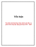
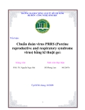
H460 and A549 (WT) 2/2 (100%) 0/2 (0%) 0/2 (0%)
H1650 and PC9 (DEL 19) 2/2 (100%) 2/2 (100%) 0/2 (0%)
H1975 (L858R + T790M) 1/1 (100%) 0/1 (0%) 1/1 (100%)
Tumor Tissue (WT; N = 22) 8/22 (36%) 0/22 (0%) 0/22(0%)
Tumor Tissue (DEL 19;N = 29) 28/29 (97%) 20/29 (69%) 0/29 (0%)
Tumor Tissue (MUT 21;N = 27) 26/27 (96%) 0/27 (0%) 25/27 (93%)
Abbreviations: WT: wild-type. DEL 19: Exon 19 deletion; MUT 21: Exon 21 mutation.
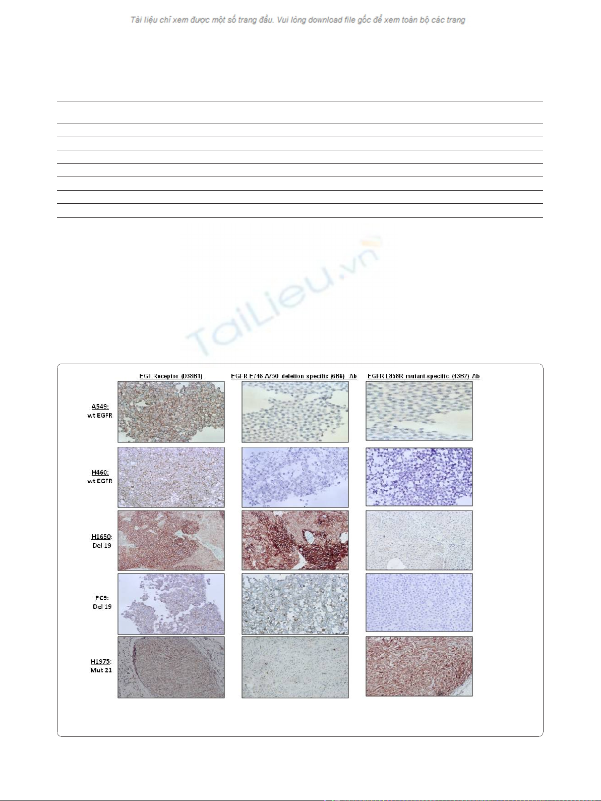
Figure 1 IHC analysis of EGFR mutations in five human NSCLC cell lines. A549 and H460 showed negativity for both mutation-specific
antibodies. EGFR E746-A750 deletion specific (6B6) antibody stained H1650 and PC9 harboring the exon 19 deletion, and EGFR L858R mutant-
specific (43B2) antibody stained the H1975 cell line with exon 21 mutation.
Simonetti et al.Journal of Translational Medicine 2010, 8:135
http://www.translational-medicine.com/content/8/1/135
Page 4 of 8
In 2009 Yu et al [12] first generated two mAbs against
E746-A750del and L858R point mutation from New
Zealand rabbits and evaluated them by Western blot-
ting, immunofluorescence and IHC. They tested these
antibodies in a series of cell lines and in tumor tissues
from patients with primary NSCLC, with known and
unknown EGFR mutations, comparing the IHC results
with DNA sequencing. They found that IHC with these
mutation-specific antibodies for EGFR mutations
showed a sensitivity of 92% and a specificity of 99%.
Recently, five studies [14,19-22] examined the presence
of EGFR mutations in NSCLC by IHC using the same
Table 3 Correlation of IHC expression of mutation-specific antibodies and EGFR exon 19 deletion subtype analyzed by
GeneScan, TaqMan and direct sequencing
EGFR EXON 19 DELETION SUBTYPE 0 1+ 2+ 3+
15 bp
N=17
0/17 (0%) 0/17 (0%) 2/17 (11%) 15/17 (89%)
9bp
N=4
2/4 (50%) 2/4(50%) 0/4 (0%) 0/4 (0%)
12 bp
N=1
1/1 (100%) 0/1 (0%) 0/1 (0%) 0/1 (0%)
18 bp
N=5
4/5 (80%) 1/5 (20%) 0/5 (0%) 0/5 (0%)
21 bp
N=1
1/1 (100%) 0/1 (0%) 0/1 (0%) 0/1 (0%)
24 bp
N=1
1/1 (100%) 0/1 (0%) 0/1 (0%) 0/1 (0%)
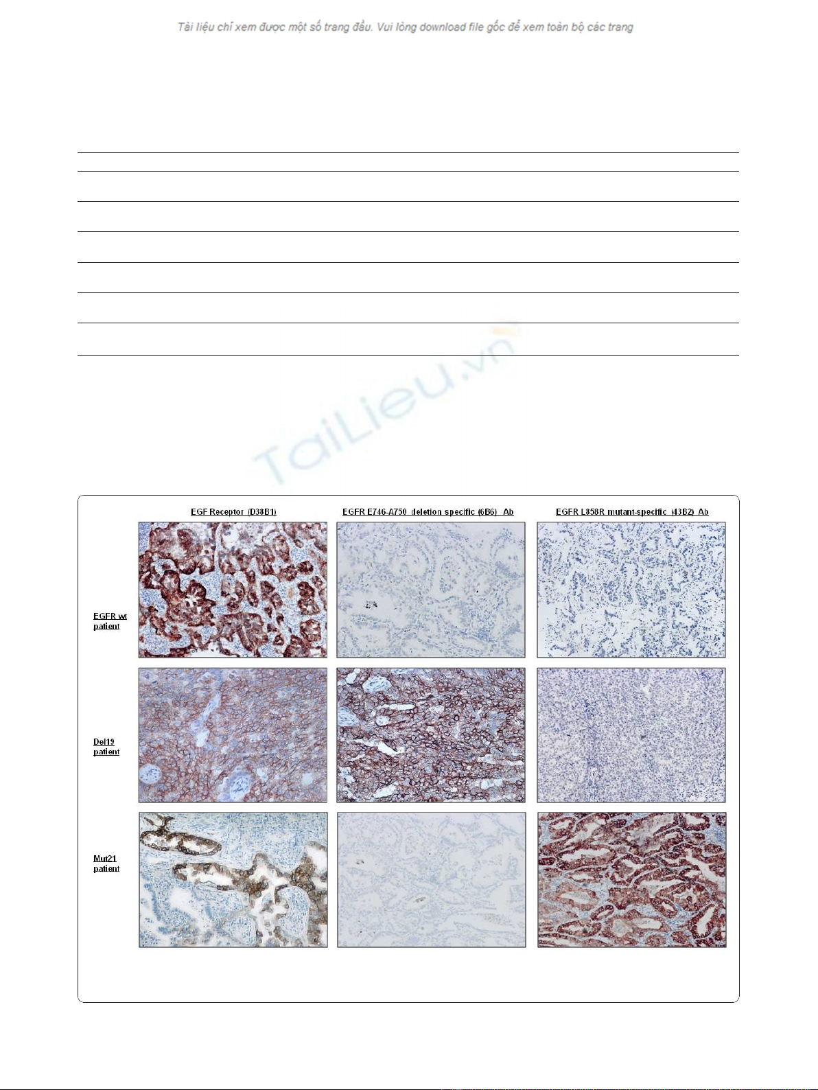
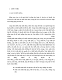
Figure 2 IHC staining of tumor samples from lung cancer patients. EGFR E746-A750 deletion specific (6B6) antibody detected 100% of cases
with the 15-bp exon 19 deletion, and EGFR L858R mutant-specific (43B2) antibody detected 100% of cases harboring L858R mutation of exon
21.
Simonetti et al.Journal of Translational Medicine 2010, 8:135
http://www.translational-medicine.com/content/8/1/135
Page 5 of 8


%20--%3e%3cdefs%3e%3cstyle%3e%20.st0%20{%20fill:%20%23fff;%20}%20.st1%20{%20fill:%20%237800fa;%20}%20%3c/style%3e%3c/defs%3e%3cpath%20class='st1'%20d='M117.78,12.18H43.11c2.9,3.47,4.65,7.94,4.65,12.82,0,5.6-2.3,10.66-6.01,14.29h76.02l7.22-13.56-7.22-13.56Z'/%3e%3cg%3e%3cpath%20class='st0'%20d='M53.58,26.17h-.59v-1.46h.59v-4.96h2.83c1.78,0,2.67.94,2.67,2.82v5.76c0,1.87-.89,2.81-2.67,2.81h-2.83v-4.96ZM55.36,21.37v3.34h1.1v1.46h-1.1v3.34h1.01c.61,0,.91-.37.91-1.1v-5.93c0-.74-.3-1.1-.91-1.1h-1.01Z'/%3e%3cpath%20class='st0'%20d='M65.99,31.14h-1.8l-.31-2.07h-2.19l-.31,2.07h-1.64l1.82-11.39h2.62l1.82,11.39ZM65.28,18.04c-.25.46-.51.77-.75.94-.21.15-.47.22-.79.22-.26,0-.57-.07-.92-.22l-.38-.15c-.14-.05-.26-.07-.37-.07-.3,0-.53.18-.71.54l-.91-.68c.25-.46.51-.77.75-.94.21-.14.48-.21.79-.21.26,0,.57.07.92.21l.38.15c.14.05.26.07.37.07.3,0,.53-.18.71-.54l.91.68ZM61.91,27.52h1.73l-.87-5.76-.87,5.76Z'/%3e%3cpath%20class='st0'%20d='M74.53,26.89v1.52c0,1.91-.89,2.86-2.67,2.86s-2.67-.95-2.67-2.86v-5.93c0-1.91.89-2.86,2.67-2.86s2.67.95,2.67,2.86v1.11h-1.69v-1.22c0-.75-.31-1.12-.93-1.12s-.93.37-.93,1.12v6.15c0,.74.31,1.11.93,1.11s.93-.37.93-1.11v-1.63h1.69Z'/%3e%3cpath%20class='st0'%20d='M81.4,31.14h-1.8l-.31-2.07h-2.19l-.31,2.07h-1.64l1.82-11.39h2.62l1.82,11.39ZM75.9,19.2l1.52-1.91h1.71l1.51,1.91h-1.61l-.76-.95-.75.95h-1.61ZM77.32,27.52h1.73l-.87-5.76-.87,5.76ZM83.1,15.99l-1.76,1.91h-1.26l1.17-1.91h1.86Z'/%3e%3cpath%20class='st0'%20d='M84.86,19.75c1.78,0,2.67.94,2.67,2.82v1.48c0,1.87-.89,2.81-2.67,2.81h-.85v4.28h-1.79v-11.39h2.64ZM84.01,21.37v3.86h.85c.58,0,.87-.36.87-1.08v-1.71c0-.71-.29-1.07-.87-1.07h-.85Z'/%3e%3cpath%20class='st0'%20d='M93.51,19.75c1.78,0,2.67.94,2.67,2.82v1.48c0,1.87-.89,2.81-2.67,2.81h-.85v4.28h-1.79v-11.39h2.64ZM92.66,21.37v3.86h.85c.58,0,.87-.36.87-1.08v-1.71c0-.71-.29-1.07-.87-1.07h-.85Z'/%3e%3cpath%20class='st0'%20d='M98.8,31.14h-1.79v-11.39h1.79v4.88h2.03v-4.88h1.83v11.39h-1.83v-4.88h-2.03v4.88Z'/%3e%3cpath%20class='st0'%20d='M105.36,24.55h2.46v1.62h-2.46v3.34h3.09v1.63h-4.88v-11.39h4.88v1.63h-3.09v3.18ZM108.17,17.29l-1.76,1.91h-1.26l1.17-1.91h1.86Z'/%3e%3cpath%20class='st0'%20d='M112.2,19.75c1.78,0,2.67.94,2.67,2.82v1.48c0,1.87-.89,2.81-2.67,2.81h-.85v4.28h-1.79v-11.39h2.64ZM111.35,21.37v3.86h.85c.58,0,.87-.36.87-1.08v-1.71c0-.71-.29-1.07-.87-1.07h-.85Z'/%3e%3c/g%3e%3ccircle%20class='st1'%20cx='25'%20cy='25'%20r='20'/%3e%3cpath%20class='st0'%20d='M32.78,19.27c2.92,0,4.43,2.55,5.28,5.33l.71,2.17c.14.38-.33.75-.71.75h-5.61c.19-.33.24-.71.09-1.08l-.75-2.45c-.43-1.32-.99-2.64-1.79-3.77.75-.57,1.65-.94,2.78-.94h0ZM25,18.38c3.25,0,4.9,2.78,5.89,5.89l.76,2.45c.14.42-.33.8-.8.8h-11.69c-.42,0-.94-.38-.8-.8l.75-2.45c.99-3.11,2.64-5.89,5.89-5.89h0ZM25,11.35c1.74,0,3.11,1.37,3.11,3.11s-1.37,3.11-3.11,3.11-3.11-1.41-3.11-3.11,1.41-3.11,3.11-3.11h0ZM17.27,19.27c1.08,0,1.98.38,2.73.94-.8,1.13-1.37,2.45-1.74,3.77l-.8,2.45c-.14.38-.05.75.09,1.08h-5.56c-.42,0-.9-.38-.75-.75l.71-2.17c.9-2.78,2.41-5.33,5.33-5.33h0ZM17.27,12.91c1.51,0,2.78,1.27,2.78,2.83s-1.27,2.83-2.78,2.83-2.83-1.27-2.83-2.83,1.27-2.83,2.83-2.83h0ZM32.78,12.91c1.56,0,2.78,1.27,2.78,2.83s-1.23,2.83-2.78,2.83-2.83-1.27-2.83-2.83,1.27-2.83,2.83-2.83h0ZM27.07,28.56v.09c0,.57-.24,1.08-.61,1.46h0v.05c-.38.33-.9.57-1.46.57s-1.08-.24-1.46-.61h0c-.38-.38-.61-.9-.61-1.46v-.09h1.41v.09c0,.19.05.38.19.47v.05c.09.09.28.19.47.19s.38-.09.47-.19v-.05c.14-.09.24-.28.24-.47t-.05-.09h1.41ZM30.99,28.56v.09c0,1.65-.66,3.16-1.74,4.24-1.08,1.08-2.59,1.79-4.24,1.79s-3.16-.71-4.24-1.79l-.05-.05c-1.04-1.08-1.7-2.55-1.7-4.2v-.09h1.41v.09c0,1.27.47,2.4,1.27,3.25h.05c.85.85,1.98,1.37,3.25,1.37s2.4-.52,3.25-1.37c.85-.8,1.37-1.98,1.37-3.25v-.09h1.37ZM34.99,28.56v.09c0,2.78-1.13,5.28-2.92,7.07-1.79,1.79-4.29,2.92-7.07,2.92s-5.23-1.13-7.07-2.92c-1.79-1.79-2.92-4.29-2.92-7.07v-.09h1.41v.09c0,2.4.94,4.53,2.5,6.08,1.56,1.56,3.72,2.5,6.08,2.5s4.52-.94,6.08-2.5c1.56-1.56,2.5-3.68,2.5-6.08v-.09h1.41Z'/%3e%3c/svg%3e)