RESEARCH Open Access
EGFR and COX-2 protein expression in
non-small cell lung cancer and the
correlation with clinical features
Feng Li
1
, Yongmei Liu
1
, Huijiao Chen
2
, Dianying Liao
2
, Yali Shen
1
, Feng Xu
1*†
, Jin Wang
1*†
Abstract
Background: To evaluate the expression of EGFR and COX-2 and their correlation with prognosis in NSCLC
Methods: The paraffin embedded tumor samples of 50 NSCLC patients receiving radical resection were analyzed
immunohistochemically for EGFR and COX-2 expression and their prognostic values were explored.
Results: The positive rate of EGFR protein in NSCLC tumor cells was 46%, which was significantly higher than its
expression in normal lung (p = 0.0234) and paracancerous tissues (p = 0.020). EGFR expression was significantly
higher in nodal positive than in nodal negative patients (p = 0.04). The mean survival time for EGFR positive patients
(31 months) was significantly lower than that for patients with EGFR negative expression (48 months) (p = 0.008,). In
patients receiving post-operation thoracic irradiation, the mean survival time for EGFR positive patients was
significantly lower than that for patients without EGFR positive expression (25 vs. 48 months, P = 0.004). The positive
rate of COX-2 protein expression in NSCLC tumor cells was 90%, which was significantly higher than that in normal
tissue(p = 0.00) and paracancerous tissue (p = 0.00). There was no correlation between COX-2 expression and patient
survival, and no correlation between COX-2 and EGFR protein expression (P = 0.555).
Conclusions: COX-2 and EGFR are over-expressed in NSCLC. EGFR is an independent prognostic factor and a
predictive factor for radiotherapy response in NSCLC.
Background
Lung cancer is the leading cause of death world wide.
The non-small cell lung cancer (NSCLC) accounts for
75-85% among all lung cancers. The conventional treat-
ment e.g. surgery, radiotherapy and chemotherapy yields
a dismal overall 5-year survival of 14% which necessi-
tates the development of new treatment options [1].
With advances in cytogenetic and molecular biology, the
detection and analysis of tumor suppressor gene and
oncogene may provide predictive values for prognosis
and treatment choice for NSCLC. Among these molecu-
lar markers, the epidermal growth factor receptor
(EGFR) and cyclooxygenase-2 (COX-2) over expression
are common in NSCLC [2-9].
EGFR (HER1, ErbB) is a transmembrane glycoprotein
with three functional domains: an extracellular domain
containing two EGF binding sites; a hydrophobic trans-
membrane domain and a cytoplasmic domain (tyrosine
kinase (TK) and a carboxyl autophosphorylation region)
[10,11]. EGFR is abnormally upregulated and activated
in a variety of tumors [12]. Deregulation of receptor tyr-
osine kinases as a result of overexpression or activating
mutations leads to the promotion of cell proliferation or
migration, inhibition of celldeath,ortheinductionof
angiogenesis [13,14].
The expression and activity of EGFR are determinants of
response to target therapy and radiosensitivity in several
tumour types [15]. EGFR overexpression in non-small cell
lung cancer (NSCLC) is variable ranging from 19% to 89%
and its prognostic value remains controversial [16,17].
COX-2 over expression is also found in many tumor types
[18]. The carcinogenic effect of COX-2 mainly exerted
through the increase of prostaglandin levels (PGE2,
PGF2a, PGD2, TXA2, PGI2 and PGJ2). In lung cancer,
* Correspondence: Fengxuster@gmail.com; jinwang593@yahoo.com.cn
†Contributed equally
1
Radiation Oncology, Tumor Center, West China Hospital, Sichuan University, China
Full list of author information is available at the end of the article
Li et al.Journal of Experimental & Clinical Cancer Research 2011, 30:27
http://www.jeccr.com/content/30/1/27
© 2011 Li et al; licensee BioMed Central Ltd. This is an Open Access article distributed under the terms of the Creative Commons
Attribution License (http://creativecommons.org/licenses/by/2.0), which permits unrestricted use, distribution, and reproduction in
any medium, provided the original work is properly cited.

COX-2 expression has been reported to inhibit apoptosis
[19], promote angiogenesis [20] and metastasis [2]. It has
been reported in a recent meta-analysis that COX-2 might
be an independent prognostic factor for NSCLC [21].
COX-2 inhibitor has been investigated in both pre-clinical
and clinical study, and has shown synergistic effects with
radiation and chemtoxic drugs on tumor [3,22]. COX-2
catalyzes the conversion of arachidonic acid into prosta-
noids including prostaglandin E2, which is often associated
with oncogenesis of lung tumors. The oncogenic signals
are transducted through the MAPK/Erk pathway [23]
which therefore closely correlates EGFR with COX-2.
Anumberofin vitro studies have postulated a link
between EGFR activation and subsequent COX-2 upregu-
lation. The relationship between these factors has not
been established in patients with NSCLC.
In order to evaluate the EGFR and COX-2 expression
and their impact on prognosis of NSCLC patients
receiving post-operative adjuvant therapy, the paraffin
embedded tumor samples from 50 NSCLC were ana-
lyzed immunohistochemically for EGFR and COX-2
expression and their prognostic values were explored.
Methods
Tumor specimen
Paraffin-embedded tissue sections from 50 histopathologi-
cally proven NSCLC patients who received radical resec-
tion during June 2001 and March 2004 were collected.
Patient data
All patients were histopathologically diagnosed NSCLC
and had not received preoperative chemotherapy nor
radiotherapy. Among them there were 31 males and 19
females, aged 36-76 (mean 58) years. According to WHO
classification (2000), there were 21 squamous, 26 adeno-
matous and 3 adenosquamous carcinomas, with 40 mod-
erate and well differentiated (G1-G2) and 10 low
differentiated (G3). 15 cases were staged I-II and 35 III-IV
based on the revised AJC staging for lung cancer (1997).
Thirty-nine cases had intra-thoracic lymph node metasta-
sis (N1-N2), and 11 were negative lymph node metastasis.
The paracancerous tissues (defined as more than 5 cm
away from the carcinoma tissue) taken from 7 cases and
the normal tissues from 6 cases were used as controls. All
patients received 4 cycles of adjuvant platinum based two
drug chemotherapy. Among them, 28 patients received
post-operative combined chemotherapy and thoracic
radiotherapy and 22 patients had chemotherapy alone.
Immunohistochemistry (IHC)
The paraffin embedded tumor specimens were cut into
4-um sections for IHC staining against EGFR and COX-2
according to the manufacturer’s instructions. In brief,
after deparaffinization and rehydration, the samples were
treated with sodium citrate buffer and microwave for epi-
tope retrieval, block non-specificity antigen with normal
goat serum incubating 10 minutes; After a washing pro-
cedure with distilled water, tissue sections were covered
for 5 min with 3% H
2
O
2
to block endogenous peroxidase,
followed by an additional washing procedure with the
supplied buffer. Slides were then placed in a 37°C water
bath and incubated for 30 min with the primary mouse
anti-EGFR MAb (Chemicon International, Inc.) diluted
1:200 and anti-COX-2 MAb (Beijing Zhongsan Biological
Company) diluted 1:100. After two rinses in buffer the
slides were incubated with the detection system for
30 min. Tissue staining was visualized with a DAB sub-
strate chromogen solution. Slides were counterstained
with hematoxylin, dehydrated, and mounted. To validate
each staining, the EGFR positive colon cancer section
provided with the EGFR kit was used as positive control
in each staining run. For COX-2 staining, the positive
control used the sample itself (internal control). The
negative control for both EGFR and COX-2 used PBS to
substitute the primary antibody.
Scoring method
The EGFR positive cell is defined as having clearly shown
brownish yellow granules within cytoplasm and cell
membrane; the COX-2 positive cell having clearly shown
brown granules in cytoplasm; with clear background.
Slide evaluation was independently performed by two
investigators blinded to all subject characteristics. The
slides were first observed for staining status under low
power microscope, and then randomly selected 5 fields
under high power (200×) light microscope. For assess-
ment of staining positivity, the number of positive cells
out of 200 tumor cells in each field was counted. The
positive cell counts from all 5 fields were averaged and
then divided by the total cell number of 5 fields to get
the positivity ratio. Staining positivity was defined if the
ratio ≥10% (+), and negative if ration < 10% (-). As
EGFR and COX-2 were not expressed in normal tissues,
any observed positivity ofEGFRandCOX-2wasthus
considered as over expression [4].
Statistical analysis
The data were analyzed using SPSS 13.0 software pack-
age. The correlation of EGFR expression with different
clinical characteristics was analyzed with chi-square test.
COX proportional-hazards model was used to analyze
the correlation of survival with various clinical charac-
teristics and EGFR protein expression. The Kaplan-
Meier method and Log-rank test were used to analyze
the correlation of patient survival with EGFR expression.
A significance level of P < 0.05 was used.
Li et al.Journal of Experimental & Clinical Cancer Research 2011, 30:27
http://www.jeccr.com/content/30/1/27
Page 2 of 8
Results
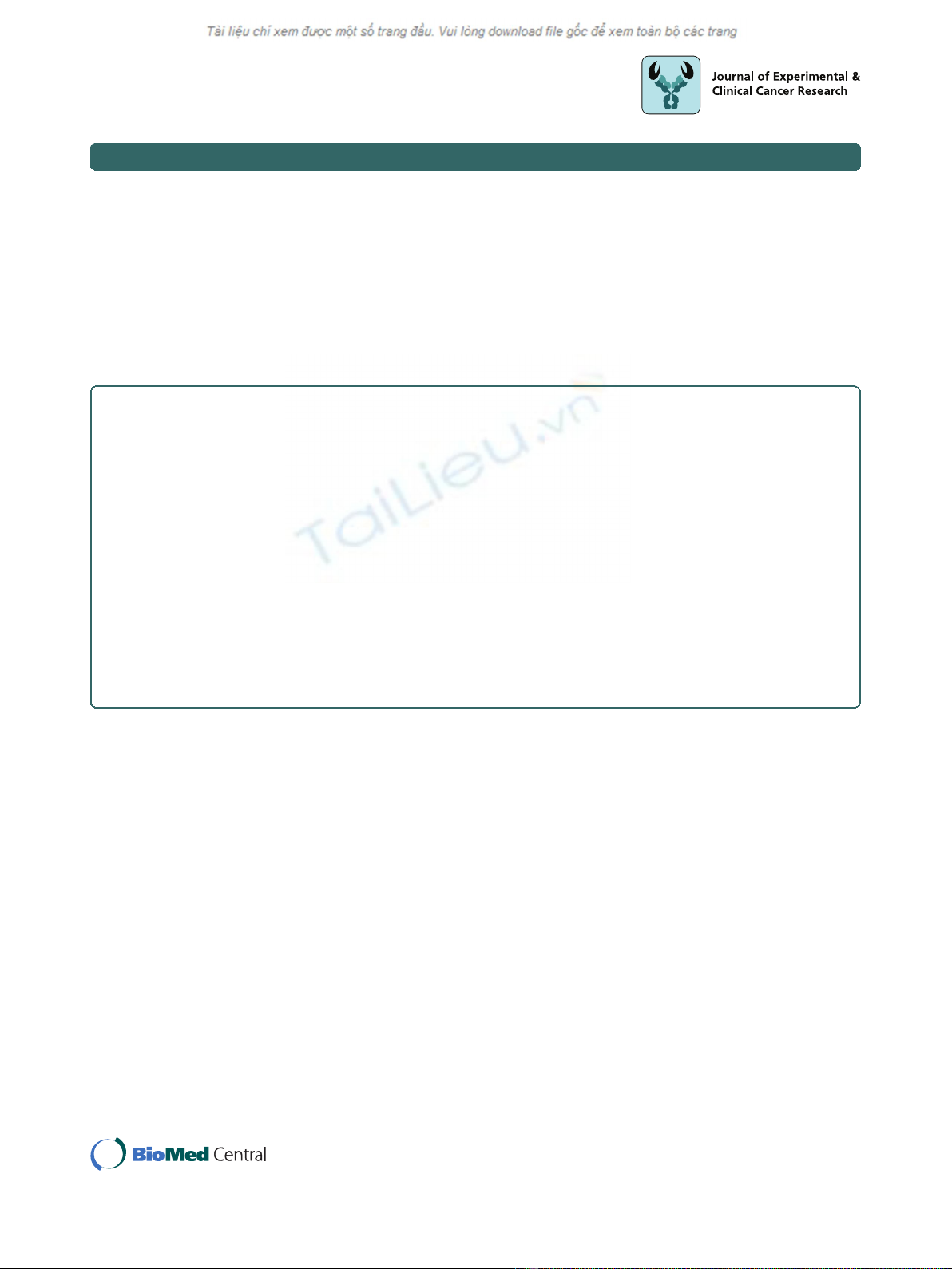
EGFR protein expression
The positive rate of EGFR protein in NSCLC tumor
cells were 46%, which was significantly higher than its
expression in normal lung (p= 0.0234) and paracancer-
ous (p= 0.020)(Figures 1A &1B, Tables 1 &2).
Correlation between EGFR expression and clinical features
The expression of EGFR in different subgroups were
compared and summarized in Table 3. It shows that the
difference of EGFR expression was only significant
between the nodal positive and negative subgroups
(56.4% vs.10%, p = 0.04). There is no significant differ-
ence between age (60 vs. under 60 ys), gender, adeno-
vs. non-adenocarcinoma, the differentiation of tumor,
and staging.
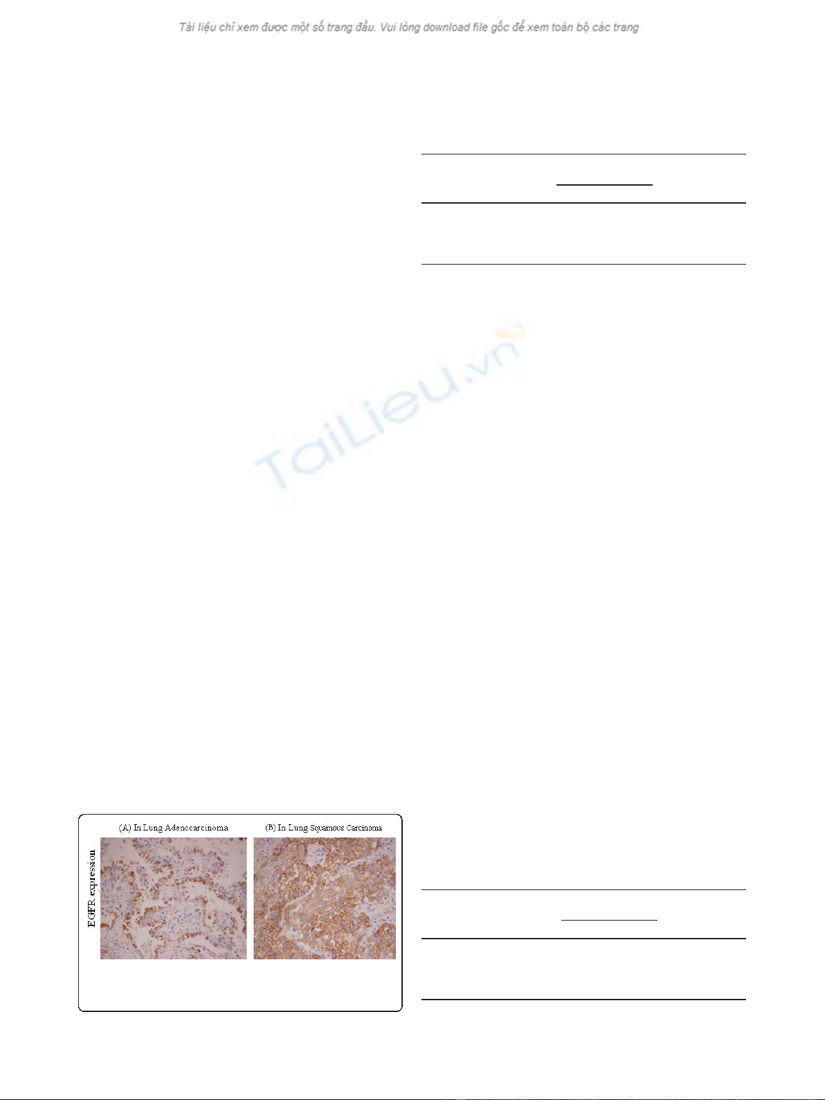
EGFR expression and overall survival
Cox proportional hazards analysis showed that EGFR
protein positive expression independently predicted
patient survival, with RR of 2.311, p= 0.038, and 95%
confidence interval (CI) of 1.049 - 5.095. The mean sur-
vival time for EGFR positive patients was 31 months,
whereasthesurvivaltimewas48monthsforpatients
with EGFR negative expression, with the latter signifi-
cantly longer than the former (p= 0.008, Log Rank
(Mantel-Cox)) (Figure 2).
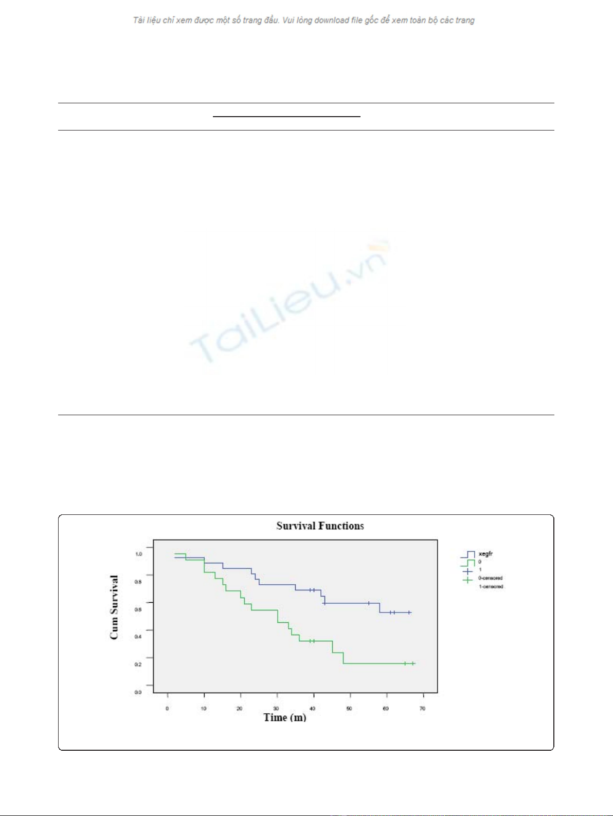
EGFR expression and outcome of radiotherapy
In patients receiving post-operation thoracic irradiation,
the mean survival time for EGFR positive patients (n =
15)was 25 months which was significantly shorter than
that (48 months)for patients (n = 13)with EGFR negative
expression (P = 0.004) (Figure 3).
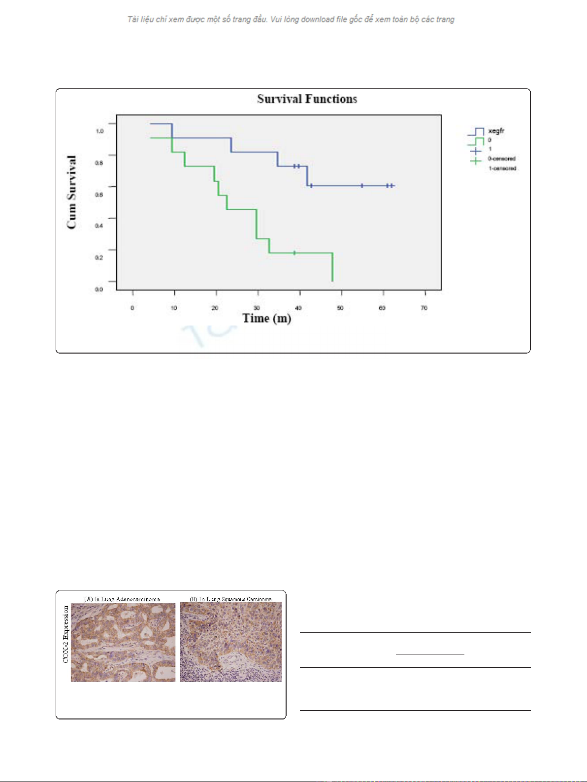
COX-2 expression
The positive rate of COX-2 protein expression in
NSCLC tumor cells was 90%, which was significantly
higher than that in normal tissue(p = 0.00) and paracan-
cerous tissue (p = 0.00) (Figure 4, Tables 4 and 5).
The COX-2 expression was 100% in adenocarcinoma
and significantly higher than that in squamous carcinoma
(76.2%) of the lung. No correlation was found between
COX-2 expression and patient survival (Figures 4, Table 6).
EGFR and COX-2 expression on chemotherapy outcome
Based on COX proportional hazards analysis which also
takes account of other clinical characteristics, there was
no correlation of EGFR and COX-2 expression with
overall survival in 22 patients receiving chemotherapy
alone (P > 0.05).
Correlation of EGFR and COX-2 expression
As shown in Table 7, no correlation was found between
COX-2 and EGFR protein expression (Χ2 = 0.112, P =
0.555).
Discussion
EGFR and COX-2 are molecular targets which have
shown importance for NSCLC. Previous studies reported
that the levels of EGFR and COX-2 expression might
correlate with poor disease prognosis and reduced survi-
val [20,24]. In this study the prognostic values of EGFR
and COX-2 were evaluated with immunohistochemical
assay.
Activation of the EGFR results in activation of down-
stream signaling pathways, including the Ras-Raf-MKK-
extracellular signal-regulated kinase (ERK) and lipid kinase
phosphatidylinositol 3-kinase/Akt pathways. Dysregulation
of these pathways can result in oncogenesis and cancer
progression [4,25-27]. Similarly, our results implied that
EGFR over-expression participated in lung cancer devel-
opment. EGFR expression was negative in paracancerous
Figure 1 EGFR protein expression in (A) adenocarcinoma and
(B) squamous carcinoma of the lung by immunohistochemical
assay (×200).
Table 1 Comparing EGFR protein expression in neoplastic
and normal tissue
Tissue type Number of
cases
EGFR Positive
rate(%)
P
value
positive negative
Neoplastic
tissue
50 23 27 46 0.034*
Normal
tissue
6060
*p < 0.05.
Table 2 Comparing EGFR protein expression in neoplastic
and paracancerous tissue
Tissue type Number of
cases
EGFR Positive
rate(%)
P
value
positive negative
Neoplastic
tissue
50 23 27 46 0.020*
Paracancerous
tissue
7070
*p < 0.05.
Li et al.Journal of Experimental & Clinical Cancer Research 2011, 30:27
http://www.jeccr.com/content/30/1/27
Page 3 of 8
and normal tissues, which was significantly lower than that
in lung cancer tissue (46%)(P < 0.05). It was similarly
reported in studies with the utilization of the immunohis-
tochemical assay that EGFR expression was very low in
normal tissue but often over-expressed in lung cancer
tissue. In normal tissue, EGFR expression was limited to
the basal layer of the epithelium where proliferation
occured. EGFR expression was significantly increased in
dysplastic cells, indicating that EGFR pathway involves in
lung cancer development [28]. Therefore, the detection of
Table 3 EGFR expression and clinical characteristics
Clinical features EGFR Positive expression rate Pvalue
positive negative
Ages 0.448
≤60 18 14 43.80%
>60 9 9 50%
Sex 0.445
Male 16 15 40.50%
Female 11 8 42.10%
Pathologic type 0.543
Squamous carcinoma 13 8 40%
Adencarcinoma 13 13 50%
Mixed type 1 2 66.70%
Tumor length 0.827
≤3 cm 9 7 43.80%
>3 cm 18 16 47.10%
Level of Differentiation 0.474
Poor Differentiated 6 4 40%
Moderate and Well Differentiated 21 19 47.50%
TNM Stage 0.129
I-II 11 5 40%
III 13 15 50.60%
IV 3 3 50%
Lymph node 0.006*
N0 9 1 10%
N1-3 17 22 56.40%
*p < 0.05.
Figure 2 Survival curves with different level of EGFR protein expression. The solid blue line indicates the survival for EGFR negative and
the green line represents survival for EGFR positive expression subgroups.
Li et al.Journal of Experimental & Clinical Cancer Research 2011, 30:27
http://www.jeccr.com/content/30/1/27
Page 4 of 8
EGFR expression in tissue sample before surgery might be
helpful in diagnosis of NSCLC.
In our study EGFR expression in NSCLC was not sig-
nificantly correlated with patients’age, gender, histo-
pathologic type, cell differentiation, tumor size and TNM
stages (P > 0.05). However, EGFR over-expression was
correlated with lymph node metastasis, the probability of
lymph node metastasis was significantly greater in
patients with EGFR over-expression than in EGFR nega-
tive group (P = 0.006). This might indicate that EGFR
was not only involved in cancer genesis but also played
an important role in cancer progression. Though EGFR
was most commonly found in squamous cell (70%) fol-
lowed by adenocarcinoma (50%) [29], and large cell carci-
nomas [28], in our study, EGFR positivity rates were
similar between squamous carcinoma (40%) and adeno-
carcinorma (50%). This discrepancy might be explained
by the small sample size of our study which could limit
the power of detection.
Our results showed that EGFR positive expression was
an independent prognostic factor for NSCLC, among
various factors including patient’sage,gender,histo-
pathology, tumor differentiation, tumor size, TNM sta-
ging and chemotherapy/radiotherapy. Based on the
COX proportional hazard analysis adjusting for other
significant variables, the mortality of patients with posi-
tive tumor EGFR expression was 2.31 times that of the
EGFR negative NSCLC (P<0.05). Nicholson et al [30]
reported a meta-analysis based on 200 studies published
in Medline between 1985 and 2000, which showed that
EGFR over-expression was correlated with patient’s
prognosis in 10 tumor types. But only 30% of the studies
considered EGFR to be associated with NSCLC prog-
nosis. However, it might not be conclusive since some
of the studies in the meta-analysis did not include treat-
ment for multivariate analysis, which might have an
impact on survival.
Figure 4 Immunohistochemical stain(×200)for COX-2
expression in (A) adenocarcinoma and (B) squamous
carcinoma of the lung.
Figure 3 Survival curves based on EGFR expression in patients receiving thoracic irradiation. The solid blue line indicates the survival for
EGFR negative and the green line represents survival for EGFR positive expression subgroups.
Table 4 COX-2 expression in neoplastic and normal tissue
Tissue type Number of
cases
COX-2 Positive
rate(%)
P
value
positive negative
Neoplastic
tissue
50 40 5 90 0.000*
Normal
tissue
6060
*p < 0.05.
Li et al.Journal of Experimental & Clinical Cancer Research 2011, 30:27
http://www.jeccr.com/content/30/1/27
Page 5 of 8


%20--%3e%3cdefs%3e%3cstyle%3e%20.st0%20{%20fill:%20%23fff;%20}%20.st1%20{%20fill:%20%237800fa;%20}%20%3c/style%3e%3c/defs%3e%3cpath%20class='st1'%20d='M117.78,12.18H43.11c2.9,3.47,4.65,7.94,4.65,12.82,0,5.6-2.3,10.66-6.01,14.29h76.02l7.22-13.56-7.22-13.56Z'/%3e%3cg%3e%3cpath%20class='st0'%20d='M53.58,26.17h-.59v-1.46h.59v-4.96h2.83c1.78,0,2.67.94,2.67,2.82v5.76c0,1.87-.89,2.81-2.67,2.81h-2.83v-4.96ZM55.36,21.37v3.34h1.1v1.46h-1.1v3.34h1.01c.61,0,.91-.37.91-1.1v-5.93c0-.74-.3-1.1-.91-1.1h-1.01Z'/%3e%3cpath%20class='st0'%20d='M65.99,31.14h-1.8l-.31-2.07h-2.19l-.31,2.07h-1.64l1.82-11.39h2.62l1.82,11.39ZM65.28,18.04c-.25.46-.51.77-.75.94-.21.15-.47.22-.79.22-.26,0-.57-.07-.92-.22l-.38-.15c-.14-.05-.26-.07-.37-.07-.3,0-.53.18-.71.54l-.91-.68c.25-.46.51-.77.75-.94.21-.14.48-.21.79-.21.26,0,.57.07.92.21l.38.15c.14.05.26.07.37.07.3,0,.53-.18.71-.54l.91.68ZM61.91,27.52h1.73l-.87-5.76-.87,5.76Z'/%3e%3cpath%20class='st0'%20d='M74.53,26.89v1.52c0,1.91-.89,2.86-2.67,2.86s-2.67-.95-2.67-2.86v-5.93c0-1.91.89-2.86,2.67-2.86s2.67.95,2.67,2.86v1.11h-1.69v-1.22c0-.75-.31-1.12-.93-1.12s-.93.37-.93,1.12v6.15c0,.74.31,1.11.93,1.11s.93-.37.93-1.11v-1.63h1.69Z'/%3e%3cpath%20class='st0'%20d='M81.4,31.14h-1.8l-.31-2.07h-2.19l-.31,2.07h-1.64l1.82-11.39h2.62l1.82,11.39ZM75.9,19.2l1.52-1.91h1.71l1.51,1.91h-1.61l-.76-.95-.75.95h-1.61ZM77.32,27.52h1.73l-.87-5.76-.87,5.76ZM83.1,15.99l-1.76,1.91h-1.26l1.17-1.91h1.86Z'/%3e%3cpath%20class='st0'%20d='M84.86,19.75c1.78,0,2.67.94,2.67,2.82v1.48c0,1.87-.89,2.81-2.67,2.81h-.85v4.28h-1.79v-11.39h2.64ZM84.01,21.37v3.86h.85c.58,0,.87-.36.87-1.08v-1.71c0-.71-.29-1.07-.87-1.07h-.85Z'/%3e%3cpath%20class='st0'%20d='M93.51,19.75c1.78,0,2.67.94,2.67,2.82v1.48c0,1.87-.89,2.81-2.67,2.81h-.85v4.28h-1.79v-11.39h2.64ZM92.66,21.37v3.86h.85c.58,0,.87-.36.87-1.08v-1.71c0-.71-.29-1.07-.87-1.07h-.85Z'/%3e%3cpath%20class='st0'%20d='M98.8,31.14h-1.79v-11.39h1.79v4.88h2.03v-4.88h1.83v11.39h-1.83v-4.88h-2.03v4.88Z'/%3e%3cpath%20class='st0'%20d='M105.36,24.55h2.46v1.62h-2.46v3.34h3.09v1.63h-4.88v-11.39h4.88v1.63h-3.09v3.18ZM108.17,17.29l-1.76,1.91h-1.26l1.17-1.91h1.86Z'/%3e%3cpath%20class='st0'%20d='M112.2,19.75c1.78,0,2.67.94,2.67,2.82v1.48c0,1.87-.89,2.81-2.67,2.81h-.85v4.28h-1.79v-11.39h2.64ZM111.35,21.37v3.86h.85c.58,0,.87-.36.87-1.08v-1.71c0-.71-.29-1.07-.87-1.07h-.85Z'/%3e%3c/g%3e%3ccircle%20class='st1'%20cx='25'%20cy='25'%20r='20'/%3e%3cpath%20class='st0'%20d='M32.78,19.27c2.92,0,4.43,2.55,5.28,5.33l.71,2.17c.14.38-.33.75-.71.75h-5.61c.19-.33.24-.71.09-1.08l-.75-2.45c-.43-1.32-.99-2.64-1.79-3.77.75-.57,1.65-.94,2.78-.94h0ZM25,18.38c3.25,0,4.9,2.78,5.89,5.89l.76,2.45c.14.42-.33.8-.8.8h-11.69c-.42,0-.94-.38-.8-.8l.75-2.45c.99-3.11,2.64-5.89,5.89-5.89h0ZM25,11.35c1.74,0,3.11,1.37,3.11,3.11s-1.37,3.11-3.11,3.11-3.11-1.41-3.11-3.11,1.41-3.11,3.11-3.11h0ZM17.27,19.27c1.08,0,1.98.38,2.73.94-.8,1.13-1.37,2.45-1.74,3.77l-.8,2.45c-.14.38-.05.75.09,1.08h-5.56c-.42,0-.9-.38-.75-.75l.71-2.17c.9-2.78,2.41-5.33,5.33-5.33h0ZM17.27,12.91c1.51,0,2.78,1.27,2.78,2.83s-1.27,2.83-2.78,2.83-2.83-1.27-2.83-2.83,1.27-2.83,2.83-2.83h0ZM32.78,12.91c1.56,0,2.78,1.27,2.78,2.83s-1.23,2.83-2.78,2.83-2.83-1.27-2.83-2.83,1.27-2.83,2.83-2.83h0ZM27.07,28.56v.09c0,.57-.24,1.08-.61,1.46h0v.05c-.38.33-.9.57-1.46.57s-1.08-.24-1.46-.61h0c-.38-.38-.61-.9-.61-1.46v-.09h1.41v.09c0,.19.05.38.19.47v.05c.09.09.28.19.47.19s.38-.09.47-.19v-.05c.14-.09.24-.28.24-.47t-.05-.09h1.41ZM30.99,28.56v.09c0,1.65-.66,3.16-1.74,4.24-1.08,1.08-2.59,1.79-4.24,1.79s-3.16-.71-4.24-1.79l-.05-.05c-1.04-1.08-1.7-2.55-1.7-4.2v-.09h1.41v.09c0,1.27.47,2.4,1.27,3.25h.05c.85.85,1.98,1.37,3.25,1.37s2.4-.52,3.25-1.37c.85-.8,1.37-1.98,1.37-3.25v-.09h1.37ZM34.99,28.56v.09c0,2.78-1.13,5.28-2.92,7.07-1.79,1.79-4.29,2.92-7.07,2.92s-5.23-1.13-7.07-2.92c-1.79-1.79-2.92-4.29-2.92-7.07v-.09h1.41v.09c0,2.4.94,4.53,2.5,6.08,1.56,1.56,3.72,2.5,6.08,2.5s4.52-.94,6.08-2.5c1.56-1.56,2.5-3.68,2.5-6.08v-.09h1.41Z'/%3e%3c/svg%3e)