JOURNAL OF MILITARY PHARMACO-MEDICINE N04 - 2025
175
PREVALENCE AND CHARACTERISTICS OF NON-ALCOHOLIC
FATTY LIVER DISEASE IN PATIENTS WITH FATTY LIVER
DIAGNOSED BY ULTRASOUND
Tran Thi Khanh Tuong1*, Tran Kinh Thanh2, Au Nhat Huy3
Abstract
Objectives: To determine the prevalence and characteristics of non-alcoholic
fatty liver disease (NAFLD), including steatosis and fibrosis, in patients with fatty
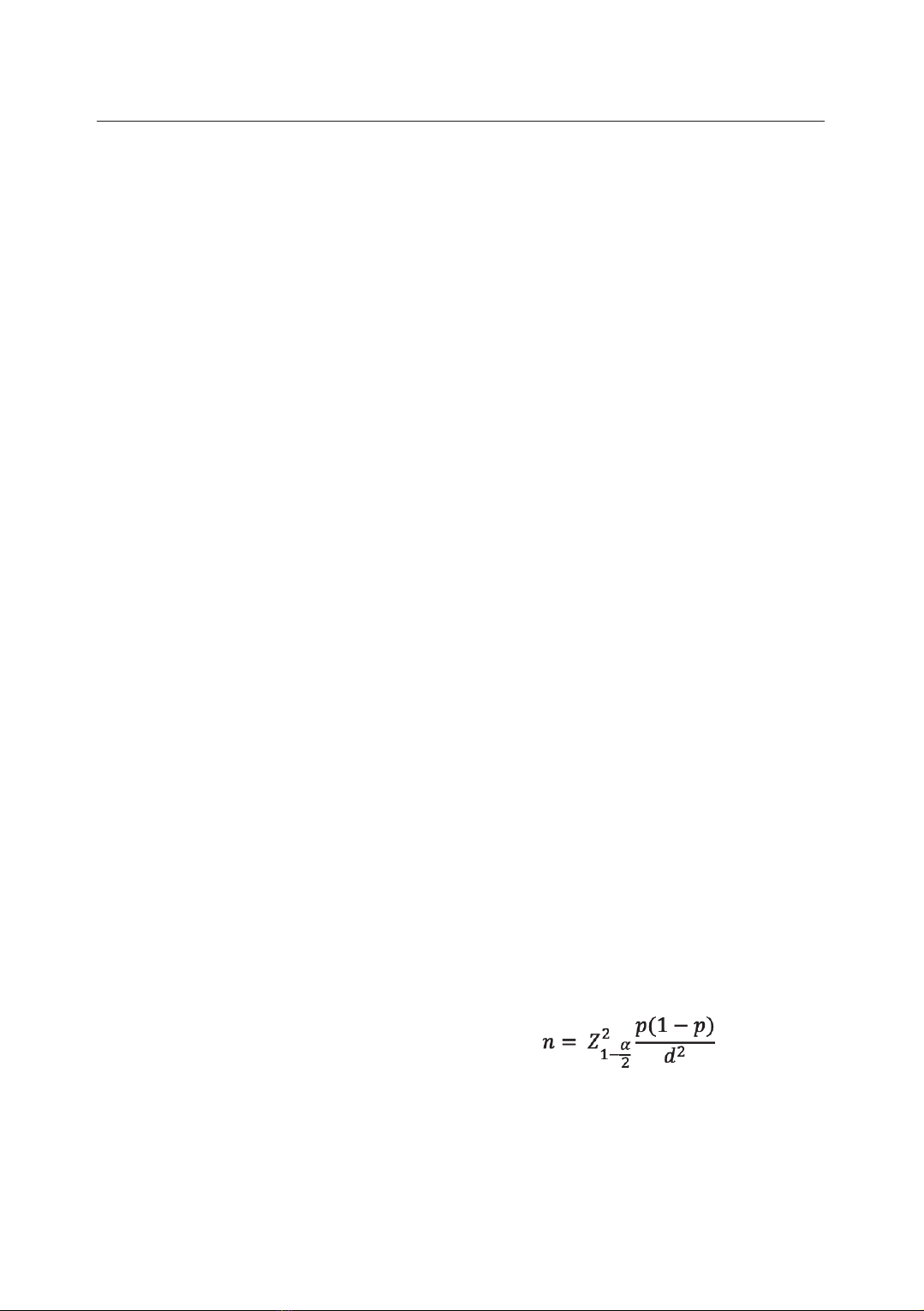
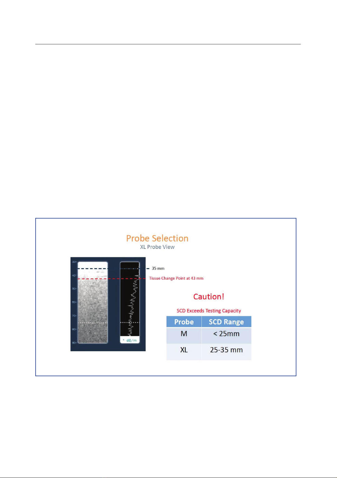
liver detected by ultrasound. Methods: A cross-sectional study was conducted on
303 patients diagnosed with fatty liver by ultrasound at People’s Hospital 115 from
August 2019 to October 2020. Steatosis and fibrosis were assessed using
FibroScan, employing the controlled attenuation parameter (CAP) and liver
stiffness measurements (LSM). Statistical analysis was performed using SPSS
version 22.0. Results: The prevalence of NAFLD in patients with fatty liver
detected by ultrasound and assessed by FibroScan using CAP probe was 66%.
Among patients with fatty liver on ultrasound who have NAFLD, the distribution
of liver fat levels was as follows: S1 = 20.5%; S2 = 27%; S3 = 52.5%. The stages
of liver fibrosis were: F0 - F1 at 74.5%; significant fibrosis at 25.5%, advanced
fibrosis at 11%, and cirrhosis at 6%. NAFLD patients exhibited higher body mass
index (BMI), waist circumference, cholesterol, triglyceride, type 2 diabetes
mellitus (T2DM), and obesity rates compared to those without NAFLD.
Conclusion: The study underscores the high prevalence of NAFLD in patients
with fatty liver detected by ultrasound, with the highest proportion of patients in
stage S3, while 25.5% of the cases are classified as significant fibrosis, which
indicates a considerable level of liver damage.
Keywords: Fatty liver; Non-alcoholic fatty liver disease; Ultrasound;
FibroScan; Steatohepatitis.
1Internal Medicine Department, Faculty of Medicine, Pham Ngoc Thach University of Medicine
2People’s Hospital 115
3Internal Medicine Department, Faculty of Medicine, Tan Tao University
*Corresponding author: Tran Thi Khanh Tuong (khanhtuong@pnt.edu.vn)
Date received: 29/8/2024
Date accepted: 23/12/2024
http://doi.org/10.56535/jmpm.v50i4.999
JOURNAL OF MILITARY PHARMACO-MEDICINE N04 - 2025
176
INTRODUCTION
Non-alcoholic fatty liver disease has
continued to rise as a predominant cause
of chronic liver disease worldwide,
particularly in light of the ongoing
global obesity and T2DM pandemics
[1]. Recent studies have shown an
alarming increase in the prevalence of
NAFLD, with an estimated 25 - 30% of
the global population affected as of
2024 [2]. The increasing burden of
NAFLD is particularly pronounced in
Asia, where urbanization and lifestyle
changes have driven higher rates of
metabolic syndrome, a key risk factor
for NAFLD [3]. Although abdominal
ultrasound is a common method for
detecting fatty liver, no studies have yet
evaluated the prevalence of NAFLD in
patients with fatty liver identified by
ultrasound.
Fatty liver is increasingly detected by
abdominal ultrasound, yet it often
receives insufficient attention from both
patients and healthcare providers. The
most common cause of this condition is
NAFLD. This disease can lead to serious
complications such as liver fibrosis,
cirrhosis, and hepatocellular carcinoma
[4]. Additionally, NAFLD is recognized
as an independent cardiovascular risk
factor, leading to cardiovascular problems
even in the absence of traditional risk
factors like hypertension or dyslipidemia
[5].
One of the new and effective non-
invasive methods for assessing liver fat
and fibrosis levels is FibroScan with the
CAP probe. This technique, similar to
conventional ultrasound, is simple and
quick to perform, but it provides
additional crucial information about the
patient's liver condition without causing
any complications. As a result, it has
been recommended by major liver
disease research associations worldwide,
including the American Association for
the Study of Liver Diseases and the
European Association for the Study of
the Liver, as a useful screening tool in
the diagnosis of NAFLD [6]. Therefore,
we conducted this study to: Determine
the prevalence and characteristics of
NAFLD, including steatosis and fibrosis,
in patients with fatty liver detected by
ultrasound.
MATERIALS AND METHODS
1. Subjects
Including 303 patients aged 18
who were diagnosed with fatty liver
by ultrasound and visited People’s
Hospital 115 from August 2019 to
October 2020.
The sample size is calculated using
the following formula:
p is the proportion of the study
variable, with d as the desired margin of
error set at 5%. Since no existing
JOURNAL OF MILITARY PHARMACO-MEDICINE N04 - 2025
177
studies determine the prevalence of
NAFLD in patients with ultrasound-
detected fatty liver, p is assumed to be
0.5 Æ n > = 196.
* Inclusion criteria: All patients
diagnosed with fatty liver by abdominal
ultrasound within the last 6 months
(Increased Echogenicity: Liver appears
brighter than right, Vascular Blurring,
Ultrasound beam weakens, making deeper
liver areas harder to see [7]); aged 18
years; able to read and sign the consent
form to participate in the study.
* Exclusion criteria: Unreliable
FibroScan results: Patients with
FibroScan results showing an
interquartile range (IQR) > 30% or a
success rate < 60%; elevated liver
enzymes > 100 U/L; cholestasis,
hepatic congestion, or presence of
ascites; SCD (Exceed testing capacity)
> 25mm (probe M) to rule out cases of
excess abdominal fat in obese patients,
an XL probe is required [8]; pregnancy;
poor health status or inability to provide
blood samples.
Figure 1. Probe selectition.
* Location and time: The study was conducted at People’s Hospital 115.
Patients who were diagnosed with fatty liver by ultrasound were recruited from
August 2019 to October 2020.
JOURNAL OF MILITARY PHARMACO-MEDICINE N04 - 2025
178
2. Methods
* Study design: A cross-sectional study.
The FibroScan machine is manufactured
by Echosens, a French medical technology
company.
The patient fasted for at least 3 hours
prior to the measurement. LSM was
performed by a hepatology and
gastroenterology specialist who has
been trained and is proficient in the
FibroScan, having conducted over 500
cases up to the time of the study. All
patients underwent the procedure with a
standard M probe.
Liver fibrosis stages based on kPa
values [4]:
- < 7 kPa: Stage F0 - F1;
- 7 kPa: F2 (Significant liver
fibrosis);
- 8.7 kPa: F3 (Advanced liver
fibrosis);
- 11.5 kPa: F4 (Cirrhosis).
Table 1. Liver Steatosis grade [5].
Steatosis
grade
Percentage of liver cells
with Steatosis
CAP (dB/m)
Remarks
S0
0 - 4%
100 - 233
Normal
S1
5 - 33%
234 - 269
Mild steatosis
S2
34 - 66%
270 - 300
Moderate steatosis
S3
67 - 100%
301
Severe steatosis
Patients are diagnosed with NAFLD
if the CAP score exceeds 233 dB/m
(and other causes of steatosis have been
excluded [5]:
- Malnutrition: BMI < 18.5 kg/m².
- Parenteral nutrition.
- Hepatitis B or C virus infection
(HBsAg (+), Anti-HCV (+)).
- Medications (e.g., amiodarone,
methotrexate, tamoxifen, corticosteroids,
valproate, antiretroviral drugs).
- Pregnancy.
- Heavy alcohol consumption:
30 g/day or 210 g/week for men and
20 g/day or 140 g/week for women
for at least two consecutive years.
* Data analysis: Statistical analysis
was performed using SPSS version 22.0.
3. Ethics
The study was approved by the Ethics
Committee of People’s Hospital 115,
Number 108/BV-NCKH, dated October 25,
2019. The authors commit to using
licensed data and take full responsibility
for it. The authors declare to have no
conflicts of interest in the study.
JOURNAL OF MILITARY PHARMACO-MEDICINE N04 - 2025
179
RESULTS
A total of 303 patients were enrolled in this study, with a mean age of 45 ± 12
years. The male-to-female ratio was 1.2:1. Among these patients, 200 (66%) were
diagnosed with NAFLD based on the CAP scores obtained via FibroScan.
Table 2. Comparison of characteristics between NAFLD
and non-NAFLD patients.
Characteristics
NAFLD
Non-NAFLD
(n = 103)
p
BMI (kg/m²)
Mean ± SD
24.7 ± 3.6
23.6 ± 3.9
0.002
Waist circumference (cm)
Mean ± SD
92.2
± 10.6
89.8 ± 8.4
0.032
Obesity (%)
44
26.2
0.007
Central obesity (%)
77
71.8
0.325
T2DM (%)
25
13.6
0.021
Dyslipidemia (%)
98
97.1
0.616
Metabolic syndrome (%)
70
59.2
0.06
Glucose (mmol/L)
Mean ± SD
6.7 ± 3.6
6.1 ± 2.4
0.084
Cholesterol (mmol/L)
Mean ± SD
5.3 ± 1.8
4.8 ± 1.5
0.009
Triglycerides (mmol/L)
Median (min - max)
2.6 (1.8 -
3.8)
2.1 (1.6 -
3.1)
0.026
HDL - C (mmol/L)
Mean ± SD
1.0 ± 0.3
1.0 ± 0.3
0.655
LDL - C (mmol/L)
Mean ± SD
3.3 ± 1.1
3.3 ± 1.2
0.939
ALT (U/L)
Median (min - max)
45.2 (27.1
-
77.5)
44.8 (28.1
-
78.5)
0.843
AST (U/L)
Median (min - max)
31.5 (23.3
-
50.6)
30.7 (24.5
-
52.8)
0.764
Patients with NAFLD have a higher BMI, larger waist circumference, and
higher obesity rates compared to those with fatty liver on abdominal ultrasound
without NAFLD. These patients also have higher levels of cholesterol and triglycerides.