Protein kinase C e phosphorylates keratin 8 at Ser8 and Ser23 in GH4C1 cells stimulated by thyrotropin-releasing hormone Yoshiko Akita1, Hiroshi Kawasaki2, Shinobu Imajoh-Ohmi3, Hiroyuki Fukuda3, Shigeo Ohno4, Hisashi Hirano2, Yoshitaka Ono5 and Hiromich Yonekawa1
1 Department of Laboratory Animal Science, The Tokyo Metropolitan Institute of Medical Science, Japan 2 Supramolecular Biology, International Graduate School of Arts and Sciences, Yokohama City University, Japan 3 Department of Basic Medical Sciences, Institute of Medical Science, University of Tokyo, Japan 4 Department of Molecular Biology, Yokohama City University School of Medicine, Japan 5 Biosignal Research Center, Kobe University, Japan
Keywords GH4C1 cells; keratin 8; phosphorylation; protein kinase C e; thyrotropin-releasing hormone
Correspondence Y. Akita, Department of Laboratory Animal Science, The Tokyo Metropolitan Institute of Medical Science, 3-18-22 Honkomagome, Bunkyo-ku, Tokyo 113-8613, Japan Fax: +81 3 3823 2130 Tel: +81 3 3823 2105 (ext. 5430) E-mail: akita@rinshoken.or.jp
(Received 7 March 2007, revised 26 April 2007, accepted 30 April 2007)
doi:10.1111/j.1742-4658.2007.05853.x
Protein kinase C e (PKCe) is activated by thyrotropin-releasing hormone (TRH), a regulator of pituitary function in rat pituitary GH4C1 cells. We analyzed the downstream mechanism after PKCe activation. Exposure of GH4C1 cells to TRH or a phorbol ester increased the phosphorylation of three p52 proteins (p52a, p52b and p52c) and decreased the phosphoryla- tion of destrin and cofilin. GF109203X, an inhibitor of protein kinases including PKC, inhibited phosphorylation of the p52 proteins by TRH sti- mulation. Peptide mapping, amino-acid sequencing, and immunochemical studies indicated that p52a, p52b, and p52c are the differentially phosphor- ylated isoforms of keratin 8 (K8), an intermediate filament protein. The unphosphorylated K8 (p52n) localized exclusively to the cytoskeleton, whereas the phosphorylated forms (especially p52c), which are increased in TRH-stimulated cells, localized mainly to the cytosol. K8 phosphorylation was enhanced in PKCe-overexpressing clones, and purified recombinant PKCe directly phosphorylated K8 with a profile similar to that observed in TRH-stimulated cells. PKCe and K8 colocalized near the nucleus under basal conditions and were concentrated in the cell periphery and cell–cell contact area after TRH stimulation. MS analyses of phospho-K8 and K8-synthesized peptide (amino acids 1–53) showed that PKCe phosphory- lates Ser8 and Ser23 of K8. Phosphorylation of these sites is enhanced in TRH-stimulated cells and PKCe-overexpressing cells, as assessed by immu- noblotting using antibodies to phospho-K8. These results suggest that K8 is a physiological substrate for PKCe, and the phosphorylation at Ser8 and Ser23 transduces, at least in part, TRH–PKCe signaling in pituitary cells.
Abbreviations IF, intermediate filament; K8, keratin 8; MARCKS, myristoylated alanine-rich C kinase substrate; PKC, protein kinase C; cPKC, Ca2+- dependent and phospholipid-dependent protein kinase, conventional PKC; nPKC, Ca2+-independent, phospholipid-dependent protein kinase, novel PKC; TPA, 12-O-tetradecanoylphorbol 13-acetate; TRH, thyrotropin-releasing hormone.
FEBS Journal 274 (2007) 3270–3285 ª 2007 The Authors Journal compilation ª 2007 FEBS
3270
that is hormonal, and immune cells [1–6]. Recent knockout, transgenic, and mutational studies have established essential roles for PKCe in diverse signaling path- ways controlling cellular proliferation, differentiation, Protein kinase C (PKC) e is a novel Ca2+-independent and phorbol ester ⁄ diacylglycerol-sensitive serine ⁄ thre- expressed in many tissues onine kinase in neuronal, and cells, and is especially abundant
Y. Akita et al.
TRH-PKCe-evoked K8 phosphorylation at Ser8 and 23
metabolism, and exocytosis, in addition to those con- trolling nervous and immune functions [1,7–9]. More- over, evidence for critical roles for PKCe in various diseases, such as malignant tumors, cardiac ischemia, is accumulating Alzheimer’s disease and diabetes, [10,11]. Despite such evidence, the nature of the signa- ling pathways downstream of PKCe that are involved in a stimulus–coupling response remains poorly under- stood. understanding PKCe-mediated of substrates Numerous potential
these substrates,
intracellular Ca2+-dependent protease [21,22]. We also reported that the overproduction of PKCe, but not an inactive mutant or other PKC isozymes, specifically enhances both resting and TRH-stimulated prolactin secretion [19]. These previous studies clearly demon- strated the specific role of PKCe in TRH-induced prolactin secretion from GH4C1 cells. Identifying the major PKCe substrate(s) and elucidating the down- stream mechanism after its activation would lead to an hormonal actions. Several phosphoproteins have been shown to be up-regulated in response to TRH stimulation in GH4C1 and GH3 cells [23,24]. However, the identity and functional roles of these proteins remain obscure, although stathmin, a regulator of cytoskeletal microtu- bules, has been studied in detail [25]. We have recently shown in GH4C1 cells that a p80 acidic substrate of PKCe (Mr (cid:2) 80 000; pI (cid:2) 4.3) is MARCKS, a regula- tor of cytoskeletal microfilaments, and furthermore that MARCKS but not stathmin acts downstream of PKCe in TRH-stimulated signaling [13].
subcellular change the
translocates it
To gain a better understanding of the mechanism by which PKCe mediates the TRH-induced signal, we evaluated the phosphorylation of three p52 proteins in TRH-stimulated GH4C1 cells and in cells overexpress- ing PKCe. We identified these proteins as differently phosphorylated isoforms of keratin 8 (K8). We also found that PKCe activated in response to TRH stimu- lation phosphorylates mainly Ser8 and Ser23 located in the N-terminal half of the head domain of K8. Implications of the phosphorylation of K8 in TRH– PKCe-mediated responses are discussed. for PKC family members have been reported, including enzymes and cytoskeletal proteins. Most of for example, the myristoylated alanine-rich C kinase sub- strate (MARCKS), are ubiquitously phosphorylated by nearly all conventional (cPKC) and novel PKC (nPKC) family members [12,13]. Substrate specificity in regulatory signaling seems to be achieved through spatial and temporal targeting of PKC to specific sub- cellular compartments. PKCe is activated by various second messengers, including diacylglycerols, phospha- tidylinositol 3,4,5-triphosphate, and fatty acids pro- duced in response to extracellular signals that activate G-protein-coupled receptors, tyrosine kinase receptors, or tyrosine kinase-coupled receptors [14]. These second messengers localization of PKCe by binding to the C1 domain. PKCe translo- cates to the plasma membrane and ⁄ or the cytoskeleton in response to diacylglycerols and tridecanoic acids, whereas to the Golgi network in response to arachidonic and linoleic acids [15]. Adap- ter proteins such as a coatomer protein b¢-COP (or RACK2) and RACK1 also regulate the localization of PKCe [4,11].
Results
Action of TRH on protein phosphorylation
FEBS Journal 274 (2007) 3270–3285 ª 2007 The Authors Journal compilation ª 2007 FEBS
3271
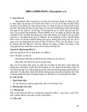
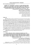
We analyzed the changes in phosphorylation of several proteins in response to treatment with TRH by in vivo labeling of GH4C1 cells with 32Pi (Fig. 1). MARCKS (p80) was phosphorylated as previously reported [13]. In addition, we observed a rapid increase in the phos- phorylation of three p52 proteins (p52a, p52b, and p52c; Mr values of 52 000 and pI values of 5.75, 5.7, and 5.6, respectively), and the dephosphorylation of p19 and p20 proteins (Mr values of 19 000–20 000 and pI values of (cid:2) 6.5 and 6.0, respectively). The TRH- induced change in p52 phosphorylation was quite rapid (Fig. 1B). p52a phosphorylation increased within 15 s and was maintained for 120 min, although it often decreased between 1 and 2.5 min. In contrast, the level of phosphorylation of p52b and p52c peaked within 15–30 s and 30–60 s, respectively, and then both rapidly Thyrotropin-releasing hormone (TRH) is a hypotha- lamic hormone that regulates the secretion of thyrotro- pin and prolactin from anterior pituitary cells. Beside its hypothalamic–pituitary function, TRH has diverse functions including the control of arousal, autonomic function, pain perception, spinal motor function, and embryonic growth in pregnancy [16]. TRH is also an antiepileptic and antidepressant agent [16]. TRH and its receptor have been reported to be produced in var- ious malignant tumors [17]. In rat pituitary GH4C1 cells, TRH stimulates the release and synthesis of prolactin by a diacylglycerol-activated PKC-mediated mechanism and inhibits proliferation of the cells through prolongation of the G2 phase of the cell cycle [18–20]. We have previously shown that treatment of the cells with TRH elicits rapid and sustained translo- cation of PKCe from the cytosolic fraction to the membrane ⁄ cytoskeletal fractions, and that this treat- ment exclusively down-regulates PKCe by a mechan- ism involving m-calpain, a ubiquitously expressed
Y. Akita et al.
TRH-PKCe-evoked K8 phosphorylation at Ser8 and 23
B
pI 6.6
4.5
25
A MW (kD)
None
20
p52a p52b p52c
97 66.2
**
**
**
**
p80
15
45
*
***
p52a b
(MARCKS)
*
10
31
*
) 4 0 1 x ( y t i
p20
**
p19
*
**
*
**
*
5
21.5
*
i
0
** * * * 1
0
2
3
4
5
20 40 60 80 100 120
TRH15sec
40
v i t c a o d a R e v
p20
97 66.2
p80
*
30
l
*
(MARCKS)
45
P52a
cb
*
**
i t a e R
*
20
**
**
31
*
10
p20
p19
21.5
0
0
1
2
3
4
5
20 40 60 80 100 120
Time (min)
Fig. 1. (A) Effect of TRH on the phosphorylation of various proteins in GH4C1 cells. Serum-starved cells were labeled with 32Pi for 3 h and incubated with (lower panel) or without (upper panel) 500 nM TRH for 15 s. The lysates derived from 7.5 · 104 cells were subjected to 2D nonequilibrium IEF gel electrophoresis ⁄ SDS ⁄ PAGE. Phosphoproteins were visualized by autoradiography. Mr values are indicated on the left. pI 6.6 (left) to 4.5 (right) is shown at the top of the gels. Arrowheads and letters indicate spots affected by TRH, as described in the text. (B) Time course of TRH action on the levels of p52 and p20 phosphoproteins. Cells were treated with 500 nM TRH for the last 15 s to 120 min of the 3.5 h labeling with 32Pi. Whole cell lysates were separated by 2D gel electrophoresis. The radioactivity of each spot corres- ponding to p52a, p52b, and p52c (upper panel) and p20 (lower panel) was quantified, and the values shown are the mean ± SE of the deter- minations in three experiments. Asterisks indicate significant differences from the values of the untreated cells (time ¼ 0): *P < 0.05, **P < 0.01, and ***P < 0.001. Note that the time scales differ before and after 5 min.
suggesting that a phosphatase
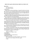
proteins showed a sustained decrease after TPA stimu- lation (lower panels in Fig. 2A) as well as TRH stimu- lation (Fig. 1), is activated in a PKC-dependent manner, which enhances the dephosphorylation of p19 and p20.
decreased. On the other hand, p19 (data not shown) and p20 (Fig. 1B) exhibited a gradual and sustained decrease in their phosphorylation between 15 s and 120 min after TRH treatment. P19 and p20 were identi- fied as phosphorylated forms of destrin and cofilin, respectively, as assessed by the profiles of 2D electro- phoresis and immunoblot analysis (data not shown).
Effect of activators and inhibitors of PKC on p52 phosphoproteins
FEBS Journal 274 (2007) 3270–3285 ª 2007 The Authors Journal compilation ª 2007 FEBS
3272
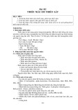
Phorbol ester (12-O-tetradecanoylphorbol 13-acetate; TPA) is a direct and potent activator of cPKC and nPKC isozymes. The 2D profile of p52 phosphopro- teins obtained upon TPA stimulation was quite similar to that obtained upon TRH activation, although the magnitude and time course of the effect was slightly different (upper panel in Fig. 2A). The phosphoryla- tion of p52a increased within 10 min of TPA treatment and decreased slightly after 15 min (data not shown). On the other hand, the phosphorylation of p52b and p52c continued to increase after 10 min. The decrease in P52a and the increases in p52b and p52c during prolonged TPA stimulation suggest that PKC activa- tion by TPA enhances predominantly the phosphoryla- tion steps from p52a to p52b, and from p52b to p52c. The phosphorylation of p19 (destrin) and p20 (cofilin) We examined the effect of bisindolylmaleimide type I (GF109203X) on the TRH-induced changes in p52 phosphorylation. It was used as an inhibitor of cPKC and nPKC, because, among several protein kinases inhibited by GF109203X [26], TRH is reported to exclusively activate cPKC and nPKC in GH4C1 cells. As shown in Fig. 2B, GF109203X blocked the TRH- stimulated phosphorylation of the three p52 proteins. Inhibition of p52a phosphorylation was evident at both 5 and 10 min after TRH exposure in the presence of GF109203X, whereas inhibition of the phosphoryla- tion of p52b and p52c was detected at 15–30 s. We also observed that GF109203X blocked TPA-stimula- ted phosphorylation and that staurosporine, a potent inhibitor of a wide variety of kinases including PKC, inhibited both TRH-induced and TPA-induced chan- ges in p52 phosphorylation (data not shown). These results support the idea that PKC is involved in the phosphorylation of p52 proteins, but cannot com- pletely exclude the possibility that other kinases that are inhibited by GF109203X, such as MAPKAP-K1b, also mediate the phosphorylation.
Y. Akita et al.
TRH-PKCe-evoked K8 phosphorylation at Ser8 and 23
None
TRH 25 sec
TPA 10 min
A
0
15 sec
5 min
15 min
60 min
A Time
S mix
p52a b
p52a
cb
p52a
cb
(d)
ε mix
P52 a b c c
p19 p20
p19 p20
p19 p20
B
Time
0
15 sec
5 min
Exp. 1
S1
TRH+GF
B
TRH
3
ε13
*
*
p52a
2.5
a b c
2
S5
Exp. 2
1.5
1
ε3
0.5
ε4
l
0 2.5
a b c
*
*
p52b
2
1.5
i
1
0.5
) d o f ( y t i v i t c a o d a R e v i t a l e R
0 2.5
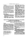
Fig. 3. Enhanced phosphorylation of p52 proteins in PKCe-overex- pressing GH4C1 cells. (A) A mixture of vector control clones (S mix) and PKCe-overexpressing clones (e mix) stably established by trans- fection were labeled with 32Pi and stimulated with 500 nM TRH for the indicated times of the total 3 h labeling. Arrowheads indicate the positions of spots of p52a, p52b, and p52c. Results shown are representative of two separate experiments. (B) Profiles of p52 phosphoproteins are visualized in the control clones, S1 and S5, and the nPKCe-overexpressing clones, e3, e4, and e13.
**
*
p52c
2
1.5
1
PKCe overexpression enhances the phosphorylation of p52 proteins
0.5
0
0
0.25
0.5
5
10
Time (min)
Fig. 2. Effects of an activator (TPA) and inhibitor (GF) of PKC on the phosphorylation of p52 proteins. (A) GH4C1 cells were treated with vehicle (dimethyl sulfoxide) alone (None), 500 nM TRH or 160 nM TPA during the last 25 s or 10 min of the total 3 h labeling reaction. Whole cell lysates were separated by 2D gel electrophor- esis. Arrowheads and letters indicate the position of the p52a, p52b, p52c, and p52d isoforms (upper panel) and the p19 and p20 proteins (lower panel). The data shown are representative of three experiments. (B) Cells were pretreated for 30 min with (black box) or without (gray box) 5 lM bisindolylmaleimide I (GF), and then sti- mulated by 500 nM TRH for the indicated time. The radioactivity corresponding to the p52 spots was normalized for each control (time ¼ 0). The values shown are the mean ± SE of the determina- tions of three experiments.
FEBS Journal 274 (2007) 3270–3285 ª 2007 The Authors Journal compilation ª 2007 FEBS
3273
We next analyzed the phosphorylation of the p52 iso- forms in the cells stably overexpressing PKCe. As com- pared with a mixture of clones expressing the control vector (S mix panel in Fig. 3A), the mixture of clones overexpressing PKCe (e mix panel) exhibited an increase in the basal level of phosphorylation of the p52a and p52b isoforms (time 0). Interestingly, p52a, p52b, and p52c exhibited a differential response to TRH stimulation of PKCe-overexpressing clones. The concentration of p52a sometimes decreased (e-mixed and e4 clones) and that of p52b increased within 15 s, resulting in a significant change in the relative propor- tions of the two species, which was maintained for 5 min after TRH stimulation. Increases in the concen- trations of p52b and p52c were observed for more than 5 min. In contrast, the phosphorylation profile of the p52 isoforms in the vector control clones after TRH stimulation (S mix panel in Fig. 3A) was similar to that observed in wild-type cells (Fig. 1). We observed a
Y. Akita et al.
TRH-PKCe-evoked K8 phosphorylation at Ser8 and 23
The three p52 phosphoproteins are differentially phosphorylated isoforms of the same polypeptide chain
sustained increase in the concentration of p52a and a more transient increase in that of p52b and p52c. Sim- ilar results were observed in three individual PKCe- overexpressing clones, e3, e4, and e13 (Fig. 3B). Among these clones, the relative change in the propor- tion of the p52a and p52b isoforms and the increase in p52c were most evident in clone e4. These results sug- gest that the activation of PKCe by TRH stimulation results predominantly in the conversion of the first phosphorylated form (p52a) to the second (p52b).
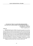
The p52 proteins have not previously been described in GH4C1 and GH3 cells [23,24]. We noticed an unphos- phorylated protein (designated p52n) with the same Mr value and a slightly higher pI value that was adjacent to p52a in the 2D gel analysis of cell lysates (left pan- els in Fig. 4A). To identify these p52 proteins, and because the amount of p52a, p52b and p52c was too low for direct purification and identification, peptide mapping analysis of p52n, p52a, p52b, and p52c, after in situ digestion with V8 protease (Fig. 4B), was performed. The phosphopeptide patterns of p52a, The level of phosphorylation of destrin and cofilin proteins did not differ significantly between the control and PKCe-overexpressing clones (data not shown), suggesting that PKCe is not involved in regulating the phosphorylation of these proteins.
A
B
CBB
acidic
32P
Silver 1 2 52n
Autoradiography Cont TRH 52a b 52a b c
basic Ly
Mr (x10-3) 97.4 66.2
a
b
n
a b c
V8
Ex
45 31 21.5
b
n a
a b c
14.4
Tp
a
b
a b
n
C
D
Blot (ks8.7)
Blot (ks8.7)
32P
32P
n
n
Cont
Lysate
a bcd
a bcd
a
b c
b
a
n
n
IP (PCK-26)
TRH
IgG
a bcd
a bcd
a
a
b c
b c
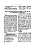
Fig. 4. p52 protein series are identified as K8. (A) Candidate unphosphorylated form (p52n) of p52 series. Whole lysates (Ly) from TRH-trea- ted cells (500 nM, 30 s) were prepared as described in Experimental procedures. Separation of Triton X-100-soluble (Ex) and Triton X-100- insoluble (Tp) fractions was performed in homogenization buffer containing 1% Triton X-100 for 5 min at 0(cid:2)C on culture dishes. The Ex fraction contained both the cytosol and Triton X-100-soluble particulate (membrane) fractions. The left panels show the profiles stained with Coomassie Brilliant Blue, and the right panels are the corresponding autoradiograms (32P). Arrowheads indicate the phosphorylated forms (a, b, and c) and arrows show the putative unphosphorylated form (n) of p52 proteins. p52c was visualized by silver staining (data not shown) but not by Coomassie Brilliant Blue staining. (B) Peptide mapping of p52 proteins after in situ digestion with V8 protease. Labeled cells were stimulated with 500 nM TRH for 30 s. The proteins in p52 spots were digested in situ with V8 protease at 50 ng ⁄ lane except for lane 2 (2.5 lg ⁄ lane) in the silver staining (left panel). Bars shown to the left indicate V8-derived bands as assessed from the electrophoretic pattern of silver staining of V8 alone (data not shown). Arrows show the p52n-derived bands comigrating with the phosphorylated peptides of p52a, p52b, and p52c. The smallest fragment in the silver-stained panel is a doublet, and its upper band coincides with the labeled peptide. Mr values are indicated on the right. Among the four fragments derived from the digestion of unphosphorylated p52n detected in lane 1 of silver stain, the two upper fragments disappear and the intensity of two smaller fragments increase with V8 digestion at a high concentration (lane 2). (C) Immunoblot analysis using antibody to K8. Left panels show the 2D profiles of p52 proteins from whole cell lysates visualized by antibody to K8 (ks8.7), and right panels show the corresponding autoradiograph (32P). Cells were treated with or without 500 nM TRH for 30 s. Arrowheads and arrows indicate the phosphorylated (a, b, and c) and the unphosphorylated form (n) of p52 proteins, respectively. (D) Immunoprecipitation of p52 proteins. 2D profiles of whole lysate (Lysate) from TPA-treated cells and the immunoprecipitate (IP) by antibody to keratins (PCK-26 reacts with multiple type II isozyme, as described in Experimental procedures) are visualized by immunostaining (ks8.7) and autoradiography (32P). Spots of heavy chains of IgG were derived from PCK-26, which cross-reacted with the secondary antibodies.
FEBS Journal 274 (2007) 3270–3285 ª 2007 The Authors Journal compilation ª 2007 FEBS
3274
Y. Akita et al.
TRH-PKCe-evoked K8 phosphorylation at Ser8 and 23
p52b, and p52c are similar, and they coincide with four bands of the silver-stained peptides resulting from the digestion of p52n (lane 1). These results suggest that p52a, p52b, and p52c are derived from the un- phosphorylated form (p52n) and that there are at least three phosphorylation sites located within the same fragments of the p52 proteins after digestion with V8 protease.
p52 proteins are identical with K8
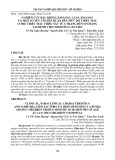
a decrease in the concentration of PKCe in the cytosolic fraction within 1 min (Fig. 5C), and that it increased in both the membrane and cytoskeletal fractions. The increase in membrane-bound PKCe peaked within 15 s (data not shown) and this was maintained for 120 min, whereas the cytoskeleton-bound form increased gradu- ally over 1 min. These data suggest that activated PKCe colocalized with unphosphorylated (p52n), monophos- phorylated (p52a) and diphosphorylated (p52b) forms of the K8 protein (Figs 4A and 5B,C). The increase in PKCe in both compartments of PKCe-overexpressing cells suggested that the increased p52 phosphorylation is due to increased PKCe activation.
(upper panels
To identify the p52 proteins, a p52n spot obtained from the detergent-insoluble fraction was subjected to digestion with lysyl endopeptidase. Peptides were puri- fied and subjected to amino-acid sequencing. The amino-acid sequences of three p52n peptide fragments were identical with those of rat K8 as deduced from the cDNA sequence (supplementary Fig. S1B) [27].
For further confirmation, immunoblot analyses were carried out. The results showed that an antibody speci- fic for K8 (Ks8.7) reacted with p52a, p52b, and p52c as well as p52n (left panels in Fig. 4C). Autoradiographic analysis showed that p52n is the unphosphorylated form of K8 (right panels in Fig. 4C). Moreover immuno- precipitation using antibody to keratins (PCK-26) again confirmed that p52n is identical with unphosphorylated K8 and that p52a, p52b, p52c and p52d are identical with the phosphorylated forms of K8 (Fig. 4D).
Immunofluorescence analysis (Fig. 5D) showed the subcellular localization of endogenous PKCe and K8. Under unstimulated conditions in Fig. 5D), PKCe staining was quite diffuse over the entire cell except the nuclear area, whereas K8 staining showed a dense area surrounding the nucleus and a weak area at the cellular periphery. After 25 s of TRH stimulation (lower panels in Fig. 5D), PKCe staining was concentrated at the cell periphery, whereas K8 staining became slightly diffuse around the nucleus and extended throughout the cytoplasm to the cell sur- face as in spreading cells. The image of the PKCe stain merged with the K8 stain suggests that PKCe and K8 colocalize at the cell periphery and cell–cell contact areas upon TRH stimulation. Both proteins seem to colocalize around the nucleus, with a dot-like staining pattern under basal conditions. The effect of TPA (data not shown) was similar to that of TRH. PKCe directly and physiologically phosphorylates K8
MS analysis of p52 phosphorylated region
three domains,
Rat K8 is composed of the head (amino acids 1–89), the rod (amino acids 90–397), and the tail (amino acids 398–482) (Fig. 6A). Eleven (S* ⁄ T*XK ⁄ R, K ⁄ putative phosphorylation motifs RXS* ⁄ T*, or K ⁄ RXXS* ⁄ T*) for PKC [29] exist within the N-terminal head domain of K8, four in the central a-helical rod domain, and five in the C-terminal tail domain. To identify the major sites phosphorylated by PKCe, we performed MS analysis.
We next immunoprecipitated K8 from unstimulated cells with the PCK-26 antibody, and subjected the to in vitro phosphorylation. As immunoprecipitates shown in Fig. 5A, phosphates were incorporated into p52a, p52b, and p52c but not p52n of K8 by purified recombinant human PKCe, indicating the direct phos- phorylation of these isoforms by PKCe. After TRH treatment of the cells, the majority of p52n remained in the cytoskeletal fraction (see Tp in the left panels of Fig. 4A), whereas p52c localized exclusively to the cytosolic fraction (sup, Fig. 5B). p52a was detected predominantly in the Triton-insoluble fraction (T-ppt in Fig. 5B) and p52b was distributed between the Triton-soluble (T-sup) and Triton-insoluble fractions. This observation suggested that p52 (K8) redistributed from the cytoskeletal to the cytosolic compartment in a manner that depended on the extent of its phos- phorylation [27,28].
FEBS Journal 274 (2007) 3270–3285 ª 2007 The Authors Journal compilation ª 2007 FEBS
3275
is The amount of PKCe associated with the membrane and cytoskeletal compartments regarded as an indicator of its activation. We observed that TRH led to First, K8 protein from TPA-treated cells was immu- noprecipitated with antibody PCK-26, and p52n, p52a, and p52b isoforms were isolated by 2D gel electro- phoresis. The phosphoserine residues of the proteins were converted into ethyl cysteine residues to enhance ionization of the phosphorylated peptide. Approxi- mately 70% of all K8 peptides were identified in each of p52n, p52a, and p52b samples by nano-LC-linked electrospray tandem MS (Q-TOF) (data not shown). Two phosphorylated peptides identified from the MS
Y. Akita et al.
TRH-PKCe-evoked K8 phosphorylation at Ser8 and 23
C
A
B
D
Fig. 5. K8 partially colocalizes with PKCe and acts as its substrate. (A) Purified PKCe directly phosphorylates K8. K8 immunoprecipitate from the unstimulated cells was phosphorylated by recombinant PKCe in the presence of TPA and [c-32P]ATP. The 2D profiles shown are visual- ized by silver staining (upper panels) and by the corresponding autoradiograph (lower panels). Arrowheads, arrows, and IgG shown are described in the legend to Fig. 4C,D. (B) Localization and solubility of three p52 phosphoproteins depend on the phosphorylated level upon TRH stimulation. Whole lysates (Lysate) from TRH-treated cells (500 nM, 30 s) were separated into the cytosol (sup), particulate (ppt), Triton X-100-soluble particulate (T-sup), and Triton X-100-insoluble particulate (T-ppt) fractions. Results shown are autoradiograms (32P). (C) Redistri- bution of PKCe from the cytosol to the membrane and cytoskeleton. Wild-type cells (W), vector-expressing control (S1), and PKCe clones (e4 and e13) were treated with TRH for the indicated times, separated by SDS ⁄ PAGE, and analyzed by immunoblotting using antibodies to PKCe. Arrowheads indicate the position of PKCe, as assessed by comigration with PKCe overproduced in COS cells (data not shown). The bands that react with PKCe antibodies are often visualized as doublets or triplets. (D) Subcellular localization of PKCe and keratin filaments. Cells were incubated with or without 500 nM TRH for 25 s, double-immunostained with antibodies to PKCe (C-15) (green) and keratins (PCK- 26) (red), and analyzed by a confocal laser scanning microscope. Focal sections, 0.3 lm apart, were scanned. Bars indicate 10 lm. Arrows and arrowheads indicate partial colocalization (yellow) of PKCe with K8 at the cellular periphery and the cell–cell contact area, respectively.
FEBS Journal 274 (2007) 3270–3285 ª 2007 The Authors Journal compilation ª 2007 FEBS
3276
analysis of the diphosphorylated isoform, p52b, were identified as corresponding to the fragments (amino acids 4–10 and 23–31) termed T2-3 and T6, respect- ively (Fig. 6B,C). The phosphorylated T6 and T5-6 (amino acids 18–31) fragments were also observed in the analyses of p52a and p52b but not p52n isolated
Y. Akita et al.
TRH-PKCe-evoked K8 phosphorylation at Ser8 and 23
A
B
C
D
Fig. 6. MS analysis of phosphorylated sites of K8. (A) Schematic presentation of K8 structure. Amino-acid sequence shown in the lowest panel indicates the head domain of K8. Double strands shown indicate the positions of the sequences identified by amino-acid sequencing. Single and double broken lines shown indicate the positions of C1, T2-3, T5-6, T6 and T8 fragments described in the text, and of the syn- thesized peptide (K853), respectively. Arrowheads (black and gray) indicate the phospho-sites detected on K853 polypeptide, and black sym- bols indicate the sites confirmed to be endogenously phosphorylated after TRH stimulation (see Fig. 7). Underlines and asterisks of letters indicate the putative motifs and sites for PKC, respectively. (B) P52n and p52b proteins derived from TPA-treated cells were analyzed as described in Experimental procedures. Mass chromatogram shown was surveyed with the theoretical mass (with permissible range of 0.1 Da) for the unphosphorylated T2-3 (T2-3) and T6 (T6), and the phosphorylated T2-3 (T2-3+P) and T6 (T6+P) peptides. Data of the signal for the phosphorylation (upper panels) were obtained in two trypsinized peptides derived from the p52b spot, whereas those for the unphosphorylated peptides (lower panels) were obtained in the p52n peptides. (C) Mass spectrum of the peaks (T2-3+P and T6+P) shown in the upper panel of (B). (D) Tandem MS analysis of PKCe in vitro phosphorylated sites of a synthesized polypeptide (K853) (amino acids 1–53) of K8. A mixture of diphosphorylated and triphosphorylated K853 peptides by PKCe was digested with chymotrypsin or trypsin. The digested fragments were separated by nano-LC and analyzed by MALDI TOF ⁄ TOF. The upper panel shows the product ion spectrum of the precursor fragment (C1+P) with 1147.58 of mass value, which was derived from chymotrypsinized phospho-K853, indicating the amino acid sequence, SVRVTQKpSY. Lower panel shows the product ion spectrum of the precursor fragment (T5-6+P) with mass value of 1507.69, which was derived from partially trypsinized phospho-K853, indicating the amino-acid sequence, AFSSRpSFTSGPGAR. The b and y ion series with asterisk and ⁄ or plus letters indicate a 98-Da neutral-loss ion and ⁄ or a 18-Da peeling sequence ion, respectively.
FEBS Journal 274 (2007) 3270–3285 ª 2007 The Authors Journal compilation ª 2007 FEBS
3277
Y. Akita et al.
TRH-PKCe-evoked K8 phosphorylation at Ser8 and 23
the phosphorylated isoforms total concentration of detected by the a-19 and a-21 antibodies by approxi- mately 2.5-fold and twofold, respectively. However, the antibodies to phospho-Ser46 (a-47) did not react with any isoforms (data not shown).
from the PKCe-overexpressing clone (e4) after treat- ment with TRH: instead, the unphosphorylated T6 fragments decreased or disappeared (data not shown). We could not detect phosphorylated peptides corres- ponding to the rod domain of K8. These results sug- gest that the p52b isoform is phosphorylated at two sites located within the amino-acid sequences 4–10 and 23–31 of K8.
that
After TRH stimulation of wild-type cells, the relat- ive proportion (p52a ? p52b) of the levels of p52 isoforms reflected recognized by a-21 antibodies closely the relative proportion of the total amount of p52 phosphorylated proteins recognized by the Ks8.7 antibody, which reacted with both the unphosphoryl- ated and phosphorylated forms of K8 protein. In contrast, the relative concentrations of the p52a and p52b isoforms recognized by the a-19 antibodies were almost equal. In other words, the concentration of p52a detected by a-19 was considerably lower than that expected from the total amount of p52a protein detected by Ks8.7 and, in contrast, the concentration of p52b was comparable to that expected. These the monophosphorylated form, results suggest p52a, has heterogeneity at the phosphorylated site and is phosphorylated mainly at Ser23 and to a lesser extent at Ser8, and that the diphosphorylated form, p52b, is phosphorylated equally at Ser8 and Ser23 (Fig. 7B).
Next, we synthesized a polypeptide (K853), amino acids 1–53 of K8, including the sequences of the T2-3 and T6 fragments, and examined the sites of the poly- peptide phosphorylated in vitro by PKCe. A mixture of diphosphorylated and triphosphorylated K853 polypep- tides purified by HPLC was digested with chymotrypsin or trypsin. The resultant fragments were analyzed by nano-LC off-line coupled MALDI tandem MS (4700 TOF ⁄ TOF). As shown in the upper panel in Fig. 6D, the product ion spectrum of the precursor fragment, termed C1+P and with a mass value of 1147.58, was obtained in the analysis of the chymotrypsinized fragments. In this spectrum, two fragment ions, y2–98 and b8–98, with a 98-Da neutral-loss of mass value corresponding to the H3PO4 ion and a 18-Da peeling sequence ion, b8+H2O, were observed, indicating the amino-acid sequence, SVRVTQKpSV, corresponding to amino acids 1–9 of phosphorylated K8 at Ser8. The lower panel in Fig. 6D showed the product ion spectrum of the precursor fragment, termed T5-6+P with a mass value of 1507.69, which was observed in the analysis of the partially trypsinized fragments. In this spectrum, two fragment ions, y9–98 and b6–98, with a 98-Da neut- ral-loss ion and two fragment ions, y9 and a6, with no loss of mass value were detected, indicating that the amino-acid sequence, AFSSRpSFTSGPGAR, corres- ponded to amino acids 18–31 of phosphorylated K8 at Ser23. Moreover, the analysis of K853 polypeptide showed that an additional major site in vitro phosphor- ylated by PKCe was Ser46 and that the minor site was Ser12 (data not shown).
PKCe phosphorylates Ser8 and Ser23 on K8 protein
In the PKCe-overexpressing clone (e4) under basal conditions (right panels in Fig. 7A), the 2D profiles of p52 isoforms detected by the a-19 and a-21 antibodies closely mimicked those observed in THR-stimulated wild-type cells, and the characteristic equal proportion of p52a and p52b recognized by a-19 was also clear. These data suggest that increased activation of PKCe as a result of its overexpression results in the phosphoryla- tion of Ser8 and Ser23 (preferentially Ser8) on K8. After TRH stimulation of e4, the total concentrations of p52a, p52b, and p52c recognized by a-19 increased slightly (1.5-fold). However, the amount detected by a-21 did not change, suggesting that Ser23 on K8 was fully phosphorylated in the PKCe-overexpressing cells under basal conditions. The relatively high concentra- tion of p52b detected with a-21 (p52a > p52b ? p53c) suggests that the Ser23-phosphorylated form of p52a was the preferential substrate of PKCe and was conver- ted into p52b. p52c clearly increased in both cases in which a-19 and a-21 were used.
Discussion
K8 is a major physiological substrate of PKCe in vivo
FEBS Journal 274 (2007) 3270–3285 ª 2007 The Authors Journal compilation ª 2007 FEBS
3278
The direct stimulation of cPKC and nPKC isozymes by phorbol ester increased K8 phosphorylation in We examined whether endogenous K8 in the cells was phosphorylated at Ser8, Ser23, and Ser46 via PKCe activation upon TRH stimulation using phospho-speci- fic antibodies generated against phospho-Ser8 (a-19), phospho-Ser23 (a-21), and phospho-Ser46 (a-47). The specific antibodies to phospho-Ser8 (a-19) and phos- pho-Ser23 (a-21) reacted with all the phosphorylated species (p52a, p52b, and p52c) of K8 in the basal and TRH-stimulated cells (Fig. 7A). TRH stimulation of wild-type cells (left panels in Fig. 7A) increased the
Y. Akita et al.
TRH-PKCe-evoked K8 phosphorylation at Ser8 and 23
A
ε4
Wild
Ks8.7
a-19
a-21
Ks8.7
a-19
a-21
None
TRH
K8 n a b c a b c a b c
n a b c a b c a b c
TRH
TRH
B
P S8
P
S8 S
PKCε
P
P
PKCε Other kinase
P
S23
S23
K8
P
S8
PPA
PPA
P S23
S
S
P
P
p52a
p52b
p52n
Fig. 7. (A) Immunoblot analysis of TRH-stimulated cells by antibodies to phospho-Ser8 and phospho-Ser23. Wild-type (left panels) and PKCe- overexpressing (right panels) cells were treated with (TRH) or without (None) 500 nM TRH for 25 s, and K8 immunoprecipitates from each cell were separated by 2D electrophoresis. The immunoblotted membranes were probed with antibodies to phospho-Ser8 (a-19). The same membrane was reprobed with antibodies to phospho-Ser23 (a-21), and again reprobed with antibody to K8 (ks8.7). The total concentration of phospho-Ser8 and phospho-Ser23 normalized to the total amount of protein detected by ks8.7 increased by 2.5 ± 0.2-fold (mean ± SE, n ¼ 4) and 2.0 ± 0.1-fold (n ¼ 4), respectively, in the wild-type cells and 1.5 ± 0.2-fold (n ¼ 6) and 0.9 ± 0.1-fold (n ¼ 5), respectively, in the PKCe-overexpressing cells after TRH stimulation. (B) Schematic representation of the model of K8 protein phosphorylation via activation of PKCe. In GH4C1 cells, phosphorylated K8 isoforms, at least p52a and p52b, have heterogeneous phosphorylated sites. The monophosphoryl- ated form (p52a) is composed of a major and a minor phosphorylated site, Ser23 and Ser8, respectively, and probably even the rare Ser12 or other site(s) as yet undetected. PKCe has the ability to phosphorylate Ser8 and Ser23 in the unphosphorylated (p52n) and phosphorylated (p52a and p52b) isoforms of K8. However, activation of PKCe after TRH stimulation of the cells predominantly enhances the conversion of the monophosphorylated isoform (p52a) into the diphosphorylated isoform (p52b), in particular because of preferential phosphorylation of Ser8 on the Ser23-phosphorylated p52a species. Besides PKCe activation, TRH seems to activate phosphatase(s) and thereby dephosphory- lates p52b and p52c, as the transient increase in the concentrations of p52b and p52c upon TRH stimulation was sustained in the presence of okadaic acid, an inhibitor of serine ⁄ threonine phosphatase (data not shown).
from the cytosol
although
FEBS Journal 274 (2007) 3270–3285 ª 2007 The Authors Journal compilation ª 2007 FEBS
3279
several intact cells [27,30]. In unstimulated epithelial HT29 cells, PKCe or PKCe-like kinase also phos- phorylates K8 [27]. Recently, it has been proposed that, after shear stress generated in a mechanically ventilated lung, PKCd is activated and phosphorylates K8 at Ser73 [31]. However, little is so far known of the physiological stimulus–coupling phosphorylation of K8 via specific activation of PKC isozymes. In this study, we have shown that K8 phosphorylation is regulated through the activation of PKCe in response to extracellular hormonal stimuli under physiological conditions, as follows. First, PKCe immediately and to the sustainedly redistributes plasma membrane and cytoskeleton upon exposure of GH4C1 cells to TRH (Fig. 5C), resulting in exclusive down-regulation, other PKC isozymes (PKCa, PKCbII and PKCd) expressed in the cells transiently redistribute to the membrane and ⁄ or cytoskeleton [19,22]. This phenomenon suggests that PKCe is strongly activated and mainly involved in TRH-evoked signaling [13,19]. Secondly, the time course of the increase in K8 phosphorylation upon TRH stimulation is consistent with that of PKCe acti- vation. Thirdly, the direct activator, TPA, and inhib- itor, GF, of PKC increase and decrease, respectively, phosphorylation of K8, p52a, p52b, and p52c (Fig. 2). Fourthly, purified PKCe directly phosphory- lates endogenous K8 and the Ser8 and Ser23 residues on the K8 polypeptide (K853) in vitro (Figs 5A and 6). Fifthly, PKCe and K8 coimmunoprecipitate (data not shown) and colocalize (Fig. 5D) under basal and TRH-stimulated conditions with different patterns. Sixthly, the 2D profile of the monophospho (p52a) and diphospho (p52b) isoforms of K8 in TRH-stimu- lated wild-type cells and the increases in the relative proportions of phospho-Ser8 and phospho-Ser23 are quite similar to those in PKCe-overproducing cells (Figs 1, 3 and 7A).
Y. Akita et al.
TRH-PKCe-evoked K8 phosphorylation at Ser8 and 23
PKCe primarily phosphorylates Ser8 of K8 upon TRH stimulation
[38]. Reorganization of suggests antibodies
catalyzes the
The relative concentration of p52b, the diphosphoryl- ated form of K8, showed the greatest change of the three phosphorylated forms in our PKCe-overproduc- ing cells under both basal and TRH-stimulated condi- tions (Fig. 3). A similar effect is shown in wild-type cells treated with PKC activators (Figs 1 and 2A). These observations suggest that the monophosphoryl- ated form, p52a, is the preferential substrate for PKCe. Immunoblot analysis of wild-type cells using phos- that, under phoserine-specific physiological conditions, the majority of p52a is phos- phorylated at Ser23, and the majority of p52b, which increases upon TRH stimulation, is phosphorylated at Ser8, as well as at Ser23 (Fig. 7A). On the basis of in which PKCe these results, we propose a model predominantly conversion from a monophosphorylated form (p52a) as the physiological substrate to a diphosphorylated form (p52b) (Fig. 7B). We also suggest that phosphorylation of p52a and p52b at minor sites such as Ser12 rather than Ser8 occurs under nonphysiological conditions that strongly activate PKCe, as observed in PKCe-overproducing cells upon TRH stimulation.
ted cells [19]. The time course of K8 phosphorylation shown in Fig. 1B is highly consistent with that of pro- lactin secretion shown previously; the transient rapid increase in p52b and p52c isoforms agrees with the first burst of secretion, and the sustained increase in p52a with the second sustained secretion. Secretion of bile acid from hepatocytes has been reported to decrease in K8 null mice, and this is accompanied by an abnormal pattern of F-actin [37]. The keratin inter- mediate filament (IF) network appears to cross-talk with the actin network through several common molecular components the actin network is thought to be required in various secretory processes. We have shown that TRH changes the phosphorylation levels of cofilin ⁄ destrin and MARCKS in GH4C1 cells (Fig. 1), which have been proposed to regulate the actin network in a manner that is dependent on phosphorylation [39,40]. More- over, we have observed that K8 is associated with b-actin and that PKCe is associated with K8 and 14-3-3 proteins in a coimmunoprecipitation assay of the PKCe-overproducing cells (data not shown). 14-3-3 proteins, which are able to associate with K8 ⁄ 18 filaments [28], have also been proposed to mediate reg- ulatory secretion [41]. Therefore, it is conceivable that TRH-induced phosphorylation of K8 helps to orches- trate prolactin secretion by influencing the reorganiza- tion of the actin network.
line [43]. We observed that
and morphological changes
Many kinases, such as cAMP-dependent protein kin- ase (protein kinase A) and mitogen-activated protein kinase, are able to phosphorylate a number of serine residues on K8 in vitro, mainly in the N-terminal head domain [30,32]. In vivo, mitogen-activated protein and cdc2 kinases have been proposed to phosphorylate Ser431 located in the C-terminal tail domain of human K8 in response to epidermal growth factor and during mitotic arrest, respectively [27,33]. Stress, apoptosis, and mitosis appear to induce the phosphorylation of Ser73 in human K8 [33]. Ser73 phosphorylation was also observed before the development of Mallory bod- ies in alcoholic hepatitis and cirrhosis [34]. Ser23 of K8 is conserved among all type II keratins and its phosphorylation has been proposed previously, but how signaling and kinase participate in this process remains unknown [35]. Protein kinase A has been reported to phosphorylate the site in vitro [36]. We showed that PKCe phosphorylates Ser8 and Ser23 located in the first half of the N-terminal head domain in vivo through hormonal stimulation.
Phosphorylation of K8 has been implicated in the reorganization of the intermediate cytoskeleton during cellular proliferation [27,42]. The in vitro phosphoryla- tion of K8 filaments by a mixture of PKC isozymes including PKCe from the brain has been shown to lead to the disassembly of the K8 ⁄ 18 filament structure [30]. TRH has been reported to cause transient reor- ganization of cytoskeletal keratin filaments in GH3B6 cells, a GH4C1-related line established from the same the parental GH3 cell TRH-stimulated increase in the solubility of K8 iso- forms depends on the state of phosphorylation (Figs 4A and 5B). We have also preliminarily observed that a dense zone of keratin around the nucleus gradu- ally moves away and spreads to the cell periphery in response to TRH stimulation accompanied by cell spreading (data not shown), which is consistent with previous data [43]. Therefore, the phosphorylation of K8 via TRH-medi- ated PKCe activation may play a role in dynamic keratin IF reorganization in GH4C1 cells.
K8 protein mediates TRH signaling via PKC activation
FEBS Journal 274 (2007) 3270–3285 ª 2007 The Authors Journal compilation ª 2007 FEBS
3280
PKCe and K8 proteins were shown to colocalize at the cell–cell contact area, possibly on desmosomes, upon TRH stimulation of the cells (Fig. 5). It is well known that the keratin IF network bridges desmo- Our previous study showed that PKCe is involved in the biphasic secretion of prolactin from TRH-stimula-
Y. Akita et al.
TRH-PKCe-evoked K8 phosphorylation at Ser8 and 23
of Medical Science). PKCe-overexpressing stable GH4C1 clones and vector control clones were as previously des- cribed [19].
Preparation of antibodies against phospho-K8
Phosphopeptides (CRVTQKpSYKMST, CAFSSRpSF- TSGP, and CGSSSSpSFRGSL) including the amino-acid residues 3–13, 18–28, and 41–51, respectively, of K8 were synthesized with a peptide synthesizer (Applied Biosystems Ltd, Foster City, CA, USA), and used to immunize rabbits. The antiserum obtained was affinity-purified using the phosphopeptide and absorbed with the corresponding non- phosphopeptides as described previously [47]. The speci- ficities of these phospho-specific (pS8-K8, pS23-K8, and pS46-K8) antibodies were confirmed by dot and western blotting of the nonphosphopeptides, phosphopeptides and lysates, respectively (supplementary Fig. S2). GH4C1 cell All animal experiments were approved by the committee of the Tokyo Metropolitan Institute of Medical Science.
somes under the apical plasma membrane in epithelial cells. PKCe has been implicated in the control of inter- cellular adhesion [44]. Evidence is accumulating that the K8 protein functions in targeting and positioning of various membrane-associated, cell–cell contact, and cytosolic proteins, including desmosome complex pro- teins such as trichoplein and desmoplakin [38,45]. These data, taken together, raise an interesting ques- tion: does K8 phosphorylation via PKCe activation play a role in cell–cell contact?
In vivo and in vitro phosphorylation
In simple glandular and secretory epithelial cells such as those of the pituitary gland and liver, K8 is specifically produced as a major keratin heteropolymer paired with keratin 18 which comprises a keratin cyto- skeletal network. Although valuable information on the roles of K8 in several tissues and cells is accumula- ting [27,37], there is very little knowledge about its role in the pituitary gland. Inclusion bodies of K8 known as ‘fibrous bodies’ have been observed in malignant human pituitary adenomas that produce growth hor- mone [46], and keratin IF reorganization has been reported in TRH-stimulated cells [43]. The present study shows that a hypothalamic hormone, TRH, enhanced the phosphorylation of the cytoskeletal IF protein, K8 (p52), via activation of PKCe in GH4C1 cells. This reveals a potential role for K8 in hormone- activated PKCe signaling.
Finally, to confirm the phosphorylation series of K8 by PKCe, we are trying to generate mutant molecules of K8. Further experiments using these mutants should help us to understand more precisely the dynamics of the phosphorylation and to elucidate its function in the pituitary gland.
Experimental procedures
In vivo 32P labeling of cells was as previously described [13]. Briefly, cells were cultured on 3-cm or 10-cm dishes in Ham’s F-12 medium supplemented with 0.5% fetal bovine serum for 2 days before use. After preincubation in phos- phate-free DMEM containing 0.5% dialyzed fetal bovine serum, the cells were labeled with 0.3 mCiÆmL)1 32Pi and then treated with 160 nm TPA or 500 nm TRH. Cells were harvested and sonicated in homogenization buffer (20 mm Tris ⁄ HCl, pH 7.5, 0.25 m sucrose, 2.5 mm MgCl2, 2.5 mm EGTA, 50 mm 2-mercaptoethanol, 100 lgÆmL)1 leupeptin, 2 mm phenylmethanesulfonyl fluoride, 2 lgÆmL)1 aprotinin, 50 mm NaF, and 100 lm Na3VO4) [22]. The samples were solubilized in 2D lysis buffer (9 m urea, 2% CHAPS, 2.5% 2-mercaptoethanol, and 2% ampholines).
For subcellular fractionation, cell lysates were separated into supernatant (cytosol) and particulate (membrane ⁄ cyto- skeletal) fractions by ultracentrifugation at 350 000 g for 20 min using an Optima TL rotor, TLA45, Beckman (Palo Alto, CA, USA) [13]. Particulate fractions were resuspended in homogenization buffer containing 1% Triton X-100 and incubated at 4 (cid:2)C for 1 h. After centrifugation, Triton X- 100-soluble (membrane) and Triton X-100-insoluble (cyto- skeletal) particulate fractions were prepared.
For in vitro phosphorylation, K8 immunoprecipitate was isolated from untreated cells and washed in a kinase buffer (50 mm Tris ⁄ HCl, pH 7.5, 1.25 mm EDTA, 1.25 mm EGTA, 50 mm 2-mercaptoethanol, 100 lgÆmL)1 leupeptin, 2 mm phenylmethanesulfonyl fluoride, 1 lgÆmL)1 okadaic acid, 0.1 mgÆmL)1 BSA, and 8% glycerol). Endogenous kinase activity was abolished by heating samples at 90 (cid:2)C for 1 min. The kinase reaction was carried out using a
[32P]Orthophosphoric acid (carrier-free) and [c-32P]ATP were purchased from DuPont-New England Nuclear (Boston, MA, USA). GF109203X and recombinant PKCe were purchased from Calbiochem Corp (La Jolla, CA, USA). TRH, TPA and V8 protease were purchased from Sigma (St Louis, MO, USA), Avanti Polar-Lipids (Bir- mingham, AL, USA), and Boehringer Mannheim GmbH (Mannheim, Germany), respectively. A monoclonal anti- body (PCK-26) to keratins 5, 6, and 8 and a specific anti- body (Ks8.7) to K8 were obtained from Sigma and Progen Biotechnic GmbH (Heidelberg, Germany), respectively. Antibodies to PKC isozymes and their specificities have been described previously [19]. Monoclonal antibody against cofilin or destrin was kindly provided by Drs K. Iida and K. Moriyama (The Tokyo Metropolitan Institute
FEBS Journal 274 (2007) 3270–3285 ª 2007 The Authors Journal compilation ª 2007 FEBS
3281
Materials
Y. Akita et al.
TRH-PKCe-evoked K8 phosphorylation at Ser8 and 23
(GE Healthcare, Little Chalfont, UK) PKC assay kit according to the manufacturer’s instructions. Then 5 lCi [c-32P]ATP, 100 lm ATP, 6 mm MgCl2, and 0.5 U recom- binant purified PKCe were added to the reaction mixture. After incubation for 15 min at 30 (cid:2)C, the reaction was stopped and analyzed by 2D gel electrophoresis.
Immunoprecipitation
Cells were treated similarly to the in vivo phosphorylation assay except for the addition of unlabeled H3PO4 instead of [32P]orthophosphate. Cells were solubilized (2 h, 4 (cid:2)C) with 1% Empigen in an immunoprecipitation buffer (20 mm Tris ⁄ HCl, pH 7.5, 0.15 m NaCl, 1 mm EDTA, 1 mm EGTA, 1.5 mm MgCl2, 1 mm dithiothreitol, 2 mm phenyl- methanesulfonyl fluoride, 0.01% leupeptin, 4 lgÆmL)1 apro- tinin, 50 mm NaF, 100 lm Na3VO4, and 0.5 lgÆmL)1 okadaic acid. After centrifugation (100 000 g, for 15 min using an Optima TL rotor, TLA45; Beckman), K8 was immunoprecipitated from the resultant supernatant using keratin antibodies (PCK-26) that were conjugated to Affigel 15 (Bio-Rad, Hercules, CA, USA) according to the manu- facturer’s instructions.
Two-dimensional gel electrophoresis
Nonequilibrium IEF gel electrophoresis [48] was performed because better separation and reproducibility of p52 proteins are obtained by this method than by IEF gel elec- trophoresis. The gels contained 2% total ampholines pH 5–8, 4–6, and 3.5–9.5 in the proportions 2 : 2 : 1, rang- ing from pH 4.2 to 7.5. Each sample derived from an equiv- alent number of cells was separated on mini-rectangle gels (Atto, Tokyo, Japan) at a total of 1700 V-h. The second dimension was SDS ⁄ PAGE (12.5% gels). The gels were stained with Coomassie Brilliant Blue and autoradio- graphed. Phosphoprotein was quantified using a Phosphor- Imager Bas2000 System (Fuji, Tokyo, Japan) and phoretix 2D advanced software (Nonlinear Dynamics Ltd, Newcas- tle-upon-Tyne, UK). Statistical significance was determined by Student’s t-test.
For preparative 2D gel electrophoresis, the first dimen- sion was carried out on a 3 mm diameter disk gel, and the second dimension was SDS ⁄ PAGE (9.5% gel).
Immunofluorescence staining
Cells (5 · 104) were seeded on to 3.5-cm poly lysine-coated glass-bottom dishes (MatTek Corp, Ashland, MA, USA), grown for 5 days, and then incubated in Ham’s F-12 med- ium supplemented with 0.5% fetal bovine serum for 2 days before use. Cells were treated with TPA or TRH in F-12 medium containing 0.5% dialyzed fetal bovine serum. Trea- ted cells were fixed with 4% paraformaldehyde in NaCl ⁄ Pi for 30 min at room temperature, permeabilized with 0.2% Triton X-100 and 10% goat serum in NaCl ⁄ Pi for 30 min at room temperature, double-stained with PKCe antibodies (C-15; Santa Cruz Biotechnology, Inc., Santa Cruz, CA, USA) and keratin antibody (PCK-26) at 4 (cid:2)C overnight, and then visualized with Alexa Fluor 488-conjugated anti- rabbit IgG (Molecular Probes, Carlsbad, CA, USA) for PKCe and Texas Red-conjugated anti-mouse IgG for keratin as described elsewhere [19]. Immunostained cells were examined by a confocal laser scanning microscope (LSM510META; Carl Zeiss Co., Ltd, Heidelberg, Ger- many) using a ·100 objective.
The p52 protein series were excised from the 2D gels and digested in situ with Staphylococcus aureus V8 prote- ase as previously described [49]. Briefly, gel pieces were loaded into a well in a 15% acrylamide gel, and 50 ng V8 protease was applied. When the material reached the stacking ⁄ separating gel interface, the current was turned off to allow the enzyme to digest the protein in situ, and then electrophoresis was completed. Proteolytic fragments were visualized by silver staining, and the gels were auto- radiographed.
Peptide mapping
MS analysis
K8 immunoprecipitate was subjected to preparative 2D gel electrophoresis, and the p52n, p52a, p52b, and p52c protein spots were visualized with Coomassie Brilliant Blue stain and excised from the gels. For detection of phosphorylated peptides in the proteins, phosphoserine residues were con- verted into ethyl cysteine by treatment of gel pieces with ethanethiol as described by Fadden & Haystead [50]. The increase in theoretical mass of a phosphoserine-containing peptide by this procedure as compared with a serine- containing peptide is the [M + H] mass of 44.01 Da, [M +2H] of 22.01 Da, or [M +3H] of 14.66 Da. After in-gel digestions were washing and drying the gel pieces,
Proteins separated by 2D gel electrophoresis were blotted on to a Clearblot-P membrane (Atto) [19]. The membranes were probed with K8 monoclonal antibody (ks8.7), and incubated with alkaline phosphatase-conjugated secondary antibodies. K8 was subsequently visualized with ABC kit (Vector Laboratories, Burlingame, CA, USA) as recommen- ded by the manufacturer. The membranes probed with phos- pho-K8 (pS8-K8) or (pS23-K8) antibodies were incubated with peroxidase-conjugated secondary antibody (GE Health- care), and the blots were visualized with an ECL-Plus kit (GE Healthcare).
FEBS Journal 274 (2007) 3270–3285 ª 2007 The Authors Journal compilation ª 2007 FEBS
3282
Immunoblot analysis
Y. Akita et al.
TRH-PKCe-evoked K8 phosphorylation at Ser8 and 23
6 Nishizuka Y & Kikkawa U (2003) Early studies of pro- tein kinase C: a historical perspective. Methods Mol Biol 233, 9–18.
7 Castrillo A, Pennington DJ, Otto F, Parker PJ, Owen
performed with trypsin in 50 mm ammonium bicarbonate at 37 (cid:2)C overnight. Aliquots of the digested peptides were analyzed by nano-liquid chromatography (nano-LC) coupled with an electrospray ionization mass spectrometer (nanoLC- coupled Q-Tof2; Micromass UK Ltd, Manchester, UK) as recommended by the manufacturer.
For
MJ & Bosca L (2001) Protein kinase Cepsilon is required for macrophage activation and defense against bacterial infection. J Exp Med 194, 1231–1242.
8 Hodge CW, Mehmert KK, Kelley SP, McMahon T,
synthesizer
Haywood A, Olive MF, Wang D, Sanchez-Perez AM & Messing RO (1999) Supersensitivity to allosteric GABA (A) receptor modulators and alcohol in mice lacking PKCepsilon. Nat Neurosci 2, 997–1002.
9 Khasar SG, Lin YH, Martin A, Dadgar J, McMahon
identification of phosphorylated sites of K8, a polypeptide (K853: amino acids 1–53) was synthesized with (Applied Biosystems Ltd) and a peptide phosphorylated in vitro with recombinant purified PKCe. A mixture of diphosphorylated and triphosphorylated K853 peptides was purified by HPLC and digested with trypsin or chymotrypsin. Aliquots of the digested peptides were separated by nano-LC and analyzed with a matrix-assisted laser desorption ionization tandem time-of-flight mass spectrometer (4700 MALDI TOF ⁄ TOF; Applied Biosys- tems Ltd).
T, Wang D, Hundle B, Aley KO, Isenberg W, McCarter G, et al. (1999) A novel nociceptor signaling pathway revealed in protein kinase C epsilon mutant mice. Neuron 24, 253–260.
Acknowledgements
10 Jansen AP, Verwiebe EG, Dreckschmidt NE, Wheeler DL, Oberley TD & Verma AK (2001) Protein kinase C-epsilon transgenic mice: a unique model for meta- static squamous cell carcinoma. Cancer Res 61, 808–812.
11 Dorn GW & 2nd & Mochly-Rosen D (2002) Intracellu- lar transport mechanisms of signal transducers. Annu Rev Physiol 64, 407–429.
12 Fujise A, Mizuno K, Ueda Y, Osada S, Hirai S, Taka- yanagi A, Shimizu N, Owada MK, Nakajima H & Ohno S (1994) Specificity of the high affinity interaction of protein kinase C with a physiological substrate, myr- istoylated alanine-rich protein kinase C substrate. J Biol Chem 269, 31642–31648.
13 Akita Y, Kawasaki H, Ohno S, Suzuki K &
We thank Dr Ken-ichi Arai (University of Tokyo, Japan) for valuable suggestions and encouragement, Dr T. Toda (Tokyo Metropolitan Institute of Geron- tology, Japan) for helpful discussion, and Drs Miyata (Kyoto University, Japan) and K. Mizuno (Yokohama City University School of Medicine, Japan) for techni- cal advice. We also thank Ms N. Kagi (Jasco Interna- tional Co., Tokyo, Japan) for technical support with the mass spectrometer. This work was supported by research grants from the Ministry of Education, Sci- ence, Sports, and Culture of Japan.
References
Kawashima S (2000) Involvement of protein kinase C epsilon in thyrotropin-releasing hormone-stimulated phosphorylation of the myristoylated alanine-rich C kinase substrate in rat pituitary clonal cells. Electrophoresis 21, 452–459.
1 Akita Y (2002) Protein kinase C-epsilon (PKC-epsilon): its unique structure and function. J Biochem (Tokyo) 132, 847–852.
2 Ohno S, Akita Y, Konno Y, Imajoh S & Suzuki K
(1988) A novel phorbol ester receptor ⁄ protein kinase, nPKC, distantly related to the protein kinase C family. Cell 53, 731–741.
3 Ono Y, Fujii T, Ogita K, Kikkawa U, Igarashi K &
14 Moriya S, Kazlauskas A, Akimoto K, Hirai S, Mizuno K, Takenawa T, Fukui Y, Watanabe Y, Ozaki S & Ohno S (1996) Platelet-derived growth factor activates protein kinase C epsilon through redundant and inde- pendent signaling pathways involving phospholipase C gamma or phosphatidylinositol 3-kinase. Proc Natl Acad Sci USA 93, 151–155.
15 Shirai Y, Kashiwagi K, Yagi K, Sakai N & Saito N
Nishizuka Y (1988) The structure, expression, and prop- erties of additional members of the protein kinase C family. J Biol Chem 263, 6927–6932.
(1998) Distinct effects of fatty acids on translocation of gamma- and epsilon-subspecies of protein kinase C. J Cell Biol 143, 511–521.
16 Nillni EA & Sevarino KA (1999) The biology of pro-
4 Newton AC (2001) Protein kinase C: structural and spatial regulation by phosphorylation, cofactors, and macromolecular interactions. Chem Rev 101, 2353–2364.
5 Akita Y, Ohno S, Konno Y, Yano A & Suzuki K
thyrotropin-releasing hormone-derived peptides. Endocr Rev 20, 599–648.
17 Wilber JF & Spinella P (1984) Identification of immu- noreactive thyrotropin-releasing hormone in human neoplasia. J Clin Endocrinol Metab 59, 432–435.
(1990) Expression and properties of two distinct classes of the phorbol ester receptor family, four conventional protein kinase C types, and a novel protein kinase C. J Biol Chem 265, 354–362.
FEBS Journal 274 (2007) 3270–3285 ª 2007 The Authors Journal compilation ª 2007 FEBS
3283
Y. Akita et al.
TRH-PKCe-evoked K8 phosphorylation at Ser8 and 23
conventional cPKC and novel nPKC. Adv Enzyme Regul 31, 287–303.
30 Yano T, Tokui T, Nishi Y, Nishizawa K, Shibata M,
18 Gershengorn MC (1986) Mechanism of thyrotropin releasing hormone stimulation of pituitary hormone secretion. Annu Rev Physiol 48, 515–526.
Kikuchi K, Tsuiki S, Yamauchi T & Inagaki M (1991) Phosphorylation of keratin intermediate filaments by protein kinase C, by calmodulin-dependent protein kinase and by cAMP-dependent protein kinase. Eur J Biochem 197, 281–290.
19 Akita Y, Ohno S, Yajima Y, Konno Y, Saido TC, Miz- uno K, Chida K, Osada S, Kuroki T, Kawashima S & Suzuki K (1994) Overproduction of a Ca(2+)-independ- ent protein kinase C isozyme, nPKC epsilon, increases the secretion of prolactin from thyrotropin-releasing hormone-stimulated rat pituitary GH4C1 cells. J Biol Chem 269, 4653–4660.
31 Ridge KM, Linz L, Flitney FW, Kuczmarski ER, Chou YH, Omary MB, Sznajder JI & Goldman RD (2005) Keratin 8 phosphorylation by protein kinase C delta regulates shear stress-mediated disassembly of keratin intermediate filaments in alveolar epithelial cells. J Biol Chem 280, 30400–30405.
20 Pickett CA, Manning N, Akita Y & Gutierrez-Hart- mann A (2002) Role of specific protein kinase C isozymes in mediating epidermal growth factor, thyrotropin-releasing hormone, and phorbol ester regulation of the rat prolactin promoter in GH4 ⁄ GH4C1 pituitary cells. Mol Endocrinol 16, 2840–2852.
21 Eto A, Akita Y, Saido TC, Suzuki K & Kawashima S
32 Omary MB, Ku NO, Tao GZ, Toivola DM & Liao J (2006) ‘Heads and tails’ of intermediate filament phos- phorylation: multiple sites and functional insights. Trends Biochem Sci 31, 383–394.
33 Liao J, Ku NO & Omary MB (1997) Stress, apoptosis, and mitosis induce phosphorylation of human keratin 8 at Ser-73 in tissues and cultured cells. J Biol Chem 272, 17565–17573.
(1995) The role of the calpain-calpastatin system in thyro- tropin-releasing hormone-induced selective down-regula- tion of a protein kinase C isozyme, nPKC epsilon, in rat pituitary GH4C1 cells. J Biol Chem 270, 25115–25120. 22 Akita Y, Ohno S, Yajima Y & Suzuki K (1990) Possible role of Ca2+-independent protein kinase C isozyme, nPKC epsilon, in thyrotropin-releasing hormone-stimu- lated signal transduction: differential down-regulation of nPKC epsilon in GH4C1 cells. Biochem Biophys Res Commun 172, 184–189.
34 Stumptner C, Omary MB, Fickert P, Denk H & Zatlou- kal K (2000) Hepatocyte cytokeratins are hyperpho- sphorylated at multiple sites in human alcoholic hepatitis and in a mallory body mouse model. Am J Pathol 156, 77–90.
35 Ku NO & Omary MB (1997) Phosphorylation of
23 Sobel A & Tashjian AH Jr (1983) Distinct patterns of cytoplasmic protein phosphorylation related to regula- tion of synthesis and release of prolactin by GH cells. J Biol Chem 258, 10312–10324.
human keratin 8 in vivo at conserved head domain ser- ine 23 and at epidermal growth factor-stimulated tail domain serine 431. J Biol Chem 272, 7556–7564. 36 Ando S, Tokui T, Yano T & Inagaki M (1996)
24 Drust DS & Martin TF (1984) Thyrotropin-releasing hormone rapidly activates protein phosphorylation in GH3 pituitary cells by a lipid-linked, protein kinase C-mediated pathway. J Biol Chem 259, 14520–14530.
Keratin 8 phosphorylation in vitro by cAMP-dependent protein kinase occurs within the amino- and carboxyl- terminal end domains. Biochem Biophys Res Commun 221, 67–71.
37 Toivola DM, Tao GZ, Habtezion A, Liao J & Omary
25 Charbaut E, Curmi PA, Ozon S, Lachkar S, Redeker V & Sobel A (2001) Stathmin family proteins display spe- cific molecular and tubulin binding properties. J Biol Chem 276, 16146–16154.
MB (2005) Cellular integrity plus: organelle-related and protein-targeting functions of intermediate filaments. Trends Cell Biol 15, 608–617.
38 Kitajima Y (2002) Mechanisms of desmosome assembly
and disassembly. Clin Exp Dermatol 27, 684–690. 39 Moriyama K, Iida K & Yahara I (1996) Phosphoryla-
26 Davies SP, Reddy H, Caivano M & Cohen P (2000) Specificity and mechanism of action of some com- monly used protein kinase inhibitors. Biochem J 351, 95–105.
tion of Ser-3 of cofilin regulates its essential function on actin. Genes Cells 1, 73–86.
40 Arbuzova A, Schmitz AA & Vergeres G (2002) Cross-
27 Ku NO, Liao J, Chou CF & Omary MB (1996) Impli- cations of intermediate filament protein phosphoryla- tion. Cancer Metastasis Rev 15, 429–444.
28 Ku NO, Liao J & Omary MB (1998) Phosphorylation of human keratin 18 serine 33 regulates binding to 14-3-3 proteins. EMBO J 17, 1892–1906.
29 Ohno S, Akita Y, Hata A, Osada S, Kubo K, Konno
talk unfolded: MARCKS proteins. Biochem J 362, 1–12. 41 Chamberlain LH, Roth D, Morgan A & Burgoyne RD (1995) Distinct effects of alpha-SNAP, 14-3-3 proteins, and calmodulin on priming and triggering of regulated exocytosis. J Cell Biol 130, 1063–1070.
Y, Akimoto K, Mizuno K, Saido T, Kuroki T & Suzuki K (1991) Structural and functional diversities of a family of signal transducing protein kinases, protein kinase C family: two distinct classes of PKC,
42 Ku NO & Omary MB (1994) Identification of the major physiologic phosphorylation site of human keratin 18: potential kinases and a role in filament reorganization. J Cell Biol 127, 161–171.
FEBS Journal 274 (2007) 3270–3285 ª 2007 The Authors Journal compilation ª 2007 FEBS
3284
Y. Akita et al.
TRH-PKCe-evoked K8 phosphorylation at Ser8 and 23
43 van de Moortele S, Rosenbaum E, Tixier-Vidal A &
sodium dodecyl sulfate and analysis by gel electrophor- esis. J Biol Chem 252, 1102–1106.
50 Fadden P & Haystead TA (1995) Quantitative and
Tougard C (1991) Rapid and transient reorganization of the cytoskeleton in GH3B6 cells during short-term exposure to thyroliberin. J Cell Sci 99 (1), 79–89.
selective fluorophore labeling of phosphoserine on pep- tides and proteins: characterization at the attomole level by capillary electrophoresis and laser-induced fluores- cence. Anal Biochem 225, 81–88.
44 Quittau-Prevostel C, Delaunay N, Collazos A, Vallentin A & Joubert D (2004) Targeting of PKCalpha and epsi- lon in the pituitary: a highly regulated mechanism invol- ving a GD (E) E motif of the V3 region. J Cell Sci 117, 63–72.
Supplementary material
45 Nishizawa M, Izawa I, Inoko A, Hayashi Y, Nagata K, Yokoyama T, Usukura J & Inagaki M (2005) Identifica- tion of trichoplein, a novel keratin filament-binding protein. J Cell Sci 118, 1081–1090.
46 Nishioka H, Haraoka J & Akada K (2003) Fibrous
bodies are associated with lower GH production and decreased expression of E-cadherin in GH-producing pituitary adenomas. Clin Endocrinol (Oxf) 59, 768–772.
is available
47 Imajoh-Ohmi S, Tokita K, Ochiai H, Nakamura M & Kanegasaki S (1992) Topology of cytochrome b558 in neutrophil membrane analyzed by anti-peptide antibodies and proteolysis. J Biol Chem 267, 180–184.
48 O’Farrell PZ, Goodman HM & O’Farrell PH
(1977) High resolution two-dimensional electrophoresis of basic as well as acidic proteins. Cell 12, 1133–1141.
The following supplementary material online: Fig. S1. Analysis by peptide mapping (A), amino-acid sequencing (B), and immunoblotting (C) of p52 proteins in GH4C1 cells after TPA treatment. Fig. S2. Characterization of phospho-specific (pS8-K8, pS23-K8, and pS46-K8) antibodies by dot blot analy- sis. This material is available as part of the online article from http://www.blackwell-synergy.com.
49 Cleveland DW, Fischer SG, Kirschner MW & Laemmli UK. (1977) Peptide mapping by limited proteolysis in
FEBS Journal 274 (2007) 3270–3285 ª 2007 The Authors Journal compilation ª 2007 FEBS
3285
Please note: Blackwell Publishing is not responsible for the content or functionality of any supplementary materials supplied by the authors. Any queries (other than missing material) should be directed to the corres- ponding author for the article.