
Research Article
Theme: Natural Products Drug Discovery in Cancer Prevention
Guest Editors: Ah-Ng Tony Kong and Chi Chen
Reserpine Inhibit the JB6 P+ Cell Transformation Through Epigenetic
Reactivation of Nrf2-Mediated Anti-oxidative Stress Pathway
Bo Hong,
1,2
Zhengyuan Su,
2,3
Chengyue Zhang,
2
Yuqing Yang,
2
Yue Guo,
2
Wenjing Li,
1
and Ah-Ng Tony Kong
2,4
Received 8 January 2016; accepted 2 March 2016; published online 17 March 2016
Abstract. Nuclear factor erythroid-2 related factor 2 (Nrf2) is a crucial transcription factor that
regulates the expression of defensive antioxidants and detoxification enzymes in cells. In a
previous study, we showed that expression of the Nrf2 gene is regulated by an epigenetic
modification. Rauvolfia verticillata, a traditional Chinese herbal medicine widely used in
China, possesses anticancer and antioxidant effects. In this study, we investigated how Nrf2 is
epigenetically regulated by reserpine, the main active component in R. verticillata, in mouse
skin epidermal JB6 P+ cells. Reserpine induced ARE (antioxidant response element)-
luciferase activity in HepG2-C8 cells. Accordingly, in JB6 P+ cells, it upregulated the mRNA
and protein levels of Nrf2 and its downstream target genes heme oxygenase-1 (HO-1) and
NAD(P)H:quinone oxidoreductase 1 (NQO1), while it only increased the protein level of
UDP-glucuronosyltransferase 1A1 (UGT1A1). Furthermore, reserpine decreased the TPA
(12-O-tetradecanoylphorbol-13-acetate)-induced colony formation of JB6 cells in a dose-
dependent manner. DNA sequencing and methylated DNA immunoprecipitation further
demonstrated the demethylation effect of reserpine on the first 15 CpGs of the Nrf2
promoter in JB6 P+ cells. Reserpine also reduced the mRNA and protein expression of
DNMT1 (DNA methyltransferase 1), DNMT3a (DNA methyltransferases 3a), and DNMT3b
(DNA methyltransferases 3b). Moreover, reserpine induced Nrf2 expression via an
epigenetic pathway in skin epidermal JB6 P+ cells, enhancing the protective antioxidant
activity and decreasing TPA-induced cell transformation. These results suggest that reserpine
exhibits a cancer preventive effect by reactivating Nrf2 and inducing the expression of target
genes involved in cellular protection, potentially providing new insight into the chemopre-
vention of skin cancer using reserpine.
KEYWORDS: epigenetics; JB6 P+; Nrf2; reserpine; skin cancer.
INTRODUCTION
Skin cancer is one of the most commonly diagnosed
cancers, accounting for at least 40% of cases globally,
particularly among fair-skinned people (1–3). The pathogen-
esis of skin cancer might be associated with many factors,
such as exposure to ultraviolet radiation, chemical
carcinogens, and inflammation. Among these factors, it has
been reported that more than 90% of cases are induced by
exposure to ultraviolet radiation (UVR) from the sun (4–7).
UVR (between 200 and 400 nm) increases free radical
production in human skin, causing DNA damage in skin cells
and resulting in skin cancer (8–12). Free radicals produce
oxidative stress, an important factor associated with many
diseases and aging (13). Oxidative stress and inflammation
are closely related, and once one process occurs in the body,
the other will generally follow. The idea that oxidative stress
leads to cancer has been confirmed in many studies (14,15).
Nrf2 is a basic helix-loop-helix leucine zipper transcription
factor that plays a key role in reducing cellular oxidative
stress through regulation of the defense system (16,17).
Nuclear translocation of Nrf2 activates the expression of
anti-oxidative stress/detoxifying enzymes such as heme
oxygenase-1 (HO-1), NAD(P)H: quinone oxidoreductase 1
1
Department of Pharmacy, Qiqihar Medical University, 161006,
Qiqihar, Heilongjiang, China.
2
Department of Pharmaceutics, Ernest Mario School of Pharmacy,
Rutgers, The State University of New Jersey, 160 Frelinghuysen
Road, Piscataway, New Jersey 08854, USA.
3
Department of Bioscience Technology, Chung Yuan Christian
University, Taoyuan City, 32023, Taiwan, Republic of China.
4
To whom correspondence should be addressed. (e-mail:
KongT@pharmacy.rutgers.edu)
The AAPS Journal, Vol. 18, No. 3, May 2016 ( #2016)
DOI: 10.1208/s12248-016-9901-6
659 1550-7416/16/0300-0659/0 #2016 American Association of Pharmaceutical Scientists

(NQO-1), and UDP-glucuronosyl transferase (UGT) by
binding to the antioxidant response element (ARE) in the
promoter region of target genes. In addition, we recently
demonstrated that Nrf2 is downregulated during 12-O-
tetradecanoylphorbol-13-acetate (TPA)-induced neoplastic
transformation of mouse skin epidermis JB6 P+ cells (18).
The deregulation of the antioxidant defense system has
received increased attention because this complication pro-
motes susceptibility and neoplastic progression (18–21).
Previous studies have reported that carcinogenesis can
be modulated by epigenetic alterations, such as DNA
methylation, of tumor suppressor genes (22,23). DNA
methylation represents an early molecular event preceding
the observation of actual neoplastic lesions on the epidermis
(24). In addition to genetic changes, accumulating evidence
suggests that carcinogenesis is associated with aberrant
epigenetic alterations, defined as gene expression that can
be regulated without alteration of DNA sequences, in tumor
suppressor genes or oncogenes (25,26). The regulation of
DNA methylation by DNA methyltransferases (DNMTs)
maintains cellular DNA stability and integrity and is the one
of the major epigenetic mechanisms regulating the transcrip-
tional activity of genes. DNMT inhibitors such as 5-
azadeoxycytidine (5-aza) have been introduced as cancer
therapeutics (27,28). However, the severe toxic effects and
lack of gene specificity limit the application of these drugs.
However, phytochemicals with DNA methylation-modulating
properties are promising alternatives for cancer chemopre-
vention, as these compounds have minor side effects (29,30).
In this study, we examined the anticancer effect of reserpine
on a JB6 P+ cell transformation model and the epigenetic
reactivation of the Nrf2 signaling pathway.
Rauvolfia verticillata (Lour.) Baill. (Luo Fu Mu in
Chinese), which belongs to the family Apocynaceae, has
been commonly used as a traditional Chinese medicine
(TCM) for centuries to treat hypertension, snake bites,
inflammation, and pruritus, among other diseases (31–33).
R. verticillata is primarily distributed in the Yunnan and
Guangxi provinces of China, India, and other tropical regions
worldwide. A major group of compounds in R. verticillata,
indole alkaloids, has been identified to include reserpine,
yohimbine, and ajmalicine. Among these components, reser-
pine is the major active ingredient officially used as a quality
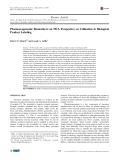
control marker in the Chinese Pharmacopoeia (Fig. 1). To
expand the clinical application of R. verticillata, we examined
the potent effects of this compound against skin diseases. In
China, as early as the 1950s, R. verticillata extract was used to
effectively cure skin diseases that cause various degrees of
itchiness and rash, with few side effects. However, few reports
exist about the therapeutic mechanism underlying R.
verticillata action. Therefore, we proposed that the produc-
tion of free radicals in human skin induces skin disease, and
the main components in R. verticillata extract exert an
antioxidant response to the free radicals produced. Al-Qirim
et al. (34) reported that R. verticillata extract protects mouse
cardiomyocytes from damage caused by elevated levels of
oxidative free radicals. In another study, Li et al. (35)showed
that a water-soluble alkaloid extract from R. verticillata
demonstrated strong antioxidant activity through scavenging
1,1-Diphenyl-2-picrylhydrazyl radical 2,2-Diphenyl-1-(2,4,6-
trinitrophenyl) hydarazyl (DPPH) in vitro.
Therefore, we hypothesized that reserpine (the most
abundant and main active compound in R. verticillata extract)
might protect skin cells from ROS (reactive oxygen species)
injury by activating the Nrf2 pathway via epigenetic modulation.
In this study, we examined the underlying epigenetic changes
caused by reserpine that protect cells from TPA-induced
carcinogenesis by restoring Nrf2 expression through DNA
methylation in a preneoplastic epidermal JB6 P+ cell line.
MATERIALS AND METHODS
Materials and Chemicals
Reserpine was extracted from Rauvolfia verticillata
(Lour) Baill. (identification data are shown in the
Supplementary Materials). Dimethyl sulfoxide (DMSO), 5-
aza (5-azadeoxycytidine, a DNMT inhibitor, has been used as
a potential chemotherapeutic agent for cancer), TPA,
trichostatin A (TSA, (27,28), bacteriological agar, and Eagle’s
basal medium (BME) were purchased from Sigma (CO.,
CA). JB6 P+ cells were purchased from the American Type
Culture Collection. Minimum essential media (MEM), fetal
bovine serum (FBS), and trypsin-EDTA solution were
purchased from Gibco Laboratories (Grand Island, NY).
The primary antibodies anti-Nrf2, anti-HO-1, anti-NQO-1,
anti-UGT1A1, and anti-β-actin were obtained from Santa
Cruz Biotechnology (Santa Cruz, CA). Anti-DNMT primary
antibodies (DNMT1, DNMT3a, and DNMT3b) were ob-
tained from IMGENEX (San Diego, CA).
Cell Culture and Treatment
The human hepatocellular HepG2-C8 cell line was previ-
ously established by stable transfectionwithanARE-luciferase
construct (36). The cells were cultured and maintained in DMEM
supplemented with 10% (V/V) FBS, 100 units/mL penicillin, and
100 μg/mL streptomycin. JB6 P+ cells were maintained in MEM
containing 5% (V/V) FBS in a humidified incubator with 5% CO
2
at 37°C. DMSO was used as a vehicle in all of the experiments at a
concentration of 0.1%. After incubation for 24 h, the cells were
treated with various concentrations of reserpine or 5-aza
(250 nmol/L) in MEM containing 1% FBS. For the combination
treatment of 5-aza and TSA, TSA (50 nmol/L) was added to the
medium on the sixth treatment day. The treated cells were
harvested on day 7 for additional assays.
Cell Viability Assay
JB6 P+ cells were seeded in 96-well plates containing
MEM at a density of 1 × 10
4
cells/mL (100 μL/well) for 1, 3,
and 5 days, and HepG2-C8 cells were seeded in plates
containing DMEM. After incubation for 24 h, the cells were
treated with either DMSO or various concentrations of
reserpine. For JB6 P+ cells, the medium was changed every
2 days for the 3-day and 5-day treatments. Cell viability was
assessed using a CellTiter 96 Aqueous One Solution Cell
Proliferation (MTS) assay kit (Promega, Madison, WI)
according to the manufacturer’s instructions. The absorbance
of the formazan product was read at 490 nm, and the cell
viability was calculated and compared with the DMSO
control group.
660 Hong et al.

Luciferase Reporter Activity Assay
The effects of reserpine on Nrf2-ARE activation were
examined using HepG2-C8 cells stably expressing the ARE-
luciferase construct. HepG2-ARE-C8 cells (1.0 × 10
5
cells/
well) were seeded into 12-well plates in 1 mL of medium
containing 10% FBS, incubated for 24 h and were subse-
quently treated with various concentrations of compounds.
ARE-luciferase activity was determined using a luciferase
assay kit according to the manufacturer’s instructions
(Promega, Madison, WI). The reporter lysis buffer was used
to lyse the cells, and 10 μL of cell lysate and 50 μLof
luciferase solution were combined to analyze luciferase
activity using a Sirius luminometer (Berthold Detection
System Gmbh, Pforzheim, Germany). We used a
bicinchoninic acid (BCA) protein assay (Pierce Biotech,
Rockford, IL, USA) to normalize the luciferase activity to
protein concentrations. The data were obtained from three
independent experiments and expressed as the inducible fold
change compared with the DMSO control group.
Anchorage-Independent Cell Growth Assay
An agar mixture was divided into control (DMSO), TPA,
and reserpine (2.5–10 μM) groups. BME containing 0.5%
agar with 10% FBS without cells was added to the bottom of
6-well plates (3 mL/well) and maintained at room tempera-
ture for 1 h. Subsequently, the JB6 P+ cells (8 × 10
3
/well) were
transferred to 1 mL of BME in 0.33% soft agar containing
TPA or various concentrations of reserpine layered on top of
the agar. The cells were cultured with TPA (20 ng/mL) and
other compounds at room temperature for an additional hour
and subsequently incubated in a 5% CO
2
incubator at 37°C
for 14 days. The cell colonies in soft agar were photographed
using a computerized microscope system with the Nikon
ACT-1 program (Version 2.20; LEAD Technologies) and
counted using ImageJ (Version 1.40 g; NIH).
RNA Isolation and Quantitative Real-Time PCR
JB6 P+ cells were seeded into 10-cm dishes at a density
of 1 × 10
4
cells/mL. The cells were treated with different
concentrations of reserpine for 5 days after incubation for
24 h. Total RNA was extracted from the treated cells using an
RNeasy Mini kit (Qiagen, Valencia, CA), and a Superscript
III First-Strand cDNA Synthesis system (Invitrogen) was
used to synthesize first-strand cDNA from total RNA. The
mRNA expression of specific genes (β-actin, Nrf2, HO-1,
NQO1, UGT1A1, DNMT1, DNMT3a, and DNMT3b) was
subsequently determined by quantitative real-time PCR
(qPCR) using first-strand cDNA as the template and Power
SYBR Green PCR Master Mix (Applied Biosystems). The
primer pairs have been previously described (37), and β-actin
mRNA expression level was used as an internal loading
control.
Whole Lysate Preparation and Western Blotting
After incubation for 24 h, JB6 P+ cells (1 × 10
5
cells/10-
cm dish) were treated with various concentrations of reser-
pine. Whole cell lysates were prepared from the treated cells
using radioimmunoprecipitation assay buffer (Cell Signaling
Technology, Danvers, MA) supplemented with a protease
inhibitor cocktail (Sigma), and a BCA kit was used to
determine protein concentrations. The proteins were sepa-
rated using 4–15% SDS-polyacrylamide gel electrophoresis
(Bio-Rad) and transferred to a polyvinylidene difluoride
(PVDF) membrane (Millipore, Bedford, MA). After blocking
with 5% BSA in Tris-buffered saline-0.1% Tween 20 buffer
for 1.5 h at room temperature, the membrane was sequen-
tially incubated with specific primary antibodies and horse-
radish peroxidase (HRP)-conjugated secondary antibodies.
The Super Signal enhanced chemiluminescence (ECL) detec-
tion and Gel Documentation 2000 system (Bio-Rad) were
used to detect and record the antibody-bound proteins on the
membrane. The densitometry of the bands was analyzed
using ImageJ (Version 1.40 g; National Institutes of Health,
NIH).
DNA Isolation and Bisulfite Genomic Sequencing
Genomic DNA was isolated from treated cells using a
QIAamp DNA Mini kit (Qiagen). After incubation for 24 h,
the cells were treated with reserpine at various concentrations
or with 5-aza (250 nM) in combination with TSA (50 nM) in
MEM containing 1% FBS for 7 days, and the medium was
Fig. 1. Chemical structure of reserpine
661Reserpine Inhibit the JB6 P+ Cell Transformation

refreshed every 2 days. TSA was added to the medium on
day 6, and the cells were harvested on day 7. The bisulfite
conversion of genomic DNA was performed using a EZ
DNA Methylation Gold kit (Zymo Research Corp.)
according to the manufacturer’s instructions, as previously
described (38). The DNA fragment containing the first 15
CpGs, located between −863 and −1226 in the murine
Nrf2 gene with the translation start site defined as
position +1, was amplified from the converted DNA with
PCR using Platinum Taq DNA polymerase (Invitrogen).
The following primer sequences were used: sense, 5′-AGT
TAT GAA GTA GTA GTA AAA A-3′and anti-sense,
5′-ACC CCA AAA AAA TAA ATA AAT C-3′.The
PCR products were cloned into the PCR 4 TOPO vector,
and ten colonies from each treatment group were ran-
domly selected. The plasmids were prepared using a
QIAprep Spin Miniprep kit (Qiagen) and analyzed by
sequencing (GeneWiz, South Plainfield, NJ).
Methylation DNA Immunoprecipitation Assay
Methylation DNA immunoprecipitation (MeDIP) analy-
sis was performed using a EpiQuik™MeDIP Ultra kit
according to the manufacturer’s instructions as previously
described (23,39). The extracted DNA from treated cells was
suspended in nuclease-free water and sonicated on ice to
generate fragments of approximately 100–800 bp. The
fragmented DNA was denatured at 95°C for 5 min and
immunoprecipitated overnight at 4°C. The primers 5′-TTT
CTA GTT GGA GGT CAC CAC A-3′(sense) and 5′-CCC
AGG GAG ATG GAT GAG T-3′(anti-sense) were used to
probe the DNA sequence containing the 15 CpG sites in
murine Nrf2. The enriched MeDIP DNA content was
calculated based on calibration using the serial dilution of
input DNA, and the relative methylated DNA ratios were
calculated based on the control, which was defined as 100%
methylated DNA.
Fig. 2. Cell viability of JB6 P+ and HepG2-C8 cells after treatment by reserpine was determined
and calculated using the MTS assay. aJB6 P+ cells were treated by reserpine for 1, 3, and 5 days. b
HepG2-C8 cells were treated by reserpine for 1 day. The IC50 values were calculated using Origin
Pro 7.5 software. The data are expressed as the mean ± SD (n=3)
Fig. 3. The induction of ARE-luciferase activity of the treatment of
reserpine with concentration from 5–50 μM on HepG2-C8 cells
expressed with ARE-luciferase vector. The BCA protein assay was
determined to normalize the luciferase activity. The data obtained
from three independent experiments expressed the inducible fold
change compared with the vehicle control. Two asterisks indicate
significant difference p< 0.01 between the treatment and control
group
662 Hong et al.

Statistical Analysis
The data are represented as the mean ± SD of three
independent experiments with similar results. The statistical
analyses were performed using ANOVA followed by post-hoc
test (Dunnett’sttest). The means were considered signifi-
cantly different at P< 0.05 and P< 0.01.
RESULTS
Cytotoxicity of Reserpine in JB6 P+ and HepG2-C8 Cells
The viability of JB6 P+ cells after treatment with
reserpine for 1, 3, and 5 days and HepG2-C8 cells for
1 day was analyzed using an MTS assay to determine the
cytotoxic effect of reserpine. The results are shown in
Fig. 2. IC50 values of 43.9 and 54.9 μM were obtained
after 1 day of treatment in JB6 P+ and HepG2-C8 cells,
respectively. We selected a reserpine concentration (2.5–
10 μM) no greater than the IC50 value, ensuring viability
greater than 70% for subsequent studies of the epigenetic
modification of the Nrf2 promoter and avoiding substan-
tial toxicity.
Reserpine Induces ARE-Luciferase Reporter Activity
The luciferase activity in cells transfected with the
ARE-luciferase reporter vector in the treatment groups
compared with the control group is shown in Fig. 3.
Reserpine induced luciferase activity in a dose-dependent
manner at concentrations ranging from 5 to 50 μM, and
no significant induction was observed at concentrations
lower than 5 μM.
Fig. 4. Inhibitory effects of reserpine on the TPA-induced transformation of JB6 P+ cells. The colonies exhibiting
anchorage-independent growth were counted under a microscope using ImageJ software. The data are represented as the
average of triplicate results. One asterisk and two asterisks represent P< 0.05 and P< 0.01, respectively, which indicate
significant differences between the reserpine-treated group and cells treated with TPA alone in soft agar
663Reserpine Inhibit the JB6 P+ Cell Transformation


![Phác đồ sử dụng MMF trong dị ghép tế bào gốc [chuẩn nhất]](https://cdn.tailieu.vn/images/document/thumbnail/2025/20250508/antrongkim0609/135x160/7241746691763.jpg)
![Hướng dẫn chẩn đoán và điều trị ung thư tế bào gan [chuẩn nhất]](https://cdn.tailieu.vn/images/document/thumbnail/2025/20250508/antrongkim0609/135x160/4691746691992.jpg)


%20--%3e%3cdefs%3e%3cstyle%3e%20.st0%20{%20fill:%20%23fff;%20}%20.st1%20{%20fill:%20%237800fa;%20}%20%3c/style%3e%3c/defs%3e%3cpath%20class='st1'%20d='M117.78,12.18H43.11c2.9,3.47,4.65,7.94,4.65,12.82,0,5.6-2.3,10.66-6.01,14.29h76.02l7.22-13.56-7.22-13.56Z'/%3e%3cg%3e%3cpath%20class='st0'%20d='M53.58,26.17h-.59v-1.46h.59v-4.96h2.83c1.78,0,2.67.94,2.67,2.82v5.76c0,1.87-.89,2.81-2.67,2.81h-2.83v-4.96ZM55.36,21.37v3.34h1.1v1.46h-1.1v3.34h1.01c.61,0,.91-.37.91-1.1v-5.93c0-.74-.3-1.1-.91-1.1h-1.01Z'/%3e%3cpath%20class='st0'%20d='M65.99,31.14h-1.8l-.31-2.07h-2.19l-.31,2.07h-1.64l1.82-11.39h2.62l1.82,11.39ZM65.28,18.04c-.25.46-.51.77-.75.94-.21.15-.47.22-.79.22-.26,0-.57-.07-.92-.22l-.38-.15c-.14-.05-.26-.07-.37-.07-.3,0-.53.18-.71.54l-.91-.68c.25-.46.51-.77.75-.94.21-.14.48-.21.79-.21.26,0,.57.07.92.21l.38.15c.14.05.26.07.37.07.3,0,.53-.18.71-.54l.91.68ZM61.91,27.52h1.73l-.87-5.76-.87,5.76Z'/%3e%3cpath%20class='st0'%20d='M74.53,26.89v1.52c0,1.91-.89,2.86-2.67,2.86s-2.67-.95-2.67-2.86v-5.93c0-1.91.89-2.86,2.67-2.86s2.67.95,2.67,2.86v1.11h-1.69v-1.22c0-.75-.31-1.12-.93-1.12s-.93.37-.93,1.12v6.15c0,.74.31,1.11.93,1.11s.93-.37.93-1.11v-1.63h1.69Z'/%3e%3cpath%20class='st0'%20d='M81.4,31.14h-1.8l-.31-2.07h-2.19l-.31,2.07h-1.64l1.82-11.39h2.62l1.82,11.39ZM75.9,19.2l1.52-1.91h1.71l1.51,1.91h-1.61l-.76-.95-.75.95h-1.61ZM77.32,27.52h1.73l-.87-5.76-.87,5.76ZM83.1,15.99l-1.76,1.91h-1.26l1.17-1.91h1.86Z'/%3e%3cpath%20class='st0'%20d='M84.86,19.75c1.78,0,2.67.94,2.67,2.82v1.48c0,1.87-.89,2.81-2.67,2.81h-.85v4.28h-1.79v-11.39h2.64ZM84.01,21.37v3.86h.85c.58,0,.87-.36.87-1.08v-1.71c0-.71-.29-1.07-.87-1.07h-.85Z'/%3e%3cpath%20class='st0'%20d='M93.51,19.75c1.78,0,2.67.94,2.67,2.82v1.48c0,1.87-.89,2.81-2.67,2.81h-.85v4.28h-1.79v-11.39h2.64ZM92.66,21.37v3.86h.85c.58,0,.87-.36.87-1.08v-1.71c0-.71-.29-1.07-.87-1.07h-.85Z'/%3e%3cpath%20class='st0'%20d='M98.8,31.14h-1.79v-11.39h1.79v4.88h2.03v-4.88h1.83v11.39h-1.83v-4.88h-2.03v4.88Z'/%3e%3cpath%20class='st0'%20d='M105.36,24.55h2.46v1.62h-2.46v3.34h3.09v1.63h-4.88v-11.39h4.88v1.63h-3.09v3.18ZM108.17,17.29l-1.76,1.91h-1.26l1.17-1.91h1.86Z'/%3e%3cpath%20class='st0'%20d='M112.2,19.75c1.78,0,2.67.94,2.67,2.82v1.48c0,1.87-.89,2.81-2.67,2.81h-.85v4.28h-1.79v-11.39h2.64ZM111.35,21.37v3.86h.85c.58,0,.87-.36.87-1.08v-1.71c0-.71-.29-1.07-.87-1.07h-.85Z'/%3e%3c/g%3e%3ccircle%20class='st1'%20cx='25'%20cy='25'%20r='20'/%3e%3cpath%20class='st0'%20d='M32.78,19.27c2.92,0,4.43,2.55,5.28,5.33l.71,2.17c.14.38-.33.75-.71.75h-5.61c.19-.33.24-.71.09-1.08l-.75-2.45c-.43-1.32-.99-2.64-1.79-3.77.75-.57,1.65-.94,2.78-.94h0ZM25,18.38c3.25,0,4.9,2.78,5.89,5.89l.76,2.45c.14.42-.33.8-.8.8h-11.69c-.42,0-.94-.38-.8-.8l.75-2.45c.99-3.11,2.64-5.89,5.89-5.89h0ZM25,11.35c1.74,0,3.11,1.37,3.11,3.11s-1.37,3.11-3.11,3.11-3.11-1.41-3.11-3.11,1.41-3.11,3.11-3.11h0ZM17.27,19.27c1.08,0,1.98.38,2.73.94-.8,1.13-1.37,2.45-1.74,3.77l-.8,2.45c-.14.38-.05.75.09,1.08h-5.56c-.42,0-.9-.38-.75-.75l.71-2.17c.9-2.78,2.41-5.33,5.33-5.33h0ZM17.27,12.91c1.51,0,2.78,1.27,2.78,2.83s-1.27,2.83-2.78,2.83-2.83-1.27-2.83-2.83,1.27-2.83,2.83-2.83h0ZM32.78,12.91c1.56,0,2.78,1.27,2.78,2.83s-1.23,2.83-2.78,2.83-2.83-1.27-2.83-2.83,1.27-2.83,2.83-2.83h0ZM27.07,28.56v.09c0,.57-.24,1.08-.61,1.46h0v.05c-.38.33-.9.57-1.46.57s-1.08-.24-1.46-.61h0c-.38-.38-.61-.9-.61-1.46v-.09h1.41v.09c0,.19.05.38.19.47v.05c.09.09.28.19.47.19s.38-.09.47-.19v-.05c.14-.09.24-.28.24-.47t-.05-.09h1.41ZM30.99,28.56v.09c0,1.65-.66,3.16-1.74,4.24-1.08,1.08-2.59,1.79-4.24,1.79s-3.16-.71-4.24-1.79l-.05-.05c-1.04-1.08-1.7-2.55-1.7-4.2v-.09h1.41v.09c0,1.27.47,2.4,1.27,3.25h.05c.85.85,1.98,1.37,3.25,1.37s2.4-.52,3.25-1.37c.85-.8,1.37-1.98,1.37-3.25v-.09h1.37ZM34.99,28.56v.09c0,2.78-1.13,5.28-2.92,7.07-1.79,1.79-4.29,2.92-7.07,2.92s-5.23-1.13-7.07-2.92c-1.79-1.79-2.92-4.29-2.92-7.07v-.09h1.41v.09c0,2.4.94,4.53,2.5,6.08,1.56,1.56,3.72,2.5,6.08,2.5s4.52-.94,6.08-2.5c1.56-1.56,2.5-3.68,2.5-6.08v-.09h1.41Z'/%3e%3c/svg%3e)