
HUE JOURNAL OF MEDICINE AND PHARMACY ISSN 3030-4318; eISSN: 3030-4326HUE JOURNAL OF MEDICINE AND PHARMACY ISSN 3030-4318; eISSN: 3030-4326
180 181
Hue Journal of Medicine and Pharmacy, Volume 15, No.2/2025 Hue Journal of Medicine and Pharmacy, Volume 15, No.2/2025
Development of an in situ gel containing tinidazole-loaded polymeric
nanoparticles for oral cavity administration
Ho Hoang Nhan* , Phan Thi Thao Ngoc
University of Medicine and Pharmacy, Hue University
Abstract
Background: Tinidazole (TNZ) demonstrates greater efficacy against anaerobic bacteria, particularly
Gram-negative strains, compared to metronidazole. Nanosizing TNZ and incorporating it into in situ gel
formulations for topical periodontitis treatment offers several advantages. Objectives: This study aimed to
formulate an in situ gel containing preformed Eudragit RSPO-based nanoparticles (NPs) of TNZ and to evaluate
its physicochemical properties. Materials and methods: Poloxamer 407 was used as a thermosensitive
gelling agent, either alone or in combination with other gelling agents. The in situ gels containing TNZ NPs
were prepared and evaluated for physicochemical properties. Results: The in situ gel containing TNZ NPs,
formulated with Poloxamer 407 and sodium alginate, exhibited a smooth texture, a gelation temperature
of 31.33 ± 0.24 °C, a gelation time of less than one minute, a pH of 6.72 ± 0.03, and a stable gel state over
an extended period. Compared to the in situ gel with TNZ material, the TNZ NP-loaded gel prolonged drug
release. The drug release mechanism was best described by the Higuchi model (with F0). Conclusion: This
TNZ NP-loaded in situ gel formulation shows promise for further research in periodontitis treatment.
Keywords: tinidazole, in situ gel, nanoparticle, poloxamer 407.
*Corresponding author: Ho Hoang Nhan. Email: hhnhan@hueuni.edu.vn
Received: 19/12/2024; Accepted: 15/4/2025; Published: 28/4/2025
DOI: 10.34071/jmp.2025.2.25
1. INTRODUCTION
Periodontitis is a chronic inflammation of the
soft tissues that support teeth, causing damage to
periodontal structures, alveolar bone loss and even
tooth loss [1]. The principal agent primarily involved
in the formation and progression of periodontitis
is Porphyromonas gingivalis (P. gingivalis), a
gram-negative anaerobic bacterium. Therefore,
eradication of P. gingivalis is essential in the
treatment of periodontitis [2].
Topical antibiotics are the preferred choice in the
treatment of periodontitis because they are a simple
method and limit unwanted side effects commonly
encountered when using systemic antibiotics.
However, their effectiveness is limited because most
clinically used antibiotics can only remain effective
for a short period of time. At the same time, drugs in
periodontal pockets are easily washed away by saliva
in the gingival pocket, making it difficult to maintain
therapeutic concentrations at the site of impact [3].
Besides, resistance can easily occur when using
antibiotics continuously because bacteria have the
ability to produce biofilm around them to protect
them from the host’s defense mechanism. These
biofilms also act as a biological barrier to prevent the
penetration of antibiotics, protecting bacteria from
being destroyed by the treatment process, thereby
reducing the effectiveness of the drug and leading
to drug resistance [2]. Therefore, developing a drug
that can penetrate the biofilm and provide long-lasting
local effects is a big challenge.
With the development of nanotechnology in
medicine and pharmacy, nanomaterials have been
developed with many advantages such as small size,
increased ability to penetrate cells, reduced toxicity,
and biocompatibility,… [2].
Tinidazole (TNZ) exhibits higher susceptibility
to anaerobic bacteria, especially Gram-negative
bacteria. Moreover, systemic TNZ offers several
advantages compared to metronidazole for the
oral treatment of periodontitis [4]. Quantum dots
containing nanoscale TNZ have been reported
to effectively penetrate biofilm layers, thereby
inhibiting the growth of P. gingivalis [2]. In vitro
studies have demonstrated sustained drug release
for up to 20 days, along with significant antibacterial
activity achieved through TNZ-loaded nanofibers [5].
In situ gels are liquid preparations that can
be easily injected into periodontal pockets and
then form a gel with a specific shape, capable of
releasing drug at a controlled rate, maintaining drug
concentration in the gingival crevicular fluid for a
long time to achieve the desired clinical benefit [6].
Poloxamer 407 (PLX407) was widely used to form
in situ gels or in combination with other gelling
agents [7]. Carbopol 934P (CBP934), when dispersed
in water, forms a colloidal dispersion with acidic
properties. Upon neutralization, it transforms into a

HUE JOURNAL OF MEDICINE AND PHARMACY ISSN 3030-4318; eISSN: 3030-4326HUE JOURNAL OF MEDICINE AND PHARMACY ISSN 3030-4318; eISSN: 3030-4326
180 181
Hue Journal of Medicine and Pharmacy, Volume 15, No.2/2025 Hue Journal of Medicine and Pharmacy, Volume 15, No.2/2025
high-viscosity gel and is a pH-sensitive polymer [8].
Alginate with a high guluronic acid content enhances
gel properties and, upon contact with cations, forms
cross-links leading to a sol-gel transition [8, 9].
In our previous study [10], Eudragit ESPO-
based polymeric nanoparticles (NPs) of TNZ
were successfully prepared. Nanosizing TNZ and
incorporating it into in situ gel formulations for
topical periodontitis treatment offers several
advantages, including improved bioavailability,
controlled drug release directly at the site of action,
and optimized dosing and duration. To the best of
our knowledge, this is the first time polymeric TNZ
NPs have been incorporated into an in situ gel.
Therefore, the aim of this study was to develop an
in situ gel containing TNZ NPs and to evaluate its
physicochemical properties.
2. MATERIALS AND METHODS
2.1. Materials
TNZ (European Pharmacopoeia 10) was
purchased from Zhejiang Supor Pharmaceuticals
Co. Ltd. (Zhejiang, China). Standard TNZ (99.7%,
batch number C0120363.01) was obtained from the
National Institute of Drug Quality Control (Hanoi,
Vietnam). Eudragit RSPO was obtained from Evonik
(Essen, Germany). PLX407 was purchased from BASF
(Ludwigshafen am Rhein, Germany). CBP934 and
sodium alginate (SA) were obtained from China. All
other chemicals were of analytical grade.
Equipments
The following instruments were used in this
study: a Zetasizer Lab instrument (Malvern,
England), a NovaSEM microscope (Hitachi S-4800,
Japan), a centrifuge (Z326K, Hermlé, Germany), a
sonicator (Ultrasonic Processor VCX130, USA), an
evaporator (Buchi Rotavapor R-300, Switzerland), a
magnetic stirrer (IKA C-MAG HS 7 digital, Germany),
a pH meter (Sension PH3, HACH, Spain), a balance
(Sartorius Quintix 125D–1S, Germany), a diffusion
device (Hanson Research, USA), a high-performance
liquid chromatography system (HPLC, Shimadzu
LC–20A, Japan), centrifugal filters (10 kDa, Sartorius,
England), a dialysis bag (MWCO 12–14 kDa, Visking
Tubes, Medicell Membranes Ltd, London, UK), and
other equipment used for the preparation and
characterization of NPs and in situ gels.
2.2. Methods
2.2.1. Formulation of in situ gels containing
tinidazole nanoparticles
Eudragit RSPO-based TNZ NPs were prepared
using the emulsification-solvent evaporation
method, as described in our previous research [10].
In situ gels were formulated using gelling excipients
such as PLX407, either alone or in combination with
CBP934 and SA at various concentrations. Briefly,
the gel-forming excipients were soaked in the NP
suspension at 4°C in a refrigerator overnight to
ensure complete swelling. The resulting gels were
then mechanically homogenized. An in situ gel
containing TNZ material was prepared using the
same procedure described above.
2.2.2. Characterization of in situ gel containing
polymeric TNZ NPs
Appearance and pH
The resulting in situ gels were visually observed
for color and physical properties. The pH of in situ
gels were evaluated using a pH meter (pH Sension
PH3, HACH, Loveland, CO) at 25 °C.
Gelation temperature and time of gelation
Two milliliters of the in situ gels were added into
glass tubes sealed with paraffin, and immersed in
a beaker of water at 4 °C. The temperature of the
beaker was gradually increased at a rate of 1.0 °C/
min for temperatures below 20 °C, or 0.5 °C/min for
temperatures above 20 °C, using a combination of
magnetic stirring and heating on a magnetic stirrer
(IKA C-MAG HS 7 digital, Staufen, Germany). Following
each temperature increment, the system will be
allowed to equilibrate for 15 minutes at the new
setpoint. The gelation temperature was recorded
at the point where the gel exhibited no movement
when tilted 90°. All measurements were performed
in triplicate [11].
The time of gelation was determined by the
inversion method. Briefly, two milliliters of the
formulation were transferred into glass tubes. These
tubes were placed in water beakers at 37 ± 0.5 °C
and time of gelation was recorded [11].
In vitro gelling capacity
Briefly, 1 mL of the in situ gel formulation
was added into a glass tube containing 2 mL of
simulated saliva (pH 6.8) maintained at 37 ± 0.5 °C
using a water bath with the help of a 1 mL pipette.
The pipette was placed at the fluid surface, and
the formulation was slowly released. Changes in
the visual appearance of the gel solution were
observed. The in vitro gelling capacity was evaluated
and categorized into three groups based on gelation
time and the duration of the gel state: (+) Gelling
after a few minutes, dispersing rapidly; (++) Gelling
immediately, maintaining the gel state for a few
hours; (+++) Gelling immediately, maintaining the
gel state for an extended period [12].

HUE JOURNAL OF MEDICINE AND PHARMACY ISSN 3030-4318; eISSN: 3030-4326HUE JOURNAL OF MEDICINE AND PHARMACY ISSN 3030-4318; eISSN: 3030-4326
182 183
Hue Journal of Medicine and Pharmacy, Volume 15, No.2/2025 Hue Journal of Medicine and Pharmacy, Volume 15, No.2/2025
Particle size, size distribution, morphology of
TNZ NPs
TNZ NPs before being introduced into gels were
diluted with distilled water for size measurement
using a Zetasizer Lab (Malvern, England).
In addition, the particle size of TNZ NPs after
being introduced into gels was measured. Briefly,
about 1.5g of gel was weighed and diluted with 15 ml
of distilled water. The dispersion was then stirred to
completely dissolve the gel-forming excipients. The
mixture was then centrifuged at 5000 rpm (Z326K,
Hermlé, Wehingen, Germany) using a centrifugal
filter (Molecular weight cut-off [MWCO] 10 kDa,
Sartorius, Switzerland) for 10 min, and washed three
times with distilled water (3 ml each time). The TNZ
NPs in the upper part of the centrifugal filter was
used to determine particle size and polydispersity
index (PDI) [13].
To observe the morphology of the particles, TNZ
NPs were deposited onto aluminum foil and allowed
to dry naturally. Once dried, the foil was coated
with a conductive material, such as platinum or
silver, before being subjected to scanning electron
microscopy (SEM) analysis [13].
TNZ assay by high performance liquid
chromatography
The concentration of TNZ in the NP suspension,
prior to its incorporation into gels, was quantified
using HPLC. The test sample was diluted with the
mobile phase to achieve a final concentration
in the range of 5 to 25 μg/mL. Chromatographic
conditions included a mobile phase comprising
Acetonitrile:MeOH:Water (10:20:70), a C8 column
(3 mm × 250 mm, particle size 5.0 µm), a flow rate
of 0.5 mL/min, an injection volume of 20 µL, and a
diode array detector set at a wavelength of 320 nm
[14].
TNZ in the in situ gel (0.1 g, equivalent to 0.1 mg
of TNZ) was dispersed in an appropriate volume of
distilled water and diluted with the mobile phase to
achieve a final concentration of 5 to 25 μg/mL. The
solution was then filtered through a 0.45 µm nylon
filter and quantified using the HPLC method.
In vitro drug release
In vitro drug release was performed using
membrane diffusion through Franz cells [13]. The drug
release conditions included a dialysis bag (MWCO
12-14 kDa, Visking Tubes, Medicell Membranes Ltd,
London, UK), a release medium of simulated saliva
(pH 6.8), a release volume of 7 mL, a temperature
of 37 ± 0.5 °C, a diffusion area of 1.76 cm², a stirring
speed of 250 rpm, and a sample amount of 250–300
mg of in situ gel. At specified time intervals, 1 mL
of the release medium was withdrawn and replaced
with an equal volume of fresh medium. The in situ
gel containing TNZ material was used as a control.
The released drug was quantified using HPLC.
The f2 similarity factor was calculated to
compare the two drug release profiles. The drug
release profile of TNZ NP in situ gel was analyzed
using various mathematical models to elucidate the
release mechanism with support from the DDSolver
Add-In in Microsoft Excel [15].
2.2.3. Statistical analysis
The data were statistically analyzed using
Microsoft Excel (Microsoft 365 MSO, Microsoft
Corp., Redmond, WA) , and a p-value less than 0.05
was considered statistically significant.
3. RESULTS
3.1. Formulation of in situ gel containing TNZ
NPs
TNZ NPs were prepared using the emulsification
and solvent evaporation method based on our
previous study [10]. The particle size and PDI of
TNZ NPs were 168.9 ± 1.6 nm, and 0.142 ± 0.017,
respectively. TNZ NPs were then introduced into in
situ gels to enhance their ease of local administration
at oral cavity.
The effects of gel-forming agents including
PLX407, and SA on appearance, pH, gelation
temperature and time of gelation of in situ gels
containing TNZ NPs were investigated (Table 1).
Meanwhile, the combination of PLX407 and CBP934
caused the gel to be not homogenous and physically
unsatisfactory, so CBP934 was not selected for
further investigations. The results demonstrated
that the formulation combining PLX407 with CBP934
failed to meet the required physical properties and
uniformity standards, resulting in a cloudy in situ gel
with precipitation.
HUE JOURNAL OF MEDICINE AND PHARMACY ISSN 3030-4318; eISSN: 3030-4326HUE JOURNAL OF MEDICINE AND PHARMACY ISSN 3030-4318; eISSN: 3030-4326
182 183
Hue Journal of Medicine and Pharmacy, Volume 15, No.2/2025 Hue Journal of Medicine and Pharmacy, Volume 15, No.2/2025
Table 1. The effect of gelling agents on pH, gelation temperature and time of gelation
PLX407 conc.
(%, w/v)
SA conc.
(%, w/v) pH Gelation temperature
(°C) Time of gelation(s)
15 6.32 ± 0.01 > 38
X
16 6.41 ± 0.03 > 38
X
17 6.44 ± 0.01 35.16 ± 0.24 70.33 ± 0.47
18 6.52 ± 0.02 31.33 ± 0.47 36.67 ± 0.47
19 6.55 ± 0.03 27.50 ± 0.41 14.33 ± 0.47
15
1.5 6.65 ±0.02 > 38
X
16
1.5 6.68 ±0.01 38.17 ± 0.23
X
17
1.5 6.72 ±0.03 31.33 ± 0.24 36.3 ± 1.25
18
1.5 6.75 ±0.02 27.50 ± 0.40 19.33 ± 0.47
19
1.5 6.78 ±0.02 24.17 ± 0.23 10.33 ± 0.47
Notes: X: not forming gels at 37 °C
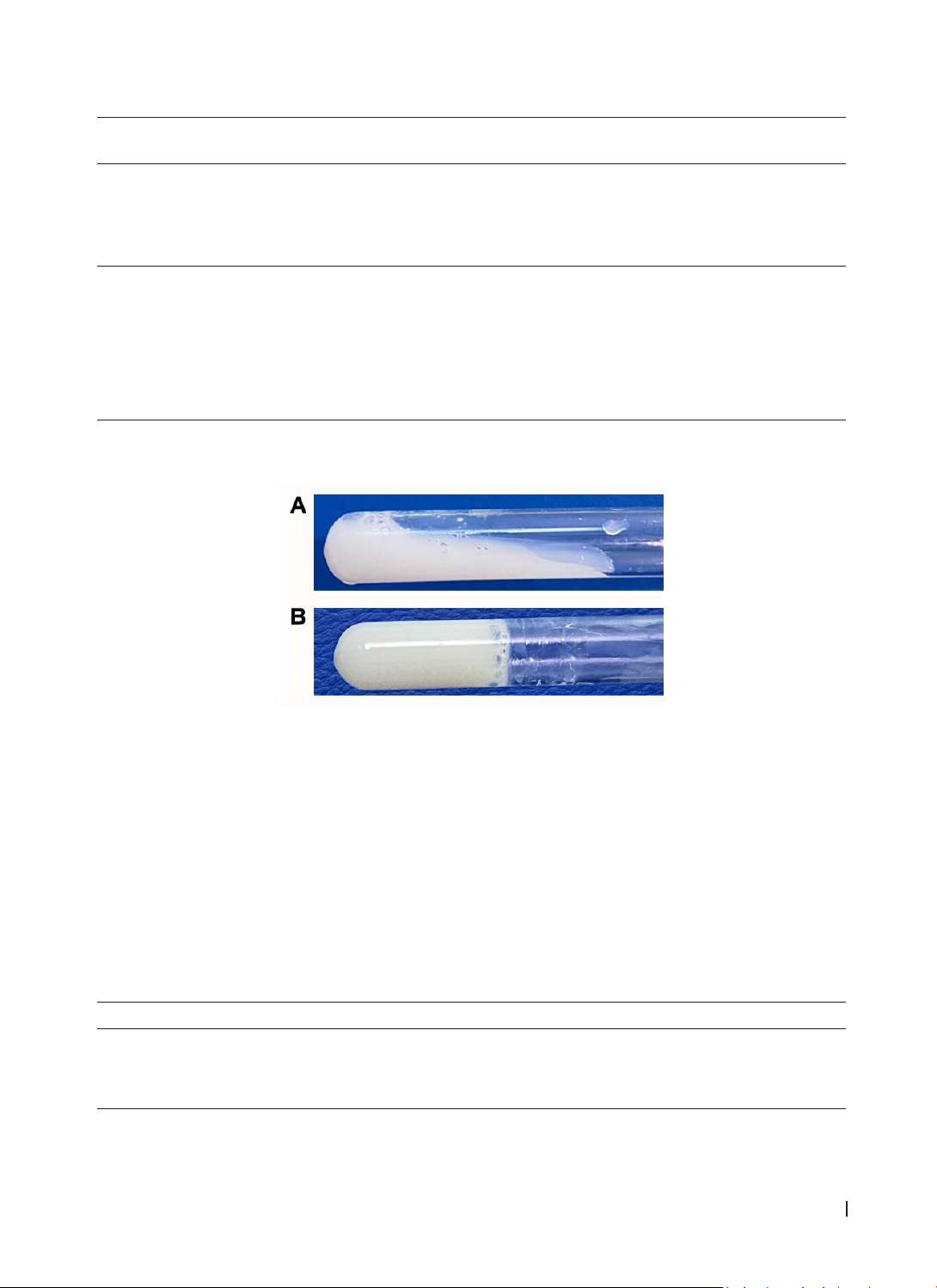
Physically, in situ gels prepared with PLX407 had a uniform, smooth texture (Fig. 1). When combining
PLX407 with SA, the gel was more viscous.
Figure 1. The appearance of in situ gels in (A) solution state, (B) gel state when investigating gelation
temperature
The pH of in situ gels formed using different
concentrations of PLX407 alone or in combination
with SA was within the required range of 6.2−7.4.
Gelation temperature and time gradually decreased
as PLX407 concentration increased and in the
presence of SA. For the optimal range of gelation
temperature of 30–36 °C, the formulations including
PLX407 17%, PLX407 18%, the combination of
PLX407 17% and SA 1.5% were selected for further
investigations.
The gelling capacity of the selected formulations
was displayed in Table 2. The formulation using the
combination of PLX407 17% and SA 1.5% had good
gel-forming capacity and maintained gel state for a
long time. Hence, the composition of the best in situ
gel containing TNZ NPs was TNZ NPs (0.1 %, w/v), 17
% (w/v) of PLX407, 1.5% (w/v) of SA, supplemented
with 0.18% (w/v) of nipagin, 0.02% (w/v) of nipasol
as preservatives and deionized water (q.s).
Table 2. The in vitro gelling capacity of gelling agents
Excipients Gelling capacity
PLX407 17% (+)
PLX407 18% (+)
PLX407 17% + SA 1.5% (+++)
(+) gelling after a few min, dispersing rapidly; (++) gelling immediately, remaining gel state in a few hours;
(+++) gelling immediately, remaining gel state for a long time
HUE JOURNAL OF MEDICINE AND PHARMACY ISSN 3030-4318; eISSN: 3030-4326HUE JOURNAL OF MEDICINE AND PHARMACY ISSN 3030-4318; eISSN: 3030-4326
184 185
Hue Journal of Medicine and Pharmacy, Volume 15, No.2/2025 Hue Journal of Medicine and Pharmacy, Volume 15, No.2/2025
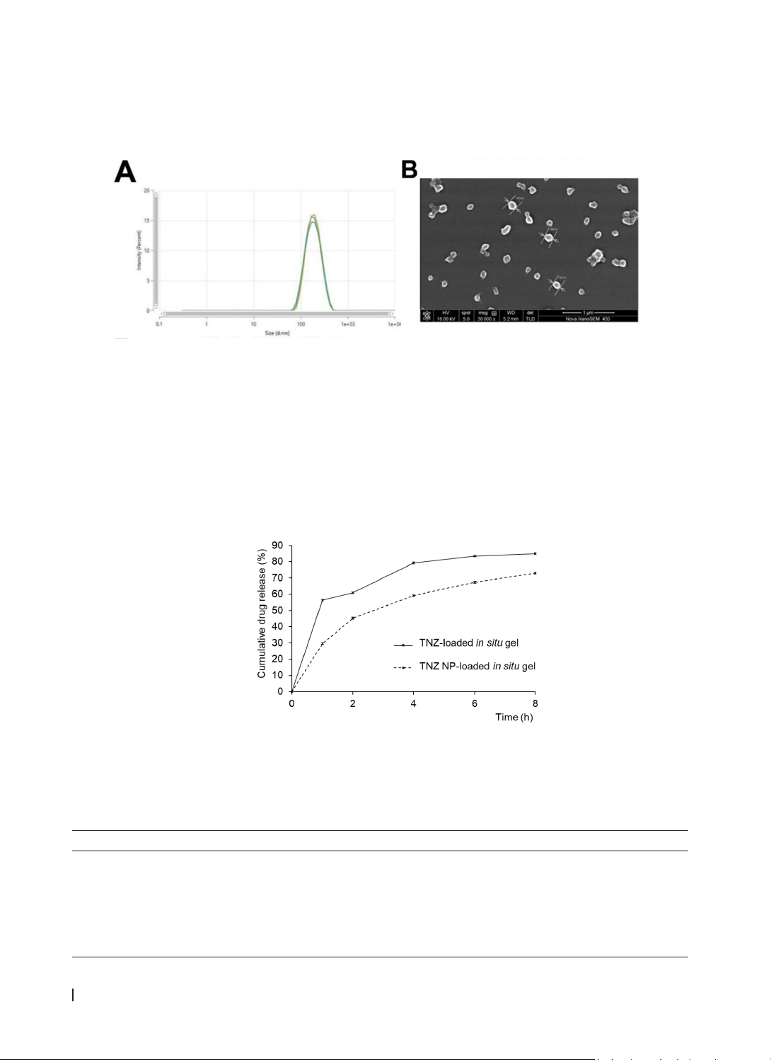
3.2. Particle size, PDI and morphology of TNZ NPs in in situ gel
The particle size and PDI of TNZ NPs in the in situ gel were 181.7 ± 2.4 nm, 0.183 ± 0.020, respectively (Fig. 2A).
The SEM image of polymeric TNZ NPs were consistent with the results obtained by dynamic light scattering
method (Fig. 2B). The TNZ content in in situ gel was 0.105 ± 0.003 % using HPLC method.
Figure 2. (A) The particle size of TNZ NPs in the in situ gel by DLS method; (B) The SEM image of TNZ NPs
in the in situ gel
3.3. In vitro drug release
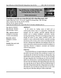
Fig. 3 showed the in vitro drug release of TNZ in in situ gels containing TNZ.
The results obtained in Fig. 3 showed that the drug release of TNZ from the in situ gel containing TNZ
material was faster than that of the in situ gel containing TNZ NPs at all time points (f2 = 36.21 < 50). In both
samples, rapid drug release through the dialysis membrane was seen within the first 4 h and slow release
in the following 4 h. After 8 h, the amount of TNZ released from the in situ gel containing TNZ NPs reached
72.96 ± 0.67%, while the amount of TNZ released from the in situ gel containing TNZ material was 85.06 ±
0.50% (p<0.05).
Figure 3. The drug release profiles of in situ gels containing TNZ
In order to predict the mechanism of the drug release from the in situ gel containing TNZ NPs, drug
release data were applied to different mathematical models. The AIC values from different models were
presented in Table 3. As shown in Table 3, the Higuchi with F0 model best described the drug release kinetics
from the in situ gel containing TNZ NPs.
Table 3. Drug release models of the in situ gel containing TNZ NPs
Model EquationaAIC
Zero-order with F0 F = 30.21 + 5.87 × t 27.29
Zero-order F = 11.12 × t 38.01
First-order F = 100 × (1 − e-0.218t)30.86
Higuchi with F0 F = 9.69 + 23.31 × t1/2 21.70
Higuchi F = 27.78 × t1/2 24.15
aF denote the cumulative drug release


![Giáo trình Kỹ năng Giao tiếp và Thực hành tốt nhà thuốc: Phần 2 [Full]](https://cdn.tailieu.vn/images/document/thumbnail/2026/20260319/hoatulip2026/135x160/44121774241072.jpg)


