BioMed Central
Acta Veterinaria Scandinavica
Open Access
Research Bacteria in milk from anterior and posterior mammary glands in sows affected and unaffected by postpartum dysgalactia syndrome (PPDS) Nicole Kemper* and Imke Gerjets
Address: Institute of Animal Breeding and Husbandry, Christian-Albrechts-University Kiel, Olshausenstraße 40, D-24098 Kiel, Germany
Email: Nicole Kemper* - nkemper@tierzucht.uni-kiel.de; Imke Gerjets - igerjets@tierzucht.uni-kiel.de * Corresponding author
Published: 22 June 2009
Received: 8 December 2008 Accepted: 22 June 2009
Acta Veterinaria Scandinavica 2009, 51:26
doi:10.1186/1751-0147-51-26
This article is available from: http://www.actavetscand.com/content/51/1/26
© 2009 Kemper and Gerjets; licensee BioMed Central Ltd. This is an Open Access article distributed under the terms of the Creative Commons Attribution License (http://creativecommons.org/licenses/by/2.0), which permits unrestricted use, distribution, and reproduction in any medium, provided the original work is properly cited.
Abstract Background: The performance of piglet weight gain is strongly dependent on the sow's ability to meet the demand for adequate milk. Postparturient disorders, especially those subsumed under the term postpartum dysgalactia syndrome (PPDS), can alter or reduce the milk production sensitively, resulting in starving piglets. The aim of this study was to gather further information about the prevalence of different bacterial species in the anterior and posterior mammary glands of sows with respect to the clinical appearance of PPDS.
Methods: In this study, the health status of 56 sows after farrowing was determined with special regard to mastitis and dysgalactia. Pooled milk samples from anterior and posterior glands were taken from both affected and non-affected animals and analysed bacteriologically for the presence of a wide spectrum of different pathogens.
Results: Mainly Escherichia coli, staphylococci and streptococci were detected in high percentages but without significant differences in healthy and diseased animals and anterior and posterior glands. However, the large percentages of coliform bacteria suggested a transmission route via faecal contamination.
Conclusion: In this study, the prevalence of different bacteria in anterior and posterior glands in PPDS positive and negative sows was analysed. No significant differences in bacteria of healthy and diseased sows were assessed. Therefore, the development of clinical PPDS and actual infection seems to be largely dependant on individual resistance in single sows.
Background In their first days of life, piglets are totally reliant on the sow for access to colostrum and milk. Every alteration in both milk yield and composition has highly sensitive impacts on weight gain and growth rate. Therefore, post- parturient disorders, including dysgalactia in sows, are a very important disease complex economically [1]. They are reported world-wide, but subsumed under different
terms depending on the geographical location. While mastitis, metritis and agalactia syndrome (MMA) [2,3] is the commonly used name in European countries, post- partum dysgalactia syndrome (PPDS or PDS) [4] has become widely accepted in English-speaking areas. At farm level, incidence is estimated to differ between 0.5% and 60% [5], with an average incidence of about 13% [6- 11]. The syndrome is characterised by greatly reduced
Page 1 of 7 (page number not for citation purposes)
Acta Veterinaria Scandinavica 2009, 51:26
http://www.actavetscand.com/content/51/1/26
milk production within 12 to 48 hours post-partum that rapidly leads to piglet starvation. Even though metritis is often a part of the syndrome, mastitis is the central symp- tom, as shown by several studies [12-14]. The bacteria most frequently isolated from mastitis-affected sows belong to the class of coliforms [13-17]. Bacterial genera included in the class of coliforms are represented by Escherichia, Enterobacter, Citrobacter, and Klebsiella. Several successful infection experiments leading to mastitis in sows have demonstrated the predominant role of these organisms [13,18]. Mastitis is a clear pathological entity: infected glands typically show signs of inflammation such as severe oedema and skin congestion [4], and, with many glands involved, sows develop fever (>40.3°C) and lose their appetites [18-20]. Mastitis can be a local process, restricted to one or several glands, but can also affect all mammary complexes [4]. Pathological foci of mastitis were detected as well in anterior complexes as in posterior complexes, but severe changes were more present in the latter [20]. These findings were supported by Bostedt et al. [21] and Baer and Bilkei [22], showing that posterior glands were more prone to pathological changes com- pared with anterior ones. However, detailed investiga- tions on the bacteriological findings in cranial and caudal mammary glands and the possible consequences have not yet been reported. Therefore, the presence of bacteria in mammary glands of different location was examined in this study with special regard to the clinical status of the sow post partum.
Bacteriology After assessment of the PPDS status, the teats were cleaned with soap solution and disinfected with 70% isopropyl alcohol (Universal Hospital Supplies LTD, Enfield, UK). Milk samples from all animals were milked on transport swabs with Amies medium (Transwab, Medical Wire & Equipment, Corsham, UK) after oxytocin injection (30 I.E. i.m.). This oxytocin dosage was chosen after pre-tests on the minimum dosage followed by milk ejection, and also recommended by Morkoc [23]. Piglets were removed before the washing procedure, and five minutes after injection, milking was started. The first streams of secre- tion from each teat were discarded in order to 'wash out' bacteria in the distal end of the teat canal. Specimens were transported to the laboratory within two hours and pooled. Pooling was performed by incubating all samples of one animal from the anterior mammary complex, sub- suming the first three pairs of pectoral glands, for enrich- ment in one tube (50 mL) with casobuillon (Roth GmbH&Co KG, Karlsruhe, Germany). Samples from the three to four following pairs of caudal glands were pooled likewise. Consequently, a sample pair consisting of pooled milk swabs from anterior and posterior glands had to be examined for each sow. In total, 112 pooled samples (56 from anterior and 56 from posterior glands) were ana- lysed. After 24 hours of incubation at 37°C, bacteriologi- cal routine diagnostic procedures including selective enrichment and biochemical confirmation (API20E, bioMérieux, Nürtingen, Germany) took place as described previously for Enterobacteriaceae, especially Escherichia coli (E. coli), Klebsiella species (spp.), Enterobacter spp., Citro- bacter spp., and Salmonella spp. [24]. Staphylococci were isolated via incubation in CSL-Bouillon (Roth GmbH&Co KG) (37°C, 24 h) and streaking out on Sheep Blood Agar (Oxoid GmbH, Wesel, Germany) (37°C, 24 h), followed by Gram-staining and Catalase-testing (Merck KGaA, Darmstadt, Germany) and biochemical identification (ID32Staph, bioMérieux). For Streptococci, incubation was performed in the same way and biochemical identifica- tion was done with API20Strep (bioMérieux).
Statistical analysis For statistical analysis, the Fisher's Exact Test was per- formed using the procedure PROC FREQ from the SAS statistical software (SAS, 2003). In general, a statistical sig- nificance level of p ≤ 0.05 was used.
Methods Animals In our study, 56 primiparous or multiparous Large White and cross-bred sows, taking part in an experiment on injected temperature transponders and water intake [16], were investigated. The study took place in a time period of six months between December 2007 and May 2008. Sows were housed at the institute's research farm with 120 sows in total. They were managed in a three-week rhythm with a 28-day lactation period. At average, the sampled animals were in their 2,9th parity. Sows were defined as PPDS-pos- itive due to a combination of appropriate criteria: with a rectal temperature higher than 39.5°C within 12 to 24 hours post-partum, and in addition, detectable inflamma- tion in the mammary gland, and/or diminished appetite and/or altered piglet behavior [1,12]. Inflammation was diagnosed by a veterinarian, observing the typical signs of inflammation, such as severe edema, hardening, and skin congestion. Altered piglet behavior was present when pig- lets show vigorous nursing efforts, decrease their attempts to nurse and their general activity, retreat to the warmest parts of the farrowing crate or lose weight.
Results According to the definition of PPDS affection, 27 sows (48.2%) out of the total number of 56 animals were pos- itive. Consequently, 29 milk sample pairs from negative sows (51.8%) were available for investigation. From all sows, one or more different bacteria were isolated at least in one of the two pooled samples. In total, 159 bacteria isolates were detected. From PPDS-affected sows, 84 iso-
Page 2 of 7 (page number not for citation purposes)
Acta Veterinaria Scandinavica 2009, 51:26
http://www.actavetscand.com/content/51/1/26
exceeded. This threshold is defined rather coincidentally between 39.3°C and 40.5°C [3] and the use of it might be regarded as critical since physiological hyperthermia is often observed in sows, leading to misinterpretations [4,16]. To avoid confusion with this physiological tem- perature increase and to reduce the administration of anti- biotics in non-affected animals, a threshold of 39.5°C in the time frame 12 to 24 hours post-partum is recom- mended. Furthermore, diagnosis should be performed not only due to temperature increase, but due to a combi- nation of appropriate criteria such as clinical mammary gland changes, diminished milk production and reduced appetite [26]. Other parameters like cell count, estab- lished for cows' milk, are not common for diagnosis and data on thresholds are rare. For instance, a threshold of 5 × 10⁶ cells per mL was proposed by Bertschinger and Bühl- mann [27], while Persson et al. [28] suggested 10 × 106 cells per mL.
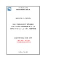
lates were proven in anterior (43 isolates) and posterior samples (41 isolates), while 75 were present in anterior (47 isolates) and posterior (28 isolates) samples from non-affected sows. In both PPDS-affected and non- affected sows, no bacteria were detected in 18.5% (PPDS+) and 13.8% (PPDS-) of pooled samples from anterior glands and in 25.9% (PPDS+) and 34.5% (PPDS- ) from posterior mammary glands (Table 1). This percent- age was increased in the posterior glands in PPDS-nega- tive sows. Furthermore, posterior mammary glands in negative sows showed the lowest variety of bacterial spe- cies. The isolated species belonged to the families Entero- and bacteriaceae, Staphylococcaceae, Streptococcaceae, Enterococcaceae (Table 2). Escherichia coli, Staphyloccocus spp. and Enterococcus spp. were the predominant bacteria species. From the genus Staphyloccocus (Staph.), Staph. aureus and Staph. hyicus were considered in the following analysis at species level. Genera found only in very low numbers were subsumed as 'others', and included Aerococ- cus spp., Enterobacter spp., Escherichia spp., Lactobacillus spp., Leuconostoc spp., Raoultella spp., and Streptococcus spp.. Due to the possible importance as a pathogen, Kleb- siella spp. were specially mentioned. After statistical anal- ysis, only the percentages of Staph. aureus and Staph. hyicus differed significantly with regard to the mammary gland location (Table 3). This is, however, related to the low prevalence of these species (Staph. aureus: n = 14; Staph. hyicus: n = 6). The comparison of the bacterial occurrence in all samples of PPDS-affected and non-affected sows showed no significant influences of the PPDS status on the bacterial flora with the exception of Staph. spp., show- ing significant differences with respect to the PPDS status (Table 4). The distribution of the different bacteria in anterior and posterior pooled samples from PPDS- affected and non-affected sows is shown in Figure 1.
The procedure of milking sows in order to obtain samples is difficult and severely restricted by practical circum- stances, but an absolutely needed prerequisite to obtain reliable results. Cleaning and disinfection are urgently required to eliminate skin flora or other contaminating microorganisms. Examinations on the skin flora on sows' teats do not exist, but studies on cows suggest, that staphy- lococci are the most common bacteria [29,30]. Pooled milking, as it is often used to get a sufficient amount of milk, most likely increases the risk of contamination. Because of this, and because of economic reasons, single teat samples from anterior and posterior glands were taken separately in our study, but pooled in the next lab- oratory step. In this way, the expected costs for bacterio- logical differentiation were reduced, while maintaining the hygienic demands. Moreover, the anatomy of the mammary gland can influence sampling: per teat, two complete gland systems end in two teat orifices without muscular sphincters [4]. Therefore, if only one gland sys- tem is affected by mastitis, the sampled milk might only consist of secretion from the other, healthy system, lead- ing to false negative results.
Discussion The aim of this study was to gather further information about the prevalence of different bacterial species in the anterior and posterior mammary glands of sows with respect to the clinical appearance of the PPDS syndrome. This clinical appearance is usually diagnosed via rectal temperature measuring post-partum [25] and sows are defined as 'PPDS-positive' when a certain threshold is
Bacteria were found in all sows, but not necessarily in both anterior and posterior samples. Compared to similar
Table 1: Number of different bacteria species isolated in samples from PPDS-affected and non-affected sows
Pooled samples from anterior mammary glands (first 3 pairs of cranial glands) Pooled samples from posterior mammary glands (3 – 4 pairs of caudal glands)
Number of isolated different bacteria species 0 1 2 3 4 5 0 1 2 3 4 5
In samples from PPDS+ sows (n = 27) (%) 18.5 29.7 25.9 25.9 0.0 0.0 25.9 25.9 25.9 18.5 0.0 3.8
Page 3 of 7 (page number not for citation purposes)
In samples from PPDS- sows (n = 29) (%) 13.8 37.9 31.0 10.3 3.5 3.5 34.5 34.5 31.0 0.0 0.0 0.0
Acta Veterinaria Scandinavica 2009, 51:26
http://www.actavetscand.com/content/51/1/26
Table 2: Numbers of isolated bacteria in all samples (n = 112)
Enterobacteriaceae Staphylococcaceae Streptococcaceae Enterococcaceae n n n n
Ec*. avium Ec. durans Ec. faecalis Ec. faecium Ec. sp. 2 16 3 9 9 Enterobacter sp. Escherichia coli Escherichia vulneris Klebsiella oxytoca Klebsiella pneumoniae Raoultella ornithinolytica 2 47 2 1 1 1
Aerococcus urinae Aerococcus viridans Aerococcus sp. Lactococcus lactis Str*. bovis Str. equinus Str. mitis Str. mutans Str. sp. 4 3 2 2 1 1 2 1 5 Staph*. aureus Staph. capites Staph. chromogenes Staph. equorum Staph. hyicus Staph. lentus Staph. saprophyticus Staph. simulans Staph. warneri Staph. sp.** 14 1 2 1 6 1 1 14 1 5
former
presence of different bacteria species in sows' milk and the comparison with investigations, conducted mainly in the 1980s and 1990s [31], is the use of appro- priate methods for identification. Most isolated bacteria in our study were representatives of the families Enterobac- teriaceae, Staphylococcaceae, Streptococcaceae, and Enterococ- caceae. This spectrum is in agreement with other studies [15,32]; for instance, in a bacteriological examination of mammary gland changes in 663 sows suffering mastitis, mainly E. coli and Klebsiella spp. were detected, but also Streptococci and Staphylococci [22].
studies, for instance Persson et al. [28] with no bacterial growth in 10% of all samples from agalactic and in 54% of all samples from healthy sows, this prevalence is high. However, in contrast to our project, no enrichment step was included in the bacteriological examination in that study, but samples were spread directly on blood agar. No selective agars were used, also lowering the probability to detect certain species. Moreover, in comparison to other studies [6,9-11], the documented incidence at farm level on the research farm was high with 48.2%. The general high incidence on farm level and the unknown back- ground of that fact were reasons to realise this study. After antibiotic treatment in combination with non-steroidal anti-inflammatory drugs, administered after sampling, all sows recovered. One important fact regarding the actual
*Staphylococcus (Staph.), Streptococcus (Str.), Enterococcus (Ec.) ** not further identified
Table 3: P-values of the effects 'PPDS-status' and 'mammary gland location' on the occurrence of different bacteria species in samples from PPDS-affected and non-affected sows (n = 112)
PPDS status Mammary gland location
E. coli 0.13 0.13
Like in other studies, the most commonly isolated bacte- ria from mastitis-affected sows belong to the class of colif- orms, covering the bacterial genera Escherichia, Klebsiella, Enterobacter and Citrobacter [13,15,17]. Subsequently, the predominant role of coliform bacteria was clearly shown by Wegmann et al. [14]; in 131 complexes with mastitis, E. coli and Klebsiella pneumonia were isolated in 79%. This importance of E. coli has been confirmed in several studies [13,33,34] and in infection experiments, provoking clini- cal and haematological changes comparable to natural infections [35,36]. To emphasize the role of coliforms and to end the confusing terminology, the term 'coliform mas- titis' was suggested for peripartal mastitis in sows [1].
Staph. aureus 0.18 0.02
Staph. hyicus 0.33 0.01
Staph. spp.* 0.04 0.14
Enterococcus spp. 0.16 0.15
Klebsiella spp. 0.23 0.51
others* 0.18 0.19
Bacteria causing or at least accompanying the syndrome of coliform mastitis may originate from the intestinal flora of the sow, from the environment or from the oral flora of the neonatal piglet. The hypothesis of a galactogenous route of infection was corroborated by experiments car- ried out by Bertschinger et al. [15], in which a reduction of PPDS prevalence could be noticed after protection of the mammary gland against faecal contamination. There- fore, faecal contamination was postulated to be of para- mount importance as a cause of puerperal mastitis. The faecal origin of E. coli isolated from sows' milk was also reported by Awad Masalmeh et al. [15]: in one quarter of
Page 4 of 7 (page number not for citation purposes)
significant effects (≤ 0.05) in bold * all Staph. species but Staph. aureus and Staph. hyicus ** Aerococcus spp., Enterobacter spp., Escherichia spp., Lactobacillus spp., Leuconostoc spp., Raoultella spp., Streptococcus spp.
Acta Veterinaria Scandinavica 2009, 51:26
http://www.actavetscand.com/content/51/1/26
Table 4: Percentage of isolated bacteria species in the total number of samples (n = 112) from PPDS-affected and non-affected sows
percentage in samples (n = 54) from PPDS+ sows (%) percentage in samples (n = 58) from PPDS- sows (%) p-values
E. coli 38.9 44.8 0.13
Staph. aureus 14.8 10.3 0.18
Staph. hyicus 11.1 10.3 0.32
Staph. spp.* 29.6 15.5 0.04
Enterococcus spp. 33.3 31.0 0.16
Klebsiella spp. 3.7 0.0 0.23
others** 20.4 17.2 0.18
significant effects (≤ 0.05) in bold * all Staph. species but Staph. aureus and Staph. hyicus ** Aerococcus spp., Enterobacter spp., Escherichia spp., Lactobacillus spp., Leuconostoc spp., Raoultella spp., Streptococcus spp.
anterior glands
posterior glands
PPDS+
PPDS+
E. coli 29.27%
E. coli 20.93%
others* 14.63%
others* 20.93%
Enterococcus spp. 31.71%
Enterococcus spp. 12.28%
Staph. spp. 20.93%
Staph. spp. 19.51%
Staph. hyicus 6.98%
Staph. aureus 13.95%
Staph. aureus 4.88%
PPDS-
PPDS-
others* 8.51%
others* 28.57%
E. coli 35.71%
E. coli 34.04%
Enterococcus spp. 27.66%
Enterococcus spp. 21.43%
Staph. spp. 14.89%
Staph. spp. 19.51%
Staph. hyicus 4.26%
Staph. aureus 3.57%
Staph. aureus 10.64%
* Aerococcus spp., Enterobacter spp., Escherichia spp., Lactobacillus spp., Leuconostoc spp., Klebsiella spp., Raoultella spp., and Streptococcus spp.
Bacterial spectrum in samples from anterior and posterior glands from PPDS-affected (n = 27) and non-affected sows (n = 29) Figure 1 Bacterial spectrum in samples from anterior and posterior glands from PPDS-affected (n = 27) and non- affected sows (n = 29).
Page 5 of 7 (page number not for citation purposes)
Acta Veterinaria Scandinavica 2009, 51:26
http://www.actavetscand.com/content/51/1/26
67 PPDS-affected sows O-serogroup-identical E. coli were detected in both milk and faecal samples. In another study comparing the bacterial flora of the uterus, the cae- cum, the ileum and the mammary gland, the prevalence of only gram-negative bacteria in the mammary glands and in the ileum of CM-affected sows was remarkable [23]. However, in this study, the faecal route of infection could not be confirmed due to the study design.
Authors' information NK, Dr. med. vet, is Certified Veterinary Specialist in Microbiology (Fachtierärztin Mikrobiologie) and in Ani- mal Hygiene (Fachtierärztin Tierhygiene) and leader of the researchgroup 'geMMA: structural and functional analysis of the genetic variation of the MMA-sydrome' (www.gemma-kiel.de). IG, MSc. Agr., is researcher in this group.
Acknowledgements This work was partially financially supported by the Federal Ministry of Edu- cation and Research (BMBF) in the research programme 'FUGATO – Func- tional Genome Analysis in Animal Organisms', project 'geMMA: structural and functional analysis of the genetic variation of the MMA-syndrome' (FKZ0315138). The authors thank Stephan Kruse for assisting this study by helping with the sows' milking, Jens Wolfmüller for excellent technical assistance and Dr. Carsten Henze for statistical advice.
References 1.
Bertschinger HU: Coliform Mastitis. In Diseases of Swine 8th edi- tion. Edited by: Straw BE, D'Allaire S, Mengeling WL, Taylor DJ. Ames: Iowa State University Press; 1999:457-464.
2. Martin CE, Hooper BE, Armstrong CH: A clinical and pathologi- cal study of the mastitis-metritis-agalactia syndrome of sows. J Am Vet Med Assoc 1967, 151:1629-1634.
3. Waldmann K-H, Wendt M: Lehrbuch der Schweinekrankheiten 4th edi-
4.
The lack of gram-negative bacterial culture growth in uter- ine samples supports the theory that uterine involvement in PPDS is of minor importance, as has been suggested in several studies [37-39]. However, infections of the urinary tract are strongly related to puerperal diseases, even though urinary infections are not apparent clinically [40]. In these infections as well, the most common organism associated with bacteriuria was found to be E. coli [41]. Therefore, not only faecal, but also urine contamination has to be considered as an infection source for PPDS. In this context, Bertschinger [1] suggested that the often rec- ommended feed reduction might not act directly in the sow's organism, but indirectly through reduced exposure of the mammary complexes due to decreased amounts of faeces and urine contaminating the lying area. Up to now, information on the actual influences leading to the mani- festation of mastitis in sows is widely lacking and further research is desirable.
5.
6.
7.
8.
9.
tion. Stuttgart: Parey Verlag; 2001. Klopfenstein C, Farmer C, Martineau GP: Diseases of the mam- mary glands and lactation problems. In Diseases of swine 9th edi- tion. Edited by: Straw BE, Zimmermann JJ. Taylor DJ: Iowa State University Press; 2006:833-860. Hirsch AC, Philipp H, Kleemannm R: Efficacy of meloxicam for treatment of the mastitis-metritis-agalactia syndrome in sows. Prakt Tierarzt 2004, 85:842-848. Bäckström L, Morkoc AC, Connor J, Larson R, Price W: Clinical study of mastitis-metritis-agalactia in sows in Illinois. J Am Vet Med Assoc 1984, 185:70-73. Hermannson I, Einarsson S, Larsson K, Bäckström L: On the agalac- tia postpartum in the sows: a clinical study. Nord Vet Med. 1978, 30(11):465-473. Jorsal SE: Epidemiological and statistical analysis of disease records from pig testing stations. PhD-Thesis University of Copenhagen; 1983. Krieter J, Presuhn U: Genetische Parameter für die Behand- lungsfrequenz beim MMA-Syndrom (Genetic parameters for MMA treatment in sows). Züchtungskd 2009, 81:149-154.
10. Madec F, Josse J: Le pathologie de la parturition chez le truie: étude épidemiologigique dans cinq élevages (Pathology of the parturition of the sow: epidemiological studies on five farms). Rec Med Vet 1992, 68:340-349.
11. Thorup F: Effect of treatment for MMA – retrospective obser- vations. 17th International Pig Veterinary Society Congress Melbourne, Australia 2000:97.
Conclusion This study examines the prevalence of different bacteria in anterior and posterior glands in PPDS positive and nega- tive sows. In conclusion, the bacterial flora of PPDS- affected and non-affected sows differs only slightly. Mainly ubiquitous bacteria were isolated and significant differences in the occurrence in anterior and posterior glands were not statistically confirmed. At the current state of knowledge, the reason for only some sows developing clinical signs of infection after contact with these ubiqui- tous bacteria remains unknown. The immune response and the actual outbreak of infection seems to depend on the immunological reactivity of the sow. Hence, one may hypothesise that developing clinical PPDS is largely dependant on the individual resistance of the sow and research is needed to define this individual resistance in detail.
12. Heinritzi K, Hagn J: Untersuchungen zur Wirksamkeit und Ver- träglichkeit des neu entwickelten Cephalosporins Cefqui- nom bei an puerperaler Septikämie und Toxämie erkrankten Sauen (Comparison of therapeutic performance of the new Cephalosporin Cefquinome with other treatment regimes in gilts with puerperal septicaemia and toxaemia syndrome). Tierärztl Prax 1999, 27:114-121.
Competing interests The authors declare that they have no competing interests.
13. Ross RF, Orning AP, Woods RD, Zimmermann BJ, Cox DF, Harris DL: Bacteriologic study of sow agalactia. Am J Vet Res 1981, 42:949-955.
14. Wegmann P, Bertschinger HU, Jecklin H: A field study on the prevalence of coliform mastitis (MMA syndrome) in Switzer- land and the antimicrobial susceptibility of the coliform bac- teria from the milk. 8th International Pig Veterinary Society Congress, Barcelona, Spain 1986:92.
Authors' contributions NK designed and coordinated the study, draft the manu- script and participated in the bacteriological analysis. IG carried out the bacteriological examinations and per- formed the statistical analysis. All authors read and approved the final manuscript.
15. Awad Masalmeh M, Baumgartner W, Passering A, Silber R, Hinterdor- fer F: Bakteriologische Untersuchungen bei an puerperaler
Page 6 of 7 (page number not for citation purposes)
Acta Veterinaria Scandinavica 2009, 51:26
http://www.actavetscand.com/content/51/1/26
Mastitis (MMA-Syndrom) erkrankten Sauen verschiedener Tierbestände Österreichs (Bacteriological studies in sows with puerperal mastitis in different herds in Austria). Tierärztl Umsch 1990, 45:526-535.
35. Bertschinger HU, Pohlenz J, Hemlep I: Mastitis metritis agalactia syndrome (milk fever) in sows. II. Bacteriological findings in spontaneous cases. Schweiz Arch Tierheilkd 1977, 119:223-233. 36. Ross RF, Harmon RL, Zimmermann BJ, Young TF: Susceptibility of sows to experimentally induced Escherichia coli mastitis. Am J Vet Res 1983, 44:949-954.
16. Gerjets I, Kruse S, Krieter J, Kemper N: Diagnosis of MMA affected sows: bacteriological differentiation, temperature measurement and water intake. 20th International Pig Veterinary Society Congress, Durban, South Africa 2008:236.
37. Armstrong CH, Hooper BE, Martin CE: Microflora associated with agalactia syndrome of sows. Am J Vet Res 1968, 29:1401-1407.
38. Martin CE: Status of the mastitis-metritis-agalactia syndrome
17. Hirsch AC, Philipp H, Kleemann R: Investigation on the efficacy of meloxicam in sows with mastitis-metritis-agalactia syn- drome. J Vet Pharmacol Ther 2003, 26:355-360.
of sows. J Am Vet Med Assoc 1970, 157:1519-1521.
39. Nachreiner RF, Ginther OJ: Induction of agalactia by adminis- tration of endotoxin (Escherichia coli) in swine. Am J Vet Res 1974, 35:619-622.
18. Bertschinger HU, Bürgi E, Eng V, Wegmann P: Lowering of the inci- dence of puerperal mastitis in the sow by protection of the mammae from contamination. Schweiz Arch Tierheilkd 1990, 132:557-566.
19. Halgaard C: Epidemiologic factors in puerperal diseases of
sow. Nord Vet Med. 1983, 35(4):161-174.
40. Mauch C, Bilkei G: The influence of prepartal bacteriuria on the reproductive performance of the sow. Dtsch Tierarztl Wochenschr. 2004, 111(4):166-169.
41. Waller CM, Bilkei G, Cameron RD: Effect of periparturient dis- eases accompanied by excessive vulval discharge and wean- ing to mating interval on sow reproductive performance. Aust Vet J. 2002, 80(9):545-549.
20. Middleton Williams DM, Pohlenz J, Lott-Stolz G, Bertschinger HU: Untersuchungen über das Mastitis-Metritis-Agalactie-Syn- drom (Milchfieber) der Sau (Investigations on the mastitis metritis agalactia syndrome (milk fever) in sows). Schweiz Arch Tierheilkd 1977, 119:214-223.
21. Bostedt H, Maier G, Herfen K, Hospen R: Clinical examinations on gilts with puerperal septicaemia and toxaemia. Tierarztl Prax Ausg G Grosstiere Nutztiere. 1998, 26(6):332-338.
22. Baer C, Bilkei G: Ultrasonographic and gross pathological find- ings in the mammary glands of weaned sows having suffered recidiving mastitis metritis agalactia. Reprod Domest Anim. 2005, 40(6):544-547.
23. Morkoc A, Backstrom L, Lund L, Smith AR: Bacterial endotoxin in blood of dysgalactic sows in relation to microbial status of uterus, milk, and intestine. J Am Vet Med Assoc 1983, 183:786-789.
25.
26.
24. Kemper N, Aschfalk A, Holler C: Campylobacter spp., Enterococ- cus spp., Escherichia coli, Salmonella spp., Yersinia spp., and Cryptosporidium oocysts in semi-domesticated reindeer (Rangifer tarandus tarandus) in Northern Finland and Nor- way. Acta Vet Scand 2006, 48:7. Furniss SJ: Measurement of rectal temperature to predict mastitis, metritis and agalactia (MMA) in sows after farrow- ing. Prev Vet Med 1987, 5:133-139. Larsen I, Thorup F: The Diagnosis of MMA. 19th International Pig Veterinary Society Congress, Copenhagen, Denmark 2006:256.
28.
27. Bertschinger HU, Bühlmann A: Absence of protective immunity in mammary glands after experimentally induced coliform mastitis. 11 International Pig Veterinary Society Congress Lausanne, Switzerland 1990:175. Persson A, Pedersen Morner A, Kuhl W: A long-term study on the health status and performance of sows on different feed allowances during late pregnancy. II. The total cell content and its percentage of polymorphonuclear leucocytes in path- ogen-free colostrum and milk collected from clinically healthy sows. Acta Vet Scand 1996, 37:279-291.
29. De Vliegher S, Laevens H, Devriese LA, Opsomer G, Leroy JLM, Barkema HW, de Kruif A: Prepartum teat apex colonization with Staphylococcus chromogenes in dairy heifers is associ- ated with low somatic cell count in early lactation. Vet Micro- biol 2003, 92:245-252.
30. Devriese LA, De Keyser H: Prevalence of different species of coagulase-negative streptococci on teats and milk samples from dairy cows. J Dairy Res 1979, 47:155-158.
31. Gerjets I, Kemper N: Coliform mastitis in sows: a review. Swine
Publish with BioMed Central and every scientist can read your work free of charge
Health Prod 2009, 17:97-105.
"BioMed Central will be the most significant development for disseminating the results of biomedical researc h in our lifetime."
Sir Paul Nurse, Cancer Research UK
33.
Your research papers will be:
available free of charge to the entire biomedical community
34.
peer reviewed and published immediately upon acceptance
cited in PubMed and archived on PubMed Central
Zentralbl Veterinarmed A.
in milk.
32. Kobera R: Vergleichende Prüfung der klinischen Wirksamkeit von Cefquinom und Enrofloxacin bei der Behandlung des Mastitis-Metritis-Agalaktie-Komlexes der Sau. Dr-Thesis Uni- versity of Leipzig; 2000. Pedersen Mörner A, Faris A, Krovacek K: Virulence determinants of Escherichia coli isolated from the milk of sows with colif- orm mastitis. Zentralbl Veterinarmed B. 1998, 45(5):287-295. Persson A: Clinical assessment of udder health status of sows at time of weaning with special reference to bacteriology 1997, and cytology 44(3):143-158.
yours — you keep the copyright
BioMedcentral
Submit your manuscript here: http://www.biomedcentral.com/info/publishing_adv.asp
Page 7 of 7 (page number not for citation purposes)