doi:10.1046/j.1432-1033.2003.03907.x
Eur. J. Biochem. 271, 1–13 (2004) (cid:1) FEBS 2003
R E V I E W A R T I C L E
Glutamate signaling in peripheral tissues
Eiichi Hinoi, Takeshi Takarada, Taichi Ueshima, Yuriko Tsuchihashi and Yukio Yoneda
Laboratory of Molecular Pharmacology, Kanazawa University Graduate School of Natural Science and Technology, Kanazawa, Ishikawa, Japan
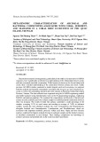
paracrine system. Emerging evidence suggests that Glu could play a dual role in mechanisms underlying the main- tenance of cellular homeostasis – as an excitatory neuro- transmitter in the central neurocrine system and an extracellular signal mediator in peripheral autocrine and/or paracrine tissues. In this review, the possible Glu signaling methods are outlined in specific peripheral tissues including bone, testis, pancreas, and the adrenal, pituitary and pineal glands.
autocrine;
glutamate
tissues; however,
Keywords: receptor; glutamate; glutamate transporter; neurotransmitter; paracrine; vesicular glutamate transporter; peripheral tissues.
The hypothesis that L-glutamate (Glu) is an excitatory amino acid neurotransmitter in the mammalian central nervous system is now gaining more support after the suc- cessful cloning of a number of genes coding for the signaling machinery required for this neurocrine at synapses in the brain. These include Glu receptors (signal detection), Glu transporters (signal termination) and vesicular Glu trans- porters (signal output through exocytotic release). Relatively little attention has been paid to the functional expression of these molecules required for Glu signaling in peripheral neuronal and non-neuronal recent molecular biological analyses show a novel function for Glu as an extracellular signal mediator in the autocrine and/or
second messengers
[4,5]. The group I
Glutamate signaling molecules
Glutamate receptors
subtype lular stimulates formation of inositol 1,4,5-triphosphate and diacylglycerol, while both group II and III subtypes induce reduction of intracellular cyclic AMP (cAMP). On the basis of sequence homology and agonist preference, the latter iGluRs are classified into N-methyl-D-aspartate (NMDA), DL-a-amino-3-hydroxy-5-methylisoxasole-4- propionate (AMPA), and kainate (KA) receptors, which are associated with ion channels permeable to particular cations [6,7].
intracellular
signal
including group I
L-Glutamate (Glu) is accepted as an excitatory amino acid neurotransmitter in the mammalian central nervous sys- tem (CNS). Receptors for Glu (GluRs) are categorized into two major classes, metabotropic (mGluRs) and ionotropic (iGluRs) receptors, according to their differen- transduction mechanisms and tial molecular homologies (Fig. 1) [1–3]. mGluRs are further divided into three distinct subtypes containing seven transmembrane domains, (mGluR1 and mGluR5), group II (mGluR2 and mGluR3) and group III (mGluR4, mGluR6, mGluR7 and mGluR8), in line with each receptor’s exogenous agonists and intracel-
NMDA receptor channels. These channels are highly permeable to Ca2+, with sensitivity to blockade by Mg2+ in a voltage-dependent manner [8,9]. Functional NMDA receptor channels are comprised of heteromeric assemblies between the essential NR1 subunit and one of four different NR2 (A–D) subunits, in addition to one of two different NR3 (A–B) subunits. Expression of the NR2 subunit alone does not lead to composition of functional ion channels in any expression system, while coexpression of each NR2 subunit with an NR1 subunit results in expression of functional channels permeable to Ca2+ in mammalian cells [10]. In contrast, NR3 subunits are a dominant-negative family that suppresses opening of functional NMDA receptor channels [11–13].
AMPA/KA receptor channels. AMPA receptor channels consist of different subunits (GluR1–4), whereas KA receptor channels are constructed using combinations of GluR5–7 subunits. Both KA1 and KA2 subunits are thought to participate in the expression of functional, high affinity KA receptor channels along with other KA receptor subunits [7]. GluR d subunits are cloned by homology screening and classified as iGluRs on the basis of sequence
Correspondence to Yukio Yoneda, Laboratory of Molecular Pharmacology, Kanazawa University Graduate School of Natural Science and Technology, 13–1 Takara-machi, Kanazawa, Ishikawa 920–0934, Japan. Tel.:/Fax: + 81 76 234 4471; E-mail: yyoneda@anet.ne.jp Abbreviations: AP1, activator protein-1; AMPA, DL-a-amino-3-hyd- roxy-5-methylisoxasole-4-propionate; Asp, aspartate; CBFA1, core binding factor a)1 ; CNS, central nervous system; EAAC1, excitatory amino acid carrier 1; EAAT, excitatory amino acid transporter; GLAST, glutamate aspartate transporter; GLT-1, glutamate trans- porter-1; Glu, glutamate; GluR, glutamate receptor; iGluR, iono- tropic glutamate receptor; KA, kainate; mGluR, metabotropic glutamate receptor; MK-801, dizocilpine; NMDA, N-methyl- D-aspartate; t-ACPD, (+/–)-1-aminocyclopentane-trans- 1,3-dicarboxylic acid; VGLUT, vesicular glutamate transporter. (Received 28 August 2003, accepted 4 November 2003)
2 E. Hinoi et al. (Eur. J. Biochem. 271)
(cid:1) FEBS 2003
Table 1. Classification of glutamate transporters and vesicular glutam- ate transporters. Glu transporters are classified into five different sub- types including GLAST (EAAT1), GLT-1 (EAAT2), EAAC1 (EAAT3), EAAT4 and EAAT5, which are responsible for the ter- mination of signal transduction mediated by Glu and for the preven- tion of neurotoxicity mediated by this endogenous excitotoxin. Vesicular Glu transports are divided into three groups, VGLUT1, VGLUT2 and VGLUT3. Expression of VGLUT suffices for definition of a glutamatergic phenotype in neurons.
Transporter Amino acid length (aa) Tissue distribution
Glutamate transporters subtype
GLAST GLT-1 EAAC1 543 576 524
EAAT4 EAAT5 564 560 Brain, retina, testis, bone Brain, retina, liver Brain, intestine, kidney, retina, liver, heart Brain, placenta Retina, liver Vesicular glutamate transporter subtypes
VGLUT1 VGLUT2 560 558
chorea, AIDS
encephalopathy
extracellular Glu released from neurons. In astrocytes, extracellular Glu is incorporated and converted into gluta- mine by glutamine synthetase, and glutamine is shuttled back to neurons for the resynthesis of Glu in neurons [30]. On this astrocyte/neuron, glutamate/glutamine hypothesis for the neurotransmitter compartment, astrocytes play an important role in mechanisms underlying the disposition of Glu at synaptic vesicles through vesicular transporters and the recycling of Glu for exocytotic release. EAAC1 is localized to neurons throughout the CNS [31], whereas EAAT4 localization is largely restricted to cerebellar Purkinje cells [27]. EAAT5 has been shown to exclusively reside in the retina [28] and more specifically on photo- receptor and bipolar rod and cone cells [32].
similarity. Using different pharmacological analyses and expression in transfected cells [14,15] researchers believe that these d subunits are thought to be orphan receptors. In the CNS, these iGluR and mGluRs are thought to mediate excitatory neurotransmission and to play key roles in synaptogenesis, formation of neuronal circuitry and to be involved in mechanisms for synaptic plasticity such as learning and memory [16,17]. Excessive activation of GluRs is thought to participate in the neurodegeneration following a wide range of neurological insults including ischemia, trauma, hypoglycemia and epileptic seizures. Chronic neurodegenerative disorders such as Alzheimer’s disease, Huntington’s and amyotrophic lateral sclerosis may also involve neuronal cell death induced by Glu [18–20].
Vesicular glutamate transporters
Glutamate transporters in the plasma membrane
The proteins responsible for vesicular glutamate transport have only recently been identified when it was recognized that putative inorganic phosphate transporters mediate Glu transport for condensation reactions in synaptic vesicles (Table 1). Within the CNS, vesicular glutamate transporter (VGLUT) isoforms-1 [33,34] and -2 [35,36] appear restricted to known glutamatergic neurons and exhibit a striking complementary pattern of expression at excitatory synapses [37,38], suggesting that they might define the excitatory neuronal phenotype. Indeed, heterol- ogous expression of VGLUT1 or VGLUT2 suffices to convert inhibitory neurons to excitatory ones [34,39,40]. The expression of VGLUT1 and VGLUT2 would account for the exocytotic release of Glu in all known glutamatergic neurons, while VGLUT3 is expressed in a number of cell types suggested previously to release Glu through exocytosis, among them, dopaminergic, GAB- Aergic and serotonergic neurons and astrocytes [41,42]. These previous findings give support to the hypothesis
In the CNS, Glu transporters are essential for the termin- ation of signal transduction mediated by Glu as well as for the prevention of neurotoxicity mediated by this endo- genous excitotoxin. These transporters maintain a 10 000- fold gradient of intracellular Glu (3–10 mM) to extracellular Glu (0.3–1 lM), which is driven by the ionic gradients generated by ion-exchanging pumps such as Na+/K+- ATPase [21,22]. These Glu transporters are classified into five different subtypes including glutamate aspartate trans- porter (GLAST) (EAAT1; excitatory amino acid transpor- ter 1) [23,24], glutamate transporter-1 (GLT-1) (EAAT2) [25], excitatory amino acid carrier (EAAC1) (EAAT3) [26], EAAT4 [27], and EAAT5 [28] to date (Table 1). These Glu transporters display heterologous regional and cellular expression profiles. Both GLAST and GLT-1 are localized to astrocytes, with GLAST predominating in the cerebellum and GLT-1 in the cortex and forebrain, respectively [29]. These transporters are believed to sequester the majority of
VGLUT3 589 Brain, bone Brain, pancreas, pineal gland Brain, retina, liver Fig. 1. Classification of glutamate receptors. GluRs are divided into two major classes, metabotropic (mGluRs) and ionotropic (iGluRs) receptors, according to their differential intracellular signal transduc- tion mechanisms as well as sequence homologies. There are three distinct subtypes of mGluRs, including groups I, II and III, classified based on sensitivity to exogenous agonists and intracellular second messengers employed. iGluRs are subclassified, on the basis of sequence homologies and agonist preference, into NMDA, AMPA and KA receptors, which are all associated with ion channels per- meable to particular cations.
Glutamate signaling in peripheral tissues (Eur. J. Biochem. 271) 3
(cid:1) FEBS 2003
that expression of either VGLUT would suffice for the definition of a glutamatergic phenotype in endocrine cells [43,44] as also shown in neurons [33,34].
fibroblast growth factor) [76,77]. Recent studies have raised the possibility that Glu may be one of the endogenous paracrine (autocrine) factors used for intercellular commu- nications in bone cells [53,54] (Fig. 2).
Fig. 2. Glutamate signaling in bone. Two dis- tinct cell types are known to coordinate to regulate bone formation and maintenance of bone; bone-forming osteoblast and bone- resorbing osteoclasts. Particular functional GluRs are identified in osteoblasts (NMDA receptors, AMPA receptors and mGluRs) as well as in osteoclasts (NMDA receptors). GLAST sensitive to mechanical stress is expressed in both osteoblasts and osteocytes, while VGLUTs are responsible for Glu release from intracellular vesicular constituents through activation of particular iGluR sub- types present in osteoblasts.
Glutamate signaling in peripheral tissues
In mammalian bone, NMDA receptors are also expressed in osteoblasts and osteoclasts as revealed by RT-PCR, in situ hybridization, immunohistochemistry, and electrophysio- logy [54,55,78–81]. Further studies reveal the expression of functional NMDA receptor channels in osteoblasts and osteoclasts. The addition of an NMDA receptor antagonist inhibits cell differentiation in cultured osteoclasts [55,82], while Glu induces elevation of intracellular free Ca2+ in a manner sensitive to antagonism by the NMDA receptor antagonist, dizocilpine (MK-801) in the human osteoblastic cell lines MG63 and SaOS-2 [83]. In our hands, however, expression of mRNA was seen for GLT-1 and EAAT4, but not for either other Glu transporters or any GluR subtypes including NMDA receptors, in mouse primary cultured osteoclasts differentiated from monocyte/macrophage pro- genitor cells purified by Ficoll density gradient centrifugation (E. Hinoi, T. Takarada, M. Inoue and Y. Yoneda, unpub- lished data).
[53–55],
Relatively little attention has been paid to functional expression of Glu signaling molecules in peripheral tissues. Evidence is emerging for a role of Glu as an extracellular signal mediator in the autocrine and/or paracrine system, in addition to an excitatory amino acid neurotransmitter role in the CNS [45]. We have shown previously that specific binding sites of [3H]Glu are localized with high affinity, selectivity and quisqualate sensitivity in rat adrenal [46] and pituitary [47] glands, in addition to the brain. Quisqualate- sensitive, Cl–/Ca2+-dependent [3H]Glu binding is also seen in bovine pineal membranes [48]. Both NMDA and L-Glu induce a rapid contraction of the guinea pig ileum in a manner sensitive to NMDA antagonists [49–51]. Contrac- tion is also induced by Glu in rat bronchial smooth muscle with pharmacological profiles different from those for any known GluRs in the brain [52]. In addition to these pioneering previous studies, recent molecular biological analyses give support to the expression of particular Glu signaling molecules in a variety of different neuronal and locations. These include bone non-neuronal peripheral (osteocyte, osteoblast, and osteoclast) testis [56,57], pancreas [44,58,59], adrenal gland [60,61], pituitary gland [62,63], pineal gland [64,65], taste buds [66], lung [67], hepatocyte [68], thymus [69], cerebral endothelium [70], megakaryocytes [71], keratinocytes [72], lymphocytes [73], platelets [74], and heart [75]. In this article, we focus on Glu signaling machinery expressed in particular tissues.
Bone
In addition to NMDA receptors, osteoblasts constitu- tively express mRNA for non-NMDA receptors such as the GluR3 subunit of AMPA receptors and KA1 and KA2 subunits of KA receptors [84]. AMPA receptors modulate the exocytotic release of Glu from cultured osteoblasts [85,86]. An antagonist for AMPA receptors is shown to significantly inhibit the release of endogenous Glu in a concentration-dependent manner in MG-63 osteosarcoma cells [85], whereas AMPA facilitates the release of endo- genous Glu from cultured osteoblasts in the presence of the inhibitor of AMPA receptor desensitization cyclothiazide [86]. Moreover, constitutive expression is also shown for particular mGluR (mGluR4 and mGluR8) that are coupled negatively to adenylyl cyclase thus inhibiting the formation of cAMP stimulated by forskolin in cultured rat calvarial osteoblasts during different developmental states [79], and prior stimulation of mGluR by (+/–)-1-aminocyclopen- tane-trans-1,3-dicarboxylic acid (t-ACPD) leads to reduc- tion of NMDA-induced whole cell current in cultured rat femoral osteoblasts [87]. Of the EAAT subtypes cloned to date, both GLAST and GLT-1 are shown to reside in bone with respect to mRNA and corresponding proteins, without expression of immunoreactive EAAC1 [53,88]. Mechanical
Two distinct cell types are known to coordinate to regulate bone formation and maintenance, these are bone-forming osteoblast and bone-resorbing osteoclasts. An imbalance between these cells leads to pathogenesis and to etiologies of certain metabolic bone diseases including osteoporosis, Paget’s disease, and osteopetrosis [76,77]. The balancing mechanism, at least in part, involves endocrine control (via estrogen and parathyroid hormone) and paracrine (auto- crine) control (via interleukin, insulin-like growth factor and
4 E. Hinoi et al. (Eur. J. Biochem. 271)
(cid:1) FEBS 2003
loading is shown to down-regulate GLAST expressed in osteocytes when determined by immunohistochemistry [53]. In GLAST knockout mice, however, no marked differences are seen in a variety of phenotypes including mandible and long bone size, morphology, trabeculation, regions of muscle attachment, resorption lacunae, and areas of formation vs. resorption of bone, compared with wild-type siblings [89]. In addition to Glu transporters expressed at the cell surface, both RT-PCR and immunohistochemical analyses reveal constitutive expression of membrane and intracellular molecular machinery generally required for vesicular release of a central neurotransmitter in osteoblastic cell lines as well as primary cultured osteoblasts [90]. We have also demonstrated constitutive expression of particular VGLUT in cultured osteoblasts irrespective of maturity of cultured osteoblasts [86].
controlled release of sex steroids [101–103]. The seminifer- ous tubules include germ cells as well as somatic cells such as Sertoli cells. Spermatogenesis in mammals is characterized by intense and continuous proliferative activity with a complex pattern of mitotic divisions in different types of spermatogonia, culminating in meiotic cleavage of primary spermatocytes to produce noncycling haploid cells (sperm- atids). Development of male germ cells in the seminiferous tubules is dependent on the action of pituitary gonando- tropins and androgens secreted from Leydig cells. The entire process of germ cell development, moreover, also relies on Sertoli cells for structural and nutritional support. For instance, germ cells are segregated from the systemic circulation because of the blood–testis barrier created by tight junctions between Sertoli cells near the basal lamina [104,105] as seen with astroglial cells for the blood–brain barrier.
Core binding factor a)1 (CBFA1) is a transcription factor essential for the growth of osteoblasts and is a master regulator at the differentiation stage [91,92]. Disruption of CBFA1 prevents skeltogenesis and heterozygous mutations lead to cleidocranial dysplasia, an autosomal dominant disorder [93,94]. CBFA1 is also shown to regulate expression of several genes such as collagenase-3, type (I) collagen, osteoprotegerin, bone sialoprotein, osteopontin, and alkaline phosphatase in addition to osteocalcin [95–97]. We have also recently demonstrated the exacerbation of osteoblastic differentiation by different NMDA receptor antagonists [55]. Sustained exposure to MK-801 invariably leads to marked inhibition of expression of both mRNA and corresponding protein for the master regulator of bone differentiation CBFA1 (in addition to its DNA binding activity) in primary cultured rat calvarial osteoblasts when determined after the stage of cellular differentiation [55]. The antagonist not only inhibits temporal marked expression of CBFA1 seen at the stage of cell differentiation but also prevents Ca2+ accumulation only when added before the period of cell differentiation. The lack of effects of MK-801 on Ca2+ accumulation and cell numbers through- out cell growth, when added after the stage of cell differen- tiation, gives support to the proposal that NMDA receptors predominantly modulate cell differentiation rather than proliferation through a mechanism associated with expres- sion of CBFA1 during cellular maturation in osteoblasts.
In rat testis tissue, mRNA and corresponding proteins are constitutively and functionally expressed for both mGluR1 and mGluR5, but not for mGluR2 and mGluR3 [56]. Immunohistochemical and molecular biological ana- lyses reveal the expression of NR1, GluR2/3, and mGlu R2/3 proteins in kidney, lung, spleen, and testis liver, [106,107]. In addition to functional expression of GluRs, Glu transporters are also expressed in testis tissue. A Northern blot study showed expression of mRNA for a novel Glu transporter later found to be identical to GLAST [22,23] in rat forebrain, cerebellum, and testis [108]. Expression of mRNA is also shown for a neuron- specific Glu transporter (later identified as EAAC1) isolated from human brain cDNA libraries in human brain, liver, muscle, ovary and testis using Northern blot analysis [109]. Figure 4 shows our recent findings on the distribution profiles of different Glu transporters expressed in rat testis. In Fig. 3A, a schematic representation is shown for histological characteristics of testis tissue. In these experiments, rat testes were removed and frozen sections dissected with subsequent fixation with parafor- maldehyde for detection of localization of each mRNA using DIG-labeled cRNA probe. In situ hybridization evaluation demonstrated clearly the localized expression of mRNA for GLAST in the interstitial space of rat testis (Fig. 3b). Constitutive expression is seen with mRNA for GLT-1 in elongated spermatids of rat testis, while expres- sion of EAAT5 mRNA is found in the basal compartment of seminiferous tubule in rat testis. However, mRNA expression is not seen for EAAC1 and EAAT4 in any particular structures of rat testis.
As mentioned above, bone cells could express all mole- cular machinery required for Glu signaling in the CNS. Glu signals in bone, as seen at glutamatergic synapses in the brain, could be dispatched through exocytotic release from vesicles expressing VGLUT for condensation in vesicular particles, transduced specifically via both iGluRs and mGluRs and terminated by Glu transporters. Moreover, bone is supposed to have sympathetic and glutamatergic innervations [98–100]. Accordingly, Glu could have a pivotal dual role in mechanisms associated with maintenance of homeostasis as an excitatory neurotransmitter (toward neurocrine pro- cesses in the CNS) and as a trophic factor (toward autocrine and/or paracrine processes in bone).
Testis
D-Aspartate (D-Asp) endogenously occurs in rat adeno- hypophysis, testis, adrenal gland, and brain in a rank order of decreasing levels, while an intraperitoneal injection of exogenous D-Asp induced a marked increase in its accu- mulation in these neuroendocrine tissues [110]. Exposure of cultured rat Leydig cells to D-Asp selectively stimulates testosterone synthesis after its incorporation into cells through GLAST but exposure to L- and D-Glu, L-Asp and L- and D-aspargine is ineffective [57]. The prevailing view, that all Glu transporters cloned to date are unable to differentiate between L- and D-Asp as a substrate, argues in favor of a role for endogenous D-Asp in testosterone synthesis following incorporation through particular Glu transporters expressed at surfaces of Leydig cells.
A testis consists of seminiferous tubules and interstitial cells, specifically Leydig cells, having unique functions in the male body – the generation of gametes and the production and
Glutamate signaling in peripheral tissues (Eur. J. Biochem. 271) 5
(cid:1) FEBS 2003
Pancreas
The islet of Langerhans, a pancreatic miniature organ for several hormones regulating the blood glucose level, is composed of four major types of endocrine cells, including glucagon-secreting alpha (a) cells, insulin-secreting beta (b) cells, somatostatin-secreting d cells, and pancreatic polypep- tide-secreting F cells. The B cells are the most numerous and are concentrated in the center of the islet, while the a cells are larger and less numerous and located at the periphery of the islet [111,112]. By molecular biological,
insulin-secreting pancreatic b cells [58], and also induced by both AMPA and KA, but not by NMDA, in isolated pancreatic islets [113,123]. Similarly, Glu stimulates glu- cagon secretion via AMPA receptors in rat isolated pancreatic islets [124]. It has been demonstrated that mGluRs also participate in hormone secretion from pancreatic islets. Agonists for group I and II mGluR subtypes increase the release of insulin in the presence of glucose at low concentrations, whereas a group III mGluR agonist inhibits insulin release determined at high glucose concentrations [119]. The activation of group III mGluR subtype expressed in pancreatic a cells leads to inhibition of glucagon release [59]. A high-affinity Glu transporter expressed in pancreatic islets of Langerhans modulates insulin secretion stimulated by glucose [121]. Besides expression in neuronal cells, moreover, both VGLUT1 and VGLUT2 are expressed preferentially in aTC6 cells, clonal pancreatic a cells, as well as a cells isolated from Langerhans islets [43,44]. Low glucose conditions result in cosecretion of stoichiometric amounts of Glu and glucagon from aTC6 cells and isolated islets,
electrophysiological, and immunohistochemical studies, it has been reported that these islet cells should express functional Glu receptors [58,59,113–119], Glu transporters [120,121] and VGLUT [43,44,122], suggesting that Glu could function as an intercellular signal mediator in the islet (Fig. 4). Several independent lines of evidence indicate that iGluR stimu- lation positively modulates secretion of both glucagon and insulin in pancreatic islets. Insulin secretion is stimulated by AMPA, KA and NMDA in MIN6 cells derived from
Fig. 3. Analyses on localization of Glu trans- porters in rat testis. (A) Schematic represen- tation of the histological characteristics of testis. (B) Rat testicular frozen sections were fixed with paraformaldehyde and then incu- bated with DIG-labeled cRNA probe for GLAST, GLT-1, EAAC1, EAAT4 and EAAT5 for in situ hybridization. Typical micrographs are shown in the figures with similar results in three independent experi- ments. Scale bars, 50 lm.
6 E. Hinoi et al. (Eur. J. Biochem. 271)
(cid:1) FEBS 2003
revealed by in situ hybridization. We could also detect both mRNA and corresponding proteins for NR1, NR2C and NR2D in rat adrenal medulla [61]. Furthermore, an intraperitoneal injection of a blocker of NMDA receptor channels, but not NMDA itself, leads to a marked increase in DNA binding activity of the nuclear transcription factor AP-1 in rat adrenal glands [61]. Single episodes of immo- bilization stress induces a significant increase in NR1 mRNA expression in adrenal glands [136], while a single dose of morphine decreases NR1 mRNA in adrenal glands but not in hippocampus [137]. The prior systemic admin- istration of NMDA prevents the marked increase in adrenal AP1 DNA binding induced by subsequent stress [138]. In rat adrenal glands, the expression of GLAST, which has an affinity for D-Asp, is transiently increased at 3 weeks of age [139] and localization patterns are almost coincident with those of endogenous D-Asp within the glands. Adrenal glands receive sympathetic efferent and afferent innerva- tions in addition to the typical cholinergic innervation [140,141]. A mechanism for Glu release is detected in bovine adrenal chromaffin cells [142] and glutamatergic innervation is also found in sympathoadrenal neurons [143,144].
Pituitary gland
which triggers GABA secretion from B cells through GluRs in the isolated islets [44].
Adrenal gland
The pituitary gland is connected to the hypothalamus at the base of the brain and consequentially important anatomical and functional relationships exist. During embryogenesis, the pituitary gland develops from the oral ectoderm and also nerve tissue. The pituitary tissue from nerve tissue is referred to as the neurohypophysis and consists of the pars nervosa and infundibulum, while the pituitary gland derived from oral ectoderm is known as the adenohypophysis and has three different areas – the pars distalis or anterior lobe, the pars tuberalis and the pars intermedia [145,146]. A portion of the pars intermedia is sometimes included in the terminology (cid:1)posterior lobe(cid:2).
Adrenal gland consists of two concentric layers; adrenal cortex which is a yellow peripheral layer, and adrenal medulla which is a reddish-brown central layer. Adrenal cortex has the characteristics of steroid-synthesizing tissues, with subdivision into the three concentric layers – the zona glomerulosa, the zona fasciculate and the zona reticularis. The zona glomerulosa secrets mineralocorticoids such as aldosterone (involved in the maintenance of balance between electrolytes and water), while the zona fasciculata and probably the zona reticularis secret the glucocorticoids corticosterone and cortisol (both relevant to the regulation of metabolism of carbohydrates, proteins, and fats). Adrenal medulla is composed of polyhedral parenchymal cells and supported by a reticular fiber network, and secretes both adrenaline and noradrenaline [125,126].
The role of Glu in adrenal glands has been investigated in independent research laboratories including ours. In addi- tion to Glu [127], iGluR agonists (NMDA, AMPA and KA) and an mGluR agonist (t-ACPD) [128] all stimulate catecholamine release from adrenal glands. We have shown previously the localization of [3H]Glu binding sites with stereoselectivity, high affinity and saturability in rat adrenal medulla [46,129]. Adrenal [3H]Glu binding sites are solubi- lized by detergents with pharmacological profiles similar to those found in membrane preparations [130,131]. On sections of rat [132] and bovine [133] adrenal glands, [3H]Glu binding sites are of high affinity, rapid, saturable, reversible, stereospecific, and specific to a single population. In addition to these early studies using conventional techniques, recent molecular biological analyses support the presence of particular iGluR subunits in adrenal glands. Rat adrenal cortex expresses AMPA receptors [134], but adrenal medulla expresses NMDA receptors [135] as
Molecular biological and immunohistochemical analyses demonstrated the presence of both non-NMDA [62,147, 148] and NMDA receptors [149,150], while studies using autoradiographic [151] and receptor binding [47,152] tech- niques showed the localization of [3H]Glu binding in the pituitary gland. Several independent lines of evidence for direct regulation by Glu of hormone secretion in isolated anterior pituitary cells are available in the literature. The secretion of prolactin is stimulated by Glu in a manner sensitive to prevention by MK-801 in primary cultures of anterior pituitary cells [153,154]. Both NMDA and KA facilitate the secretion of growth hormone in dispersed anterior pituitary cells [155], while KA is effective in stimulating gonandotropin secretion from the anterior pituitary [156]. In single primary cultured rat anterior pituitary cells, Glu induces a marked increase in cytosolic free Ca2+ concentration by a nonsynaptic mechanism [157]. These previous findings show functional expression of particular subtypes of GluRs in the pituitary gland. We have also demonstrated that the rat pituitary gland contains both mRNA and corresponding proteins for GluR6/7 subunits of KA receptors in addition to specific binding sites of [3H]KA [63]. Moreover, an intraperitoneal injection of KA results in increased AP1 DNA binding in rat pituitary gland as well as hippocampus [63].
Fig. 4. Glutamate signaling in pancreas. The islet of Langerhans, a pancreatic miniature organ, is composed of four major types of endocrine cells, including a, b, d and F cells. Particular functional GluRs, which could modulate secretion of hormones, are identified in a cells (AMPA receptors, KA receptors and mGluRs), in b cells (AMPA receptors), and in d cells (KA receptors), respectively. High- affinity Glu transporters are expressed in both a and d cells, while VGLUTs are preferentially expressed in a cells.
Glutamate signaling in peripheral tissues (Eur. J. Biochem. 271) 7
(cid:1) FEBS 2003
Pineal gland
Mammalian pineal gland consists of several types of cells, principally pinealocytes and astroglial cells. Pinealocytes are endocrine cells with the ability to synthesize and secret melatonin, a hydrophobic hormone that mediates many physiological functions such as circadian rhythms and seasonal reproduction [158,159].
[172–174].
autoradiographic binding of [3H]MK-801. Blockade of NMDA receptors, by specific antagonists, inhibits the phorbol myristate acetate-induced increases in cellular size, CD41 expression and mutual adhesion in MEG-01 clonal megakaryoblastic cells [71]. Both NMDA and non-NMDA receptors are expressed in rat heart [75], while the stimula- tion of iGluRs by Glu, NMDA, AMPA and KA invariably leads to an increase in intracellular Ca2+ oscillation frequency in cultured rat myocardial cells [182]. Moreover, mGluRs (mGluR1, mGluR2/3, and mGluR5) are also expressed in rat heart [183]. Expression of NMDA receptors has been demonstrated in lung [184,185] and NMDA elicits acute high-permeability edema in perfused rat lungs [67]. Nitric oxide synthase inhibitors and the neuropeptide vasoactive intestinal peptide protect against injuries medi- ated by NMDA receptors in lung [186]. In whole guinea-pig lungs perfused via the trachea, NMDA increases airway perfusion pressure in a manner that is totally abolished by MK-801 [187]. Expression of particular GluRs is shown in skin including keratinocytes for NMDA and AMPA receptors [72,188] and melanocytes for mGluR5 [189]. Blockade of NMDA receptors expressed in keratinocytes, induces stratification and other characteristics of differen- tiation [72]. Another excitatory amino acid, L-Asp, inhibits acid secretion stimulated by histamine, without affecting that by oxotremorine and pentagastrin in an everted preparation of isolated rat stomach [190], while an NMDA receptor antagonist blocks the inhibitory effect of L-Asp on histamine-stimulated acid secretion [190].
Conclusions
[3H]Glu binding is also seen in bovine [48] and rat [160] pineal membranes. Exogenous Glu leads to inhibition of the activities of serotonin N-acetyltransferase [161] and hydroxyindole O-methyltransferase [162], with concomit- ant suppression of both synthesis and secretion of mela- tonin [163,164]. Furthermore, expression of particular GluRs has been confirmed in the mammalian pineal gland in molecular biological and immunohistochemical studies to date. These include GluR delta [165], AMPA [166], KA [167], NMDA [167], group I mGluR [168,169], and group II mGluR [64,169]. Activation of mGluR3 negatively serotonin N-acetyl- regulates noradrenaline-dependent transferase activity and melatonin synthesis in rat pinealo- cytes [64], while GluR1 triggers exocytosis mediated by microvesicles (counterparts of the neuronal synaptic ves- icles) of Glu through activation of L-type Ca2+ channels in rat pinealocytes [65]. Immunoreactive Glu is enriched in pinealocytes of gerbil pineal gland [170], and pinealocytes accumulate Glu in microvesicles [171] for secretion through Ca2+-dependent exocytosis In mammalian pinealocytes, expression of VGLUT2 is confirmed in addition to expression in Langerhans islets [43]. Extracel- lular Glu is sequestered by pinealocytes through a GLT-1 type, Na+-dependent Glu transporter expressed in the plasma membrane [175], whilst immunoreactivity to GLAST is detected in interstitial glial cells but not in pinealocytes [176].
Other tissues
It appears that the molecular machinery required for Glu signaling, which includes iGluRs, mGluRs, Glu transport- ers and VGLUTs, are constitutively expressed to play a role crucial for maintenance of the functionality and integrity in several neuronal and non-neuronal peripheral tissues for (Table 2). Circulating Glu seems to be sufficient saturation of any of the subtype of iGluRs and mGluRs expressed at peripheral locations with intimate contacts to the blood supply. One possibility is that peripheral GluRs could be under the influence of tonic stimulation by Glu in plasma, whereas an alternative explanation is that circula- ting Glu may not easily gain access to peripheral GluRs due to particular protection mechanisms against the direct exposure to blood supply. From this point of view, the exact origin of an endogenous agonist is still unclear in contrast to the conclusive evidence for expression of mRNA for particular iGluR and mGluR subtypes as well as for VGLUTs in peripheral tissues.
In any case, Glu seems to play a dual role in mechanisms underlying maintenance of homeostasis – an excitatory neurotransmitter in the central neurocrine system and an extracellular signal mediator in the peripheral autocrine and/or paracrine process. Searches for ligands at GluRs and Glu transporters may need to be rerun with regard to permeability to the brain across blood–brain barrier. Nonpermeable compounds could be of a great benefit as therapeutic drugs without serious central side-effects for a variety of disorders associated with malfunction of Glu signaling in the periphery. In contrast to the prevailing view, a new concept could be required for classification of
In rat hepatocytes, the mGluR agonists, t-ACPD and quisqualate, not only stimulate polyphosphoinositide hydrolysis, with an antagonist for group I mGluR subtype protecting against hypoxic damage [68]. Constitutive expression of particular iGluRs, including NR1, NR2D, KA2, and GluR delta-1 subunits [177], is found in lingual epithelium obtained from rat foliate and vallate papillae, in addition to truncated a variant of mGluR4a [66,178]. These GluRs may be responsible for sensory transduction of (cid:1)umami(cid:2) taste in addition to the taste-specific receptor, T1R1/T1R3 dimer, which is coupled to G-protein for activation by dietary Glu [179]. The expression of both group I and group II mGluRs is demonstrated in mouse thymus, isolated thymocytes and thymic stromal cell line [69]. The quisqualate-sensitive binding of [3H]Glu is detected in T lymphocytes [180], whereas in human lymphocytes, Glu and other iGluR agonists potentiate the rise of intracellular free Ca2+ concentrations induced by the mAb anti-CD(3) or phytohaemagglutinin [73]. In human platelets, Glu or NMDA is antagonistic for the aggregating activity of arachidonic acid, with detection of both NMDA-displaceable [3H]Glu binding and [3H]MK-801 binding [74,181]. In megakaryocytes, both mRNA and proteins for NMDA receptors are expressed in addition to
8 E. Hinoi et al. (Eur. J. Biochem. 271)
(cid:1) FEBS 2003
8. Reynolds, I.J. & Miller, R.J. (1988) Multiple sites for the reg- ulation of the N-methyl-D-aspartate receptor. Mol. Pharmacol. 33, 581–584. 9. MacDonald, J.F. & Nowak, L.M.
(1990) Mechanisms of blockade of excitatory amino acid receptor channels. Trends. Pharmacol. Sci. 11, 167–172. Table 2. Expression of Glu signaling molecules in several neuronal and non-neuronal peripheral tissues. iGluR (A, AMPA receptors; K, KA receptors; N, NMDA receptors); mGluR (I, group I mGluR; II, group II mGluR; III, group III mGluR); GluT (1, GLAST; 2, GLT-1; 3, EAAC1; 4, EAAT4; 5, EAAT5); VGLUT (1, VGLUT1; 2, VGLUT2; 3, VGLUT3). ND, not determined; ?, unidentified.
Tissue/cell iGluR mGluR GluT VGLUT 10. Lynch, D.R. & Guttmann, R.P. (2001) NMDA receptor phar- macology: perspectives from molecular biology. Curr. Drug Targets 2, 215–231.
ND ND + (1) + (1) ND ND + (1) ND + (1,2,5) ND 11. Das, S., Sakaki, Y.F., Rothe, T., Premkumar, L.S., Takasu, M., Crandall, J.E., Dikkes, P., Conner, D.A., Rayudu, P.V., Cheung, W., Chen, H.S., Lipton, S.A. & Nakanishi, N. (1998) Increased NMDA current and spine density in mice lacking the NMDA receptor subunit NR-3A. Nature 393, 377–381. ? + (1,2) ND ND
12. Nishi, M., Hinds, H., Lu, H.P., Kawata, M. & Hayashi, Y. (2001) Motoneuron-specific expression of NR-3B, a novel NMDA-type glutamate receptor subunit that works in a domi- nant-negative manner. J. Neurosci. 21, 1–6. + (A,K,N) + (I, II) + (N) ND + (A,K,N) + (I, II) + (A,K,N) + (I, II, III) + (2) + (1) ND + (1,2) + (2) ND ND ND ND + (A,K,N) + (I, II) ND + (K,N) + (I) + (III)
13. Matsuda, K., Kamiya, Y., Matsuda, S. & Yuzaki, M. (2002) Cloning and characterization of a novel NMDA receptor subunit NR3B: a dominant subunit that reduces calcium permeability. Mol. Brain Res. 100, 43–52.
ND ? ? + (I, II) ND ND ND 14. Araki, K., Meguro, H., Kushiya, E., Takayama, C., Inoue, Y. & Mishina, M. (1993) Selective expression of the glutamate receptor channel delta 2 subunit in cerebellar Purkinje cells. Biochem. Biophys. Res. Commun. 197, 1267–1276. + (A,K,N) + (I, II) + (N)
Osteoblast Osteoclast Osteocyte Testis Pancreas Adrenal gland + (A,K,N) Pituitary gland + (A,K,N) ND Pineal gland Hepatocyte Lingual epithelium Thymus T lymphocyte Platelet Megakaryocyte + (N) Heart Lung Keratinocyte + (A,N) Melanocyte ND ND ND + (I) ND ND ND ND + (1,3) ND + (2,3) ND ND ND ND ND ND ND ND ND 15. Lomeli, H., Sprengel, R., Laurie, D.J., Kohr, G., Herb, A. & Seeburg, P. (1993) The rat delta-1 and delta-2 subunits extend the excitatory amino acid receptor family. FEBS Lett. 315, 318– 322.
extracellular signal mediators between different cells for neurocrine, endocrine, autocrine, and paracrine communi- cations.
16. Scheetz, A.J. & Constantine-Paton, M. (1994) Modulation of NMDA receptor function: implications for vertebrate neural development. FASEB J. 8, 745–752.
17. Riedel, G., Platt, B. & Micheau, J. (2003) Glutamate receptor function in learning and memory. Behav. Brain Res. 140, 1–47. 18. Choi, D.W. (1988) Glutamate neurotoxicity and diseases of the nervous system. Neuron 1, 623–634.
Acknowledgements
This work was supported in part by Grants-in-Aids for Scientific Research to E.H. and Y.Y. from the Ministry of Education, Culture, Sports, Science and Technology, and to Y.Y. from the Ministry of Health, Labour and Welfare, Japan, respectively. 19. Goff, D.C. & Wine, L. (1997) Glutamate in schizophrenia: clin- ical and research implications. Schizophr. Res. 27, 157–168. 20. Cull-Candy, S., Brickley, S. & Farrant, M. (2001) NMDA receptor subunits: diversity, development and disease. Curr. Opin. Neurobiol. 11, 327–335.
References
21. Schousboe, A. & Divac, I. (1979) Difference in glutamate uptake in astrocytes cultured from different brain regions. Brain Res. 177, 407–409. 22. Danbolt, N.C. (2001) Glutamate uptake. Prog. Neurobiol. 65, 1–105. 1. Hollmann, M., O’Shea-Greenfield, A., Rogers, S.W. & Heine- mann, S. (1989) Cloning by functional expression of a member of the glutamate receptor family. Nature 342, 643–648.
23. Storck, T., Schulte, S., Hofmann, K. & Stoffel, W. (1992) Structure, expression, and functional analysis of a Na+- dependent glutamate/aspartate transporter from rat brain. Proc. Natl Acad. Sci. USA 89, 10955–10959. 2. Nakanishi, N., Shneider, N.A. & Axel, R. (1990) A family of glutamate receptor genes: evidence for the formation of hetero- multimeric receptors with distinct channel properties. Neuron 5, 569–581.
3. Yoneda, Y., Kuramoto, N., Kitayama, T. & Hinoi, E. (2001) Consolidation of transient ionotropic glutamate signals through nuclear transcription factors in the brain. Prog. Neurobiol. 63, 697–719. 24. Tanaka, K. (1993) Cloning and expression of a glutamate transporter from mouse brain. Neurosci. Lett. 159, 183–186. 25. Pines, G., Danbolt, N.C., Bjoras, M., Zhang, Y., Bendahan, A., Eide, L., Koepsell, H., Storm-Mathisen, J., Seeberg, E. & Kan- ner, B.I. (1992) Cloning and expression of a rat brain L-glutamate transporter. Nature 360, 464–467.
4. Masu, M., Tanabe, Y., Tsutida, K., Shigemoto, R. & Nakanishi, S. (1991) Sequence and expression of a metabotropic glutamate receptor. Nature 349, 760–765. 26. Kanai, Y. & Hediger, M.A. (1992) Primary structure and func- tional characterization of a high-affinity glutamate transporter. Nature 360, 467–471.
5. Tanabe, Y., Masu, M., Ishii, T., Shigemoto, R. & Nakanishi, S. (1992) A family of metabotropic glutamate receptors. Neuron 8, 169–179. 6. Wisden, W. & Seeburg, P.H. (1993) Mammalian ionotropic 27. Fairman, W.A., Vandenberg, R.J., Arriza, J.L., Kavanaugh, M.P. & Amara, S.G. (1995) An excitatory amino-acid transporter with properties of a ligand-gated chloride channel. Nature 375, 599–603. glutamate receptors. Curr. Opin. Neurobiol. 3, 291–298. 7. Hollmann, M. & Heinemann, S. (1994) Cloned glutamate 28. Arriza, J.L., Eliasof, S., Kavanaugh, M.P. & Amara, S.G. (1997) Excitatory amino acid transporter 5, a retinal glutamate trans- receptors. Ann. Rev. Neurosci. 17, 31–108.
Glutamate signaling in peripheral tissues (Eur. J. Biochem. 271) 9
(cid:1) FEBS 2003
porter coupled to a chloride conductance. Proc. Natl Acad. Sci. USA 94, 4155–4160.
Moriyama, Y. (2003) Secretory granule-mediated co-secretion of L-glutamate and glucagon triggers glutamatergic signal transmission in islets of Langerhans. J. Biol. Chem. 278, 1966– 1974. transporters rat brain: quantitative in the 45. Skerry, T.M. & Genever, P.G. (2001) Glutamate signalling in
non-neuronal tissues. Trend Pharmacol. Sci. 22, 174–181. 46. Yoneda, Y. & Ogita, K. (1986) Localization of [3H]glutamate binding sites in rat adrenal medulla. Brain Res. 383, 387–391. 47. Yoneda, Y. & Ogita, K. (1986) [3H]Glutamate binding sites in the 29. Lehre, K.P., Levy, L.M., Ottersen, O.P., Storm-Mathisen, J. & Danbolt, N.C. (1995) Differential expression of two glial gluta- mate and immunocytochemical observations. J. Neurosci. 15, 1835–1853. 30. Hamberger, A., Chiang, G.H., Sandoval, E. & Cotman, C.W. (1979) Glutamate as a CNS transmitter. II. Regulation of synthesis in the releasable pool. Brain Res. 168, 531–541. rat pituitary. Neurosci. Res. 3, 430–435.
31. Kanai, Y., Bhide, P.G., DiFiglia, M. & Hediger, M.A. (1995) Neuronal high-affinity glutamate transport in the rat central nervous system. Neuroreport 6, 2357–2362. 48. Govitrapong, P., Ebadi, M. & Murrin, L.C. (1986) Identification of a Cl–/Ca2+-dependent glutamate (quisqualate) binding site in bovine pineal organ. J. Pineal Res. 3, 223–234.
32. Pow, D.V. & Barnett, N.L. (2000) Developmental expression of excitatory amino acid transporter 5: a photoreceptor and bipolar cell glutamate transporter in rat retina. Neurosci. Lett. 280, 21–24. 49. Moroni, F., Luzzi, S., Franchi-Micheli, S. & Zilletti, L. (1986) The presence of N-methyl-D-aspartate-type receptors for glu- tamic acid in the guinea-pig myenteric plexus. Neurosci. Lett. 68, 57–62.
33. Bellocchio, E.E., Reimer, R.J., Fremeau, R.T. Jr & Edwards, R.H. (2000) Uptake of glutamate into synaptic vesicles by an inorganic phosphate transporter. Science 289, 957–960.
50. Luzzi, S., Zilletti, L., Franchi-Micheli. A.M. & Moroni, F. (1988) Agonists, antagonists, and modulators of excitatory amino acid receptors in the guinea-pig myenteric plexus. Br. J. Pharmacol. 95, 1271–1277.
51. Shannon, H.E. & Sawyer, B.D. (1989) Glutamate receptors of N-methyl-D-aspartate subtype in the myenteric plexus of guinea- pig ileum. J. Pharmacol. Exp. Ther. 251, 518–523. 34. Takamori, S., Rhee, J.S., Rosenmund, C. & Jahn, R. (2000) Identification of a vesicular glutamate transporter that defines a glutamatergic phenotype in neurons. Nature 407, 189–194. 35. Bai, L., Xu, H., Collins, J.F. & Ghishan, F.K. (2001) Molecular and functional analysis of a novel neuronal vesicular glutamate transporter. J. Biol. Chem. 276, 36764–33679.
52. Aas, P., Tanso, R. & Fonnum, F. (1989) Stimulation of peri- pheral cholinergic nerves by glutamate indicates a new peripheral glutamate receptor. Eur. J. Pharmacol. 164, 93–102.
36. Varoqui, H., Schafer, M.K., Zhu, H., Weihe, E. & Erickson, J.D. (2002) Identification of the differentiation-associated Na+/PI transporter as a novel vesicular glutamate transporter expressed in a distinct set of glutamatergic synapses. J. Neurosci. 22, 142–155.
53. Mason, D.J., Suva, L.J., Genever, P.G., Patton, A.J., Steuckle, S., Hillam, R.A. & Skerry, T.M. (1997) Mechanically regulated expression of a neural glutamate transporter in bone: a role for excitatory amino acids as osteotropic agents? Bone 20, 199–205.
37. Fremeau, R.T. Jr, Troyer, M.D., Pahner, I., Nygaard, G.O., Tran, C.H., Reimer, R.J., Bellocchio, E.E., Fortin, D., Storm- Mathisen, J. & Edwards, R.H. (2001) The expression of vesicular glutamate transporters defines two classes of excitatory synapse. Neuron 31, 247–260. 54. Chenu, C., Serre, C.M., Raynal, C., Burt-Pichat, B. & Delmas, P.D. (1998) Glutamate receptors are expressed by bone cells and are involved in bone resorption. Bone 22, 295–299.
55. Hinoi, E., Fujimori, S. & Yoneda, Y. (2003) Modulation of cel- lular differentiation by N-methyl-D-aspartate receptors in osteo- blasts. FASEB J. 17, 1532–1534.
38. Herzog, E., Bellenchi, G.C., Gras, C., Bernard, V., Ravassard, P., Bedet, C., Gasnier, B., Giros, B. & El Mestikaway, S. (2001) The existence of a second vesicular glutamate transporter specifies subpopulations of glutamatergic neurons. J. Neurosci. 21, RC181.
56. Storto, M., Sallese, M., Salvatore, L., Poulet, R., Condorelli, D.F., Dell’Albani, P., Marcello, M.F., Romeo, R., Piomboni, P., Barone, N., Nicoletti, F., De Nicoletti, F. & Blasi, A. (2001) Expression of metabotropic glutamate receptors in the rat and human testis. J. Endocrinol. 170, 71–78. 39. Takamori, S., Rhee, J.S., Rosenmund, C. & Jahn, R. (2001) Identification of differentiation-associated brain-specific phos- phate transporter as a second vesicular glutamate transporter (VGLUT2). J. Neurosci. 21, RC182.
57. Nagata, Y., Homma, H., Lee, J.A. & Imai, K. (1999) D-Aspartate stimulation of testosterone synthesis in rat Leydig cells. FEBS Lett. 444, 160–164. 40. Kaneko, T. & Fujiyama, F. (2002) Complementary distribution of vesicular glutamate transporters in the central nervous system. Neurosci. Res. 42, 243–250.
58. Gonoi, T., Mizuno, N., Inagaki, N., Kuromi, H., Seino, Y., Miyazaki, J. & Seino, S. (1994) Functional neuronal ionotropic glutamate receptors are expressed in the non-neuronal cell line MIN6. J. Biol. Chem. 269, 16989–16992.
41. Schafer, M.K., Varoqui, H., Defamie, N., Weihe, E. & Erickson, J.D. (2002) Molecular cloning and functional identification of mouse vesicular glutamate transporter 3 and its expression in subsets of novel excitatory neurons. J. Biol. Chem. 277, 50734– 50748.
59. Tong, Q., Ouedraogo, R. & Kirchgessner, A.L. (2002) Locali- zation and function of group III metabotropic glutamate receptors in rat pancreatic islets. Am. J. Physiol. Endocrinol. Metab. 282, 1324–1333.
42. Fremeau, R.T. Jr, Burman, J., Qureshi, T., Tran, C.H., Proctor, J., Johnson, J., Zhang, H., Sulzer, D., Copenhagen, D.R., Storm- Mathisen, J., Reimer, R.J., Chaudhry, F.A. & Edwards, R.H. (2002) The identification of vesicular glutamate transporter 3 suggests novel modes of signaling by glutamate. Proc. Natl Acad. Sci. USA 99, 14488–14493.
60. Kristensen, P. (1993) Differential expression of AMPA glutamate receptor mRNA in the rat adrenal glands. FEBS Lett. 332, 14–18. 61. Hinoi, E., Fujimori, S., Nakamura, Y., Balcar, V.J., Kubo, K., Ogita, K. & Yoneda, Y. (2002) Constitutive expression of het- erologous N-methyl-D-aspartate receptor subunits in rat adrenal medulla. J. Neurosci. Res. 68, 36–45. inorganic
43. Hayashi, M., Otsuka, M., Morimoto, R., Hirota, S., Yatsushiro, S., Takeda, J., Yamamoto, A. & Moriyama, Y. (2001) Differ- entiation-associated Na+-dependent phosphate cotransporter (DNPI) is a vesicular glutamate transporter in endocrine glutamatergic systems. J. Biol. Chem. 276, 43400– 43406.
44. Hayashi, M., Yamada, H., Uehara, S., Morimoto, R., Muroyama, A., Yatsushiro, S., Takeda, J., Yamamoto, A. & 62. Kiyama, H., Sato, K. & Tohyama, M. (1993) Characteristic localization of non-NMDA type glutamate receptor subunits in the rat pituitary gland. Brain Res. Mol. Brain Res. 19, 262–268. 63. Hinoi, E. & Yoneda, Y. (2001) Expression of GluR6/7 subunits of kainate receptors in rat adenohypophysis. Neurochem. Int. 38, 539–547.
10 E. Hinoi et al. (Eur. J. Biochem. 271)
(cid:1) FEBS 2003
81. Gu, Y., Genever, P.G., Skerry, T.M. & Publicover, S.J. (2002) The NMDA type glutamate receptors expressed by primary rat osteoblasts have the same electrophysiological characteristics as neuronal receptors. Calcif. Tissue Int. 70, 194–203. 64. Yamada, H., Yatsushiro, S., Ishio, S., Hayashi, M., Nishi, T., Yamamoto, A., Futai, M., Yamaguchi, A. & Moriyama, Y. (1998) Metabotropic glutamate receptors negatively regulate melatonin synthesis in rat pinealocytes. J. Neurosci. 18, 2056– 2062.
65. Yatsushiro, S., Yamada, H., Hayashi, M., Yamamoto, A. & Moriyama, Y. (2000) Ionotropic glutamate receptors trigger microvesicle-mediated exocytosis of L-glutamate in rat pine- alocytes. J. Neurochem. 75, 288–297.
82. Peet, N.M., Grabowski, P.S., Laketic-Ljubojevic, I. & Skerry, T.M. (1999) The glutamate receptor antagonist MK801 mod- ulates bone resorption in vitro by a mechanism predominantly involving osteoclast differentiation. FASEB J. 13, 2179–2185. 83. Laketic-Ljubojevic, I., Suva, L.J., Maathuis, F.J., Sanders, D. & Skerry, T.M. (1999) Functional characterization of N-methyl- D-aspartic acid-gated channels in bone cells. Bone 25, 631–637. 66. Chaudhari, N., Landin, A.M. & Roper, S.D. (2000) A metabo- tropic glutamate receptor variant functions as a taste receptor. Nat. Neurosci. 3, 113–119.
84. Hinoi, E., Fujimori, S., Takemori, A., Kurabayashi, H., Nakamura, Y. & Yoneda, Y. (2002) Demonstration of expres- sion of mRNA for particular AMPA and kainate receptor sub- units in immature and mature cultured rat calvarial osteoblasts. Brain Res. 943, 112–116. 67. Said, S.I., Berisha, H.I. & Pakbaz, H. (1996) Excitotoxicity in the lung: N-methyl-D-aspartate-induced, nitric oxide-dependent, pulmonary edema is attenuated by vasoactive intestinal peptide and by inhibitors of poly (ADP-ribose) polymerase. Proc. Natl. Acad. Sci. USA 93, 4688–4692.
85. Genever, P.G. & Skerry, T.M. (2001) Regulation of spontaneous glutamate release activity in osteoblastic cells and its role in dif- ferentiation and survival: evidence for intrinsic glutamatergic signaling in bone. FASEB J. 15, 1586–1588.
68. Storto, M., de Grazia, U., Knopfel, T., Canonico, P.L., Copani, A., Richelmi, P., Nicoletti, F. & Vairetti, M. (2000) Selective blockade of mGlu5 metabotropic glutamate receptors protects rat hepatocytes against hypoxic damage. Hepatology 31, 649– 655.
86. Hinoi, E., Fujimori, S., Takarada, T., Taniura, H. & Yoneda, Y. (2002) Facilitation of glutamate release by ionotropic glutamate receptors in osteoblasts. Biochem. Biophys. Res. Commun. 297, 452–458.
69. Storto, M., de Grazia, U., Battaglia, G., Felli, M.P., Maroder, M., Gulino, A., Ragona, G., Nicoletti, F., Screpanti, I., Frati, L. & Calogero, A. (2000) Expression of metabotropic glutamate receptors in murine thymocytes and thymic stromal cells. J. Neuroimmunol. 109, 112–120. 87. Gu, Y. & Publicover, S.J. (2000) Expression of functional metabotropic glutamate receptors in primary cultured rat os- teoblasts. Cross-talk with N-methyl-D-aspartate receptors. J. Biol. Chem. 275, 34252–34259.
88. Huggett, J., Vaughan-Thomas, A. & Mason, D. (2000) The open reading frame of the Na+-dependent glutamate transporter GLAST-1 is expressed in bone and a splice variant of this molecule is expressed in bone and brain. FEBS Lett. 485, 13–18.
70. Krizbai, I.A., Deli, M.A., Pestenacz, A., Siklos, L., Szabo, C.A., Andras, I. & Joo, F. (1998) Expression of glutamate receptors on cultured cerebral endothelial cells. J. Neurosci. Res. 54, 814–819. 71. Genever, P.G., Wilkinson, D.J., Patton, A.J., Peet, N.M., Hong, Y., Mathur, A., Erusalimsky, J.D. & Skerry, T.M. (1999) Expression of a functional N-methyl-D-aspartate-type glutamate receptor by bone marrow megakaryocytes. Blood 93, 2876–2883. 72. Genever, P.G., Maxfield, S.J., Kennovin, G.D., Maltman, J., Bowgen, C.J., Raxworthy, M.J. & Skerry, T.M. (1999) Evidence for a novel glutamate-mediated signaling pathway in keratino- cytes. J. Invest. Dermatol. 112, 337–342. 89. Gray, C., Marie, H., Arora, M., Tanaka, K., Boyde, A., Jones, S. & Attwell, D. (2001) Glutamate does not play a major role in controlling bone growth. J. Bone Miner. Res. 16, 742–749. 90. Bhangu, P.S., Genever, P.G., Spencer, G.J., Grewal, T.S. & Skerry, T.M. (2001) Evidence for targeted vesicular glutamate exocytosis in osteoblasts. Bone 29, 16–23.
91. Ducy, P., Zhang, R., Geoffroy, V., Ridall, A.L. & Karsenty, G. (1997) Osf2/Cbfa1: a transcriptional activator of osteoblast dif- ferentiation. Cell 89, 747–754.
73. Lombardi, G., Dianzani, C., Miglio, G., Canonico, P.L. & Fantozzi, R. (2001) Characterization of ionotropic glutamate receptors in human lymphocytes. Br. J. Pharmacol. 133, 936–944. 74. Franconi, F., Miceli, M., De Montis, M.G., Crisafi, E.L., Ben- nardini, F. & Tagliamonte, A. (1996) NMDA receptors play an anti-aggregating role in human platelets. Thromb. Haemost. 76, 84–87.
92. Komori, T., Yagi, H., Nomura, S., Yamaguchi, A., Sasaki, K., Deguchi, K., Shimizu, Y., Bronson, R.T., Gao, Y.H., Inada, M., Sato, M., Okamoto, R., Kitamura, Y., Yoshiki, S. & Kishimoto, T. (1997) Targeted disruption of Cbfa1 results in a complete lack of bone formation owing to maturational arrest of osteoblasts. Cell 89, 755–764. 75. Gill, S.S., Pulido, O.M., Mueller, R.W. & McGuire, P.F. (1998) Molecular and immunochemical characterization of the iono- tropic glutamate receptors in the rat heart. Brain Res. Bull. 46, 429–434.
76. Ducy, P., Schinke, T. & Karsenty, G. (2000) The osteoblast: a sophisticated fibroblast under central surveillance. Science 289, 1501–1504. 77. Teitelbaum, S.L. (2000) Bone resorption by osteoclasts. Science 93. Otto, F., Thornell, A.P., Crompton, T., Denzel, A., Gilmour, K.C., Rosewell, I.R., Stamp, G.W., Beddington, R.S., Mundlos, S., Olsen, B.R., Selby, P.B. & Owen, M.J. (1997) Cbfa1, a can- didate gene for cleidocranial dysplasia syndrome, is essential for osteoblast differentiation and bone development. Cell 89, 765–771. 289, 1504–1508.
78. Patton, A.J., Genever, P.G., Birch, M.A., Suva, L.J. & Skerry, (1998) Expression of an N-methyl-D-aspartate-type T.M. receptor by human and rat osteoblasts and osteoclasts suggests a novel glutamate signaling pathway in bone. Bone 22, 645–649. 94. Mundlos, S., Otto, F., Mundlos, C., Mulliken, J.B., Aylsworth, A.S., Albright, S., Lindhout, D., Cole, W.G., Henn, W., Knoll, J.H., Owen, M.J., Mertelsmann, R., Zabel, B.U. & Olsen, B.R. (1997) Mutations involving the transcription factor CBFA1 cause cleidocranial dysplasia. Cell 89, 773–779.
95. Komori, T. (2000) A fundamental transcription factor for bone and cartilage. Biochem. Biophys. Res. Commun. 276, 813–816. 96. Karsenty, G. (2000) Role of Cbfa1 in osteoblast differentiation 79. Hinoi, E., Fujimori, S., Nakamura, Y. & Yoneda, Y. (2001) Group III metabotropic glutamate receptors in cultured rat calvarial osteoblasts. Biochem. Biophys. Res. Commun. 281, 341–346. and function. Semin. Cell Dev. Biol. 11, 343–346.
97. Yang, X. & Karsenty, G. (2002) Transcription factors in bone: developmental and pathological aspects. Trends Mol. Med. 8, 340–345. 80. Espinosa, L., Itzstein, C., Cheynel, H., Delmas, P.D. & Chenu, C. (1999) Active NMDA glutamate receptors are expressed by mammalian osteoclasts. J. Physiol. 518, 47–53.
Glutamate signaling in peripheral tissues (Eur. J. Biochem. 271) 11
(cid:1) FEBS 2003
98. Hohmann, E.L., Elde, R.P., Rysavy, J.A., Einzig, S. & Gebhard, R.L. (1986) Innervation of periosteum and bone by sympathetic vasoactive intestinal peptide-containing nerve fibers. Science 232, 868–871. 118. Morley, P., MacLean, S., Gendron, T.F., Small, D.L., Tremblay, R., Durkin, J.P. & Mealing, G. (2000) Pharmacological and molecular characterization of glutamate receptors in the MIN6 pancreatic beta-cell line. Neurol. Res. 22, 379–385.
119. Brice, N.L., Varadi, A., Ashcroft, S.J. & Molnar, E. (2002) Metabotropic glutamate and GABA (B) receptors contribute to the modulation of glucose-stimulated insulin secretion in pan- creatic beta cells. Diabetologia 45, 242–252.
99. Serre, C.M., Farlay, D., Delmas, P.D. & Chenu, C. (1999) Evi- dence for a dense and intimate innervation of the bone tissue, including glutamate-containing fibers. Bone 25, 623–629. 100. Takeda, S., Elefteriou, F., Levasseur, R., Liu, X., Zhao, L., Parker, K.L., Armstrong, D., Ducy, P. & Karsenty, G. (2002) Leptin regulates bone formation via the sympathetic nervous system. Cell 111, 305–317. 120. Manfras, B.J., Rudert, W.A., Trucco, M. & Boehm, B.O. (1994) Cloning and characterization of a glutamate transporter cDNA from human brain and pancreas. Biochim. Biophys. Acta. 1195, 185–188. 101. Fujisawa, M. (2001) Cell-to-cell cross talk in the testis. Urol. Res. 29, 144–151.
102. Cheng, C.Y. & Mruk, D.D. (2002) Cell junction dynamics in the interactions and male contraceptive 121. Weaver, C.D., Gundersen, V. & Verdoorn, T.A. (1998) A high affinity glutamate/aspartate transport system in pancreatic islets of Langerhans modulates glucose-stimulated insulin secretion. J. Biol. Chem. 273, 1647–1653. testis: Sertoli–germ cell development. Physiol. Rev. 82, 825–874. 103. Silva, F.R., Leite, L.D. & Wassermann, G.F. (2002) Rapid signal transduction in Sertoli cells. Eur. J. Endocrinol. 147, 425–433.
122. Bai, L., Zhang, X. & Ghishan, F.K. (2003) Characterization of vesicular glutamate transporter in pancreatic alpha – and beta- cells and its regulation by glucose. Am. J. Physiol. Gastrointest. Liver Physiol. 284, G808–G814. 104. Dym, M. & Fawcett, D.W. (1970) The blood–testis barrier in the rat and the physiological compartmentation of the seminiferous epithelium. Biol. Reprod. 3, 308–326.
105. Holash, J.A., Harik, S.I., Perry, G. & Stewart, P.A. (1993) Barrier properties of testis microvessels. Proc. Natl Acad. Sci. USA 90, 11069–11073. 123. Bertrand, G., Gross, R., Puech, R., Loubatieres-Mariani, M.M. & Bockaert, J. (1993) Evidence for a glutamate receptor of the AMPA subtype which mediates insulin release from rat perfused pancreas. Br. J. Pharmacol. 106, 354–359.
106. Gill, S.S. & Pulido, O.M. (2001) Glutamate receptors in periph- eral tissues: current knowledge, future research, and implication for toxicology. Toxicol. Pathol. 29, 2089–2223. 124. Bertrand, G., Gross, R., Puech, R., Loubatieres-Mariani, M.M. & Bockaert, J. (1993) Glutamate stimulates glucagon secretion via an excitatory amino acid receptor of the AMPA subtype in rat pancreas. Eur. J. Pharmacol. 237, 45–50.
107. Gill, S.S., Mueller, R.W., McGuire, P.F. & Pulido, O.M. (2000) Potential target sites in peripheral tissues for excitatory neuro- transmission and excitotoxicity. Toxicol. Pathol. 28, 277–284. 108. Tanaka, K. (1993) Expression cloning of a rat glutamate trans- 125. Rosol, T.J., Yarrington, J.T., Latendresse, J. & Capen, C.C. (2001) Adrenal gland: structure, function, and mechanisms of toxicity. Toxicol. Pathol. 29, 41–48. 126. Kemppainen, R.J. & Behrend, E.N. (1997) Adrenal physiology. porter. Neurosci. Res. 16, 149–153. Vet. Clin. North Am. Small Anim. Pract. 27, 173–186.
127. Nishikawa, T., Morita, K., Kinjo, K. & Tsujimoto, A. (1982) Stimulation of catecholamine release from isolated adrenal glands by some amino acids. Jpn. J. Pharmacol. 32, 291–297. 109. Shashidharan, P., Huntley, G.W., Meyer, T., Morrison, J.H. & Plaitakis, A. (1994) Neuron-specific human glutamate transpor- ter; molecular cloning, characterization and expression in human brain. Brain Res. 662, 245–250.
128. Gonzalez, M.P., Herrero, M.T., Vicente, S. & Oset-Gasque, M.J. (1998) Effect of glutamate receptor agonists on catecholamine secretion in bovine chromaffin cells. Neuroendocrinology 67, 181–189.
110. D’Aniello, A., Di Flore, M.M., Fisher, G.H., Milone, A., Seleni, A., D’Aniello, S., Perna, A.F. & Ingrosso, D. (2000) Occurrence of D-aspartic acid and N-methyl-D-aspartic acid in rat neuro- endocrine tissues and their role in the modulation of lutenizing hormone and growth hormone release. FASEB J. 14, 699–714. 111. Bramblett, D.E., Huang, H.P. & Tsai, M.J. (2000) Pancreatic islet development. Adv. Pharmacol. 47, 255–315. 129. Yoneda, Y. & Ogita, K. (1987) Enhancement of [3H]glutamate binding by N-methyl-D-aspartic acid in rat adrenal. Brain Res. 406, 24–31.
112. Moldovan, S. & Brunicardi, F.C. (2001) Endocrine pancreas: summary of observations generated by surgical fellows. World J. Surg. 25, 468–473. 130. Yoneda, Y. & Ogita, K. (1987) Solubilization of novel binding sites for [3H]glutamate in rat adrenal. Biochem. Biophys. Res. Commun. 142, 609–616.
131. Yoneda, Y. & Ogita, K. (1989) Characterization of quisqualate- sensitive [3H]glutamate binding activity solubilized from rat adrenal. Neurochem. Int. 15, 137–147. 113. Inagaki, N., Kuromi, H., Gonoi, T., Okamoto, Y., Ishida, H., Seino, Y., Kaneko, T., Iwanaga, T. & Seino, S. (1995) Expression and role of ionotropic glutamate receptors in pancreatic islet cells. FASEB J. 9, 686–691.
132. Nakamuta, H., Ogita, K., Fukuda, Y., Koida, M. & Yoneda, Y. (1987) Characterization of [3H]glutamate binding sites on frozen sections from rat adrenal. Neurochem. Int. 10, 565–570.
114. Molnar, E., Varadi, A., McIlhinney, R.A. & Ashcroft, S.J. (1995) Identification of functional ionotropic glutamate receptor pro- teins in pancreatic beta-cells and in islets of Langerhans. FEBS Lett. 371, 253–257.
133. O’Shea, R.D., Marley, P.D., Mercer, L.D. & Beart, P.M. (1992) Biochemical, autoradiographic and functional studies on a unique glutamate binding site in adrenal gland. J. Auton. Nerv. Syst. 40, 71–85. 115. Weaver, C.D., Yao, T.L., Powers, A.C. & Verdoorn, T.A. (1996) Differential expression of glutamate receptor subtypes in rat pancreatic islets. J. Biol. Chem. 271, 12977–12984.
134. Kristensen, P. (1993) Differential expression of AMPA gluta- mate receptor mRNAs in the rat adrenal gland. FEBS Lett. 332, 14–18.
116. Liu, H.P., Tay, S.S. & Leong, S.K. (1997) Localization of glu- tamate receptor subunits of the alpha-amino-3-hydroxy-5- methyl-4-isoxazolepropionate (AMPA) type in the pancreas of newborn guinea pigs. Pancreas 14, 360–368.
135. Watanabe, M., Mishina, M. & Inoue, Y. (1994) Distinct gene expression of the N-methyl-D-aspartate receptor channel subunit in peripheral neurons of the mouse sensory ganglia and adrenal gland. Neurosci. Lett. 165, 183–186.
117. Weaver, C.D., Partridge, J.G., Yao, T.L., Moates, J.M., Magnuson, M.A. & Verdoorn, T.A. (1998) Activation of glycine and glutamate receptors increases intracellular calcium in cells derived from the endocrine pancreas. Mol. Pharmacol. 54, 639–646. 136. Schwendt, M. & Jezova, D. (2001) Gene expression of NMDA receptor subunits in rat adrenals under basal and stress condi- tions. J. Physiol. Pharmacol. 52, 719–727.
12 E. Hinoi et al. (Eur. J. Biochem. 271)
(cid:1) FEBS 2003
155. Niimi, M., Sato, M., Murao, K., Takahara, J. & Kawanishi, K. (1994) Effect of excitatory amino acid receptor agonists on secretion of growth hormone as assessed by the reverse hemolytic plaque assay. Neuroendocrinology 60, 173–178. 137. Pirnik, Z., Schwendt, M. & Jezova, D. (2001) Single dose of morphine influences plasma corticosterone and gene expression of main NMDA receptor subunit in the adrenal gland but not in the hippocampus. Endocr. Regul. 35, 187–193.
156. Zanisi, M., Galbiati, M., Messi, E. & Martini, L. (1994) The anterior pituitary gland as a possible site of action of kainic acid. Proc. Soc. Exp. Biol. Medical 206, 431–437. 138. Hinoi, E., Fujimori, S., Yoneyama, M. & Yoneda, Y. (2002) Blockade by N-methyl-D-aspartate of elevation of activator protin-1 binding after stress in rat adrenal gland. J. Neurosci. Res. 70, 161–171.
157. Giovannucci, D.R. & Stuenkel, E.L. (1995) Glutamate receptor agonists modulate [Ca2+]i in isolated rat melanotropes. Neuro- endocrinology 62, 111–122.
158. Paquette, H. (2000) The pineal gland. Neonatal Netw. 19, 9–11. 159. Fukada, Y. & Okano, T. (2002) Circadian clock system in the 139. Lee, J.A., Long, Z., Nimura, N., Iwatsubo, T., Imai, K. & Homma, H. (2001) Localization, transport, and uptake of D-aspartate in the rat adrenal and pituitary glands. Arch. Bio- chem. Biophys. 385, 242–249. pineal gland. Mol. Neurobiol. 25, 19–30.
140. Parker, T.L., Kesse, W.K., Mohamed, A.A. & Afework, M. (1993) The innervation of the mammalian adrenal gland. J. Anat. 183, 265–276.
160. Kus, L., Handa, R.J. & McNulty, J.A. (1993) Characterization of a [3H]glutamate binding site in rat pineal gland: enhanced affinity following superior cervical ganglionectomy. J. Pineal Res. 14, 39–44.
141. Pyner, S. & Coote, J.H. (1995) Arrangement of dendrites and morphological characteristics of sympathetic preganglio- nic neurones projecting to the superior cervical ganglion and adrenal medulla in adult cat. J. Auton. Nerv. Syst. 52, 35– 41. 161. Govitrapong, P. & Ebadi, M. (1988) The inhibition of pineal arylalkylamine N-acetyltransferase by glutamic acid and its analogues. Neurochem. Int. 13, 223–230.
142. Romero, O., Figueroa, S., Vicente, S., Gonzalez, M.P. & Oset- Gasque, M.J. (2003) Molecular mechanisms of glutamate release by bovine chromaffin cells in primary culture. Neuroscience 116, 817–829. 162. Ishio, S., Yamada, H., Craft, C.M. & Moriyama, Y. (1999) Hydroxyindole-O-methyltransferase is another target for L-glu- tamate-evoked inhibition of melatonin synthesis in rat pine- alocytes. Brain Res. 850, 73–78.
143. Llewellyn-Smith, I.J., Phend, K.D., Minson, J.B., Pilowsky, P.M. & Chalmers, J.P. (1992) Glutamate-immunoreactive synapses on retrogradely-labelled sympathetic preganglionic neurons in rat thoracic spinal cord. Brain Res. 581, 67–80.
163. Kus, L., Handa, R.J. & McNulty, J.A. (1994) Glutamate inhibition of the adrenergic-stimulated production of melatonin in rat pineal gland in vitro. J. Neurochem. 62, 2241–2245. 164. van Wyk, E. & Daya, S. (1994) Glutamate inhibits the iso- prenaline-induced raise in melatonin synthesis by organ cultures of rat pineal glands. Med. Sci. Res. 22, 635–636.
144. Llewellyn-Smith, I.J., Minson, J.B., Pilowsky, P.M., Arnolda, L.F. & Chalmers, J.P. (1995) The one hundred percent hypo- thesis: glutamate or GABA in synapses on sympathetic pregan- glionic neurons. Clin. Exp. Hypertens. 17, 323–333. 145. Amar, A.P. & Weiss, M.H. (2003) Pituitary anatomy and phy- siology. Neurosurg. Clin. N. Am. 14, 11–23. 165. Yatsushiro, S., Hayashi, M., Morita, M., Yamamoto, A. & Moriyama, Y. (2000) Glutamate receptor subunit delta2 is highly expressed in a novel population of glial-like cells in rat pineal glands in culture. J. Neurochem. 75, 1115–1122. 146. Dorton, A.M. (2000) The pituitary gland: embryology, physiol- ogy, and pathophysiology. Neonatal Netw. 19, 9–17. 166. Mick, G. (1995) Non-N-methyl-D-aspartate glutamate receptors in glial cells and neurons of the pineal gland in a higher primate. Neuroendocrinology 61, 256–264.
167. Sato, K., Kiyama, H., Shimada, S. & Tohyama, M. (1993) Gene expression of KA type and NMDA receptors and of a glycine transporter in the rat pineal gland. Neuroendocrinology 58, 77–79. 168. Yatsushiro, S., Yamada, H., Hayashi, M., Tsuboi, S. & Mor- iyama, Y. (1999) Functional expression of metabotropic glutamate receptor type 5 in rat pinealocytes. Neuroreport 10, 1599–1603. 147. Petrusz, P. (1994) The glutamate receptor subunit GLuR1 is expressed in gonadotrophs of the anterior pituitary and is regu- lated by gonadal feedback. Acta. Biol. Hung. 45, 387–397. 148. Mahesh, V.B., Zamorano, P., De Sevilla, L., Lewis, D. & Brann, D.W. (1999) Characterization of ionotropic glutamate receptors in rat hypothalamus, pituitary and immortalized gonadotropin- releasing hormone (GnRH) neurons (GT1-7 cells). Neuroendo- crinology 69, 397–407.
169. Pabst, H. & Redecker, P. (1999) Interstitial glial cells of the gerbil pineal gland display immunoreactivity for the metabotropic glutamate receptors mGluR2/3 and mGluR5. Brain Res. 838, 60–68. 149. Petralia, R.S., Yokotani, N. & Wenthold, R.J. (1994) Light and electron microscope distribution of the NMDA receptor subunit NMDAR1 in the rat nervous system using a selective anti-pep- tide antibody. J. Neurosci. 14, 667–696.
170. Redecker, P. & Veh, R.W. (1994) Glutamate immunoreactivity is enriched over pinealocytes of the gerbil pineal gland. Cell Tissue Res. 278, 579–588.
150. Bhat, G.K., Mahesh, V.B., Chu, Z.W., Chorich, L.P., Zamor- ano, P.L. & Brann, D.W. (1995) Localization of the N-methyl- D-aspartate R1 receptor subunit in specific anterior pituitary hormone cell types of the female rat. Neuroendocrinology 62, 178–186. 171. Moriyama, Y. & Yamamoto, A. (1995) Microvesicles isolated from bovine pineal gland specifically accumulate L-glutamate. FEBS Lett. 367, 233–236.
151. Meeker, R.B., Greenwood, R.S. & Hayward, J.N. (1994) Glu- tamate receptors in the rat hypothalamus and pituitary. Endocrinology 134, 621–629.
172. Moriyama, Y. & Yamamoto, A. (1995) Vesicular L-glutamate transporter in microvesicles from bovine pineal glands. Driving force, mechanism of chloride anion activation, and substrate specificity. J. Biol. Chem. 270, 22314–22320. 152. Yoneda, Y. & Ogita, K. (1989) Solubilization of stereospecific and quisqualate-sensitive activity of [3H]glutamate binding in the pituitary of the rat. Neuropharmacology 28, 611–616. 153. Login, I.S. (1990) Direct stimulation of pituitary prolactin release by glutamate. Life Sci. 47, 2269–2275. 173. Yamada, H., Yamamoto, A., Takahashi, M., Michibata, H., Kumon, H. & Moriyama, Y. (1996) The L-type Ca2+ channel is involved in microvesicle-mediated glutamate exocytosis from rat pinealocytes. J. Pineal Res. 21, 165–174.
174. Yamada, H., Yamamoto, A., Yodozawa, S., Kozaki, S., Takahashi, M., Morita, M., Michibata, H., Furuichi, T., Mikoshiba, K. & Moriyama, Y. (1996) Microvesicle-mediated exocytosis of glutamate is a novel paracrine-like chemical trans- 154. Pampillo, M., Theas, S., Duvilanski, B., Seilicovich, A. & Lasaga, M. (2002) Effect of ionotropic and metabotropic glutamate agonists and D-aspartate on prolactin release from anterior pituitary cells. Exp. Clin. Endocrinol. Diabetes 110, 138–144.
Glutamate signaling in peripheral tissues (Eur. J. Biochem. 271) 13
(cid:1) FEBS 2003
non-classical glutamate receptor binding in cultured rat myo- cardial cells. Life Sci. 57, 1925–1934. duction mechanism and inhibits melatonin secretion in rat pine- alocytes. J. Pineal Res. 21, 175–191.
183. Gill, S.S., Pulido, O.M., Mueller, R.W. & McGuire, P.F. (1999) localization of the metabotropic glutamate Immunochemical receptors in the rat heart. Brain Res. Bull. 48, 143–146.
184. Dickman, K.G., Youssef, J.G., Mathew, S.M. & Said, S.I. (2003) Ionotropic glutamate receptors in lungs and airways: molecular basis for glutamate toxicity. Am. J. Respir. Cell Mol. Biol. in press. 175. Yamada, H., Yatsushiro, S., Yamamoto, A., Hayashi, M., Nishi, T., Futai, M., Yamaguchi, A. & Moriyama, Y. (1997) Functional expression of a GLT-1 type Na+-dependent glutamate trans- porter in rat pinealocytes. J. Neurochem. 69, 1491–1498. 176. Redecker, P. & Pabst, H. (2000) Immunohistochemical study of the glutamate transporter proteins GLT-1 and GLAST in rat and gerbil pineal gland. J. Pineal Res. 28, 179–184. 185. Said, S.I., Dey, R.D. & Dickman, K. (2001) Glutamate signalling in the lung. Trends Pharmacol. Sci. 22, 344–345. 177. Chaudhari, N., Yang, H., Lamp, C., Delay, E., Cartford, C., Than, T. & Roper, S. (1996) The taste of monosodium glutamate: membrane receptors in taste buds. J. Neurosci. 16, 3817–3826.
186. Said, S.I., Berisha, H.I. & Pakbaz, H. (1995) N-methyl-D-aspar- tate receptors outside the central nervous system: activation causes acute lung injury that is mediated by nitric oxide synthesis and prevented by vasoactive intestinal peptide. Neuroscience 65, 943–946. 187. Said, S.I. (1999) Glutamate receptors and asthmatic airway dis- 178. Toyono, T., Seta, Y., Kataoka, S., Harada, H., Morotomi, T., Kawano, S., Shigemoto, R. & Toyoshima, K. (2002) Expression of the metabotropic glutamate receptor, mGluR4a, in the taste hairs of taste buds in rat gustatory papillae. Arch. Histol. Cytol. 65, 91–96. ease. Trends Pharmacol. Sci. 20, 132–134.
179. Li, X., Staszewski, L., Xu, H., Durick, K., Zoller, M. & Adler, E. (2002) Human receptors for sweet and umami taste. Proc. Natl Acad. Sci. USA 99, 4692–4696.
188. Morhenn, V.B., Waleh, N.S., Mansbridge, J.N., Unson, D., Zolotorev, A., Cline, P. & Toll, L. (1994) Evidence for an NMDA receptor subunit in human keratinocytes and rat cardiocytes. Eur. J. Pharmacol. 268, 409–414. 180. Kostanyan, I.A., Merkulova, M.I., Navolotskaya, E.V. & Nur- ieva, R.I. (1997) Study of interaction between 1-glutamate and human blood lymphocytes. Immunol. Lett. 58, 177–180.
189. Frati, C., Marchese, C., Fisichella, G., Copani, A., Nasca, M.R., Storto, M. & Nicoletti, F. (2000) Expression of functional mGlu5 metabotropic glutamate receptors in human melanocytes. J. Cell Physiol. 183, 364–372. 181. Franconi, F., Miceli, M., Alberti, L., Seghieri, G., De Montis, M.G. & Tagliamonte, A. (1998) Further insights into the anti- aggregating activity of NMDA in human platelets. Br. J. Phar- macol. 124, 35–40.
182. Winter, C.R. & Baker, R.C. (1995) L-glutamate-induced changes through calcium oscillation intracellular frequency in 190. Tsai, L.H., Lee, Y.J. & Wu, J. (1999) Effect of excitatory amino acid neurotransmitters on acid secretion in the rat stomach. J. Biomed. Sci. 6, 36–44.